Effect of high-voltage electrical stimulation on the
albumin and histamine serum concentrations, edema,
and pain in acute joint inflammation of rats
Maria C. Sandoval1†, Carolina R. Ramirez1, Diana M. Camargo1,
Thiago L. Russo2, Tania F. Salvini2
ABSTRACT | Background: The mechanism by which high-voltage electrical stimulation (HVPC) acts on edema reduction is unknown. Objective: To assess the effect of HVPC with negative polarity (-) applied to the ankle of rats with acute joint inlammation. Method: Sixty-four rats were divided into four groups (n=16): inlamed+HVPC(-), 0.03 mL application
of ι-carrageenan (3%) to the tibiotarsal joint plus HVPC(-); inlamed+HVPC placebo, carrageenan application and HVPC placebo; normal+HVPC(-), HVPC application(-); and normal control, no intervention. The HVPC(-) 100 Hz at a submotor level was applied daily for 45 min on three consecutive days. The variables were pain, hind-foot volume, and serum histamine and albumin assessed before and during the 48 hours following inlammation. The variables were compared using the t test, one-way ANOVA, nested ANOVA for repeated measures, and the post hoc Bonferroni test. Analysis of covariance was applied to adjust the effects of HVPC(-) by measurements of pain, inlammation, albumin, and histamine at 24 h, and the inal weight was compared to the other groups. The signiicance level was set at p<0.05.
Results: There were no differences between the inlamed+HVPC(-) and inlamed+HVPC placebo groups in terms of
pain or edema (p>0.05). Albumin was reduced in the groups that received the intervention, but there was no differences between them. There was only a 24 hour increase in histamine with the normal+HVPC(-) (p=0.0001) and inlamed+HVPC placebo groups (p=0.01) compared to the normal control group. Conclusions: The results of the present study suggest that HVPC(-) with the parameters employed did not reduce pain or edema and did not change serum albumin or histamine levels,, which indicates the inability of this resource to have a positive effect when treating treat acute joint inlammation.
Keywords: physical therapy; electrical stimulation; sprain; inlammation.
HOW TO CITE THIS ARTICLE
Sandoval MC, Ramirez CR, Camargo DM, Russo TL, Salvini TF. Effect of high-voltage electrical stimulation on the albumin and histamine serum concentrations, edema, and pain in acute joint inlammation of rats. Braz J Phys Ther. 2015 Mar-Apr; 19(2):89-96. http://dx.doi.org/10.1590/bjpt-rbf.2014.0079
† In Memorian
1Escola de Fisioterapia, Universidade Industrial de Santander (UIS), Bucaramanga, Colômbia 2Departamento de Fisioterapia, Universidade Federal de São Carlos (UFSCar), São Carlos, SP, Brazil Received: Mar. 13, 2014 Revised: Sept. 16, 2014 Accepted: Oct. 21, 2014
Introduction
The individual’s ability to return to normal activity
in the presence of joint inlammation often depends
on the reduction of pain and edema1,2. Therefore, the control of pain and edema are essential for a quick recovery of musculoskeletal function.
High-voltage electrical stimulation (high-voltage pulsed current - HVPC) is one of the treatments used in physical therapy to control these symptoms. HVPC is a monophasic pulsed electric current that consists of double-peaked impulses with a very short
duration (5-100 μs) and longer interpulse intervals, which generates a low total current (1.5 mA), despite a voltage greater than 150 V. It has been reported that in animals, the use of the monophasic current3
with negative polarity (-) and a submotor level of stimulation4-8 reduces acute post-traumatic edema3,4.
Other studies using low-voltage current9 with positive
polarity10 and motor-level stimulation11 have shown no
reduction in edema. To our knowledge, only one study
has demonstrated clinical differences in the reduction of chronic post-traumatic hand edema in humans1.
The mechanisms by which HVPC(-) acts to decrease
edema have not been extensively studied, and several hypotheses have been proposed. One of these hypotheses
is the electrophysiological phenomenon described by Cosgrove et al.12 that assumes that the HVPC(-)
repels the proteins, causing their displacement in the interstitium of the traumatized area, accelerating protein uptake by the lymphatic capillaries, which, in turn, facilitates lymphatic low and decreases the
interstitial oncotic pressure and edema. This hypothesis
90 Braz J Phys Ther. 2015 Mar-Apr; 19(2):89-96
HVPC(-) at 30 and 50 V signiicantly decreased the permeability to the macromolecules, and then limited the edema formation, particularly in the acute phase. Another hypothesis suggested that HVPC(-) stimulated lymphatic low and then facilitated its drainage5,14. Based
on the hypothesis of Cosgrove et al.12, Cook et al.15
suggested that HVPC(-) facilitated albumin transport, increasing its movement, which then increased the
diameter of lymphatic lumen and caused contraction of lymphatic smooth muscle. Karnes et al.9 proposed
other hypotheses: 1) HVPC(-) contracted the smooth
muscle surrounding blood vessels, decreasing the diameter of arterioles; 2) because HVPC(-) is polarized, it generated the accumulation of electric charges,
which then changed the pH and decreased the edema. Taylor et al.3 also reported that HVPC(-) reduced the diameter of arterioles and thereby reduced the blood
low, which might be another mechanism of action. Therefore, HVPC(-) could either inluence histamine
release from cells or affect the histamine-binding site of the histamine receptors in the postcapillary-venule endothelial cells. These hypotheses hold that albumin and histamine were important for the positive effects of
HVPC(-). Albumin accounts for 50% of total plasma protein and is responsible for 75% of the colloid
osmotic pressure16. Histamine is a dibasic vasoactive
amine secreted at the beginning of the inlammation process. It causes arteriolar dilation, increasing the vascular permeability of the large venules, which then allows the movement of luid and proteins into
the extravascular space17. Therefore, albumin and
histamine are important inlammation markers. Based on this background, the hypothesis of the
present study was that HVPC(-) decreased edema and the production of histamine and reduced the movement
of albumin into the interstitial space, as a result of
reduced endothelial permeability. The present study aimed to assess the effect HVPC(-) applied to the rat
ankle on pain, edema, and serum levels of albumin and histamine in acute joint inlammation.
Method
Animals
Sixty-four male Wistar rats (311.5±36.1 g) were kept in plastic cages (four animals per cage), with water
and pelleted food ad libitum. They were maintained in a controlled environment with a 12:12 h light-dark
cycle and temperature of 22±2°C. At the end of the experimental protocol (48 hours), the animals were
killed by anesthetic overdose. This study was approved
by the Animal Experimentation Ethics Committee of
the Universidade Federal de São Carlos (UFSCar),
São Carlos, SP, Brazil (No. 027/2007).
Experimental groups
The animals were randomly divided into four groups (16 animals per group).
Inlamed+HVPC(-) group: animals received an
injection of ι-carrageenan in the ankle of the right paw, followed by immediate application of HVPC(-).
Inlamed+HVPC Sham group: animals received an
injection of ι-carrageenan, followed by the placement of the HVPC electrodes, but without the application
of the current.
Normal+HVPC(-) group: animals were submitted to HVPC(-) with the same parameters used in the intervention group.
Normal control group (NCG): this group received no intervention.
The animals were weighed before and after the
experiment. For all experimental procedures and euthanasia, animals were anesthetized through intraperitoneal injection of a cocktail containing xylazine (12 mg/kg)
and ketamine (95 mg/kg). The dorsal region of the
trunk and both hind legs were shaved to visualize the
sites of carrageenan injection and electrode placement.
Joint inflammation procedure
The inflammation was induced in the right
tibiotarsal joint by an injection of 0.03 mL of 3% ι-carrageenan (Sigma Chemical Company, St. Louis, USA) dissolved in saline solution (0.9% NaCl). The
administration followed the method described by
Omote et al.18. All joint inlammation was induced
in the morning.
Volume assessment: Volumetry was used to assess
the volume, which is considered a gold-standard
method with high reproducibility (intraclass correlation
coeficient (ICC)=0.99) and an error less than 1%19. A
glass cup constructed especially to it the animal paw
(5 cm high and 4 cm in diameter) was used. It had been previously evaluated to ensure reproducibility
in the volume measurements (ICC=0.95). For the measurements, the lateral region of the rat’s ankle was marked, measuring 1 cm from the base of the heel. Following the ankle marking, the animal was suspended in a sling similar to that used by Dolan et al.6,7 in their
studies. Water was placed in the cup to the maximum
level, allowing the liquid leakage to stabilize. Then,
measured, where 1 mL = 1 mg. This measurement was performed before the application of carrageenan, as well as 5, 24, and 48 hours after the induction of
inlammation.
Pain evaluation: A paw withdrawal latency to radiant heat was used20-23, which is a method with
high reproducibility (r2=0.78)23. The animal was placed in a glass box (20 cm long, 15 cm wide, 15 cm high), which was maintained on an elevated platform with a lamp under the right paw. After 10 minutes of acclimation, the lamp and the timer were simultaneously turned on, and the time at which
the animal withdrew its paw from the bottom of the
box was recorded (in seconds); this interval was noted as the paw withdrawal latency (PWL)20. This
assessment was repeated three times with 5 minutes of rest between each measurement. The median of the values obtained in the three assessments was
calculated. The PWL was assessed 12 hours before starting the experiment and 6, 24, and 48 hours after the induction of inlammation. Pain was assessed 1
hour after measuring paw volume because the animal needed to be awake for the test.
Serum levels of albumin and histamine: Two milliliters of blood was collected from the animals of the electrically stimulated and placebo groups 24
and 48 hours after injection of carrageenan. Only one collection was performed in the NCG group. Blood was
collected from 8 of the 16 animals in each group. The
irst blood sample was collected from the tail vein, and
the second sample was collected from the renal artery.
Albumin was processed using the bromocresol green
method24 and quantiied in a spectrophotometer at a
wavelength of 630 nm. Histamine was measured by
radioimmunoassay24.
Volume and pain assessment and blood collection
were performed between 8 am and 2 pm, according to the time that inlammation was induced in each animal.
HVPC protocol
HVPC was applied with a Neurodyn High Volt (Ibramed, Brazil), with current characterized by
double-peak monophasic pulses. The HVPC protocol was as
follows: pulse duration of 20 μs, interpulse interval of 100 μs, frequency of 100 pps, and amplitude at a
submotor level. The pressure was gradually increased
until a visible twitch was observed, and then it was
decreased to a threshold immediately below the
muscle contraction. Negative polarity was used with two square, active, self-adhesive electrodes (1 cm2),
attached with adhesive tape on the lateral and medial
regions of the tibiotarsal joint1. A rectangular dispersive
self-adhesive electrode (9x3 cm) was maintained in the dorsal region of the animal’s trunk. During the application of current, the animals were kept in a
warm environment to prevent hypothermia caused
by anesthesia. The inlamed+HVPC(-) group and the inlamed+HVPC Sham group were submitted to the
HVPC treatment immediately after the carrageenan
injection, while the normal+HVPC(-) group was submitted to this treatment after shaving. All animals
received a daily session of electrical stimulation or Sham for 45 minutes on three consecutive days.
Statistical analysis
The paired student’s t-test was used to determine
possible differences (i.e. initial vs. inal) in weight, histamine, and albumin between groups of animals; the
percentage of weight variation was also determined [i.e.
(inal mass x 100)/initial mass]. One-way analysis of variance (ANOVA) was used to describe and compare the variables between the groups, and the post hoc
Bonferroni test was applied when differences were observed. The Kruskal-Wallis test was used to determine the differences in the variable pain between groups.
Repeated-measures ANOVA (i.e. nested ANOVA) was used to analyze the volume and pain values obtained at different times. In addition, analysis of covariance was used for the output variables (i.e. pain, inlammation, histamine, and albumin) 48 hours after the induction of inlammation to evaluate the differences between the inlamed+HVPC(-) group and the other groups. With this analysis, the results were adjusted by the measurements performed at 0, 5, 6, and 24 hours, as well as by the initial and inal weight of the animals. The data were processed with STATA 9.0 software. The signiicance level was set at p<0.05.
Results
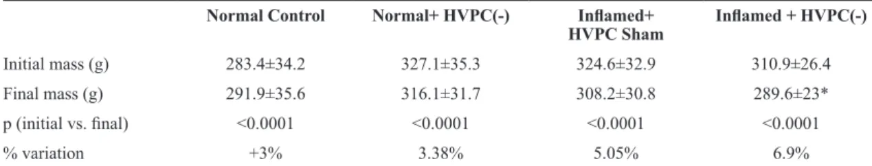
Body weight
In all groups, there was a difference between the initial and the inal weight. The three groups that received intervention presented weight loss, which was greater in the inlamed+HVPC(-) group (Table 1).
Pain
No signiicant differences were observed in PWL
between groups at any time-point (Table 2). Similarly,
no signiicant differences were observed in the PWL
92 Braz J Phys Ther. 2015 Mar-Apr; 19(2):89-96
differences by treatment or by time-point (p=0.09 and p=0.33, respectively).
Volume
All groups showed similar paw volume at baseline (p=0.65). There was a signiicant increase in paw volume 24 hours after the induction of inlammation compared with baseline in the inlamed+HVPC(-) and inlamed+HVPC placebo groups, and this difference was also observed at 48 hours. However, there was
no difference between these two groups in any of
the analyzed time-points. There were no differences by treatment (p=0.22) or by time-point (p=0.057).
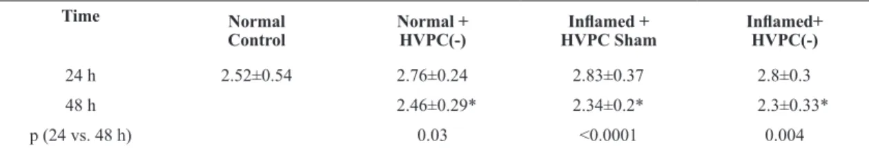
Albumin
There was no difference in serum albumin at 24 hours
between the groups (p=0.34). Serum albumin was lower at 24 than at 48 hours in the normal+HVPC(-), inlamed+HVPC placebo, and inlamed+HVPC(-) groups, with no difference between these groups (p=0.89; Table 3).
Histamine
Serum histamine was different between the groups
at 24 hours (p=0.006; Table 4). However, only the
normal+HVPC(-) and inlamed+HVPC placebo groups presented a signiicant increase in histamine compared
with NCG (p=0.001 and p=0.01, respectively). At 48 hours, a reduction in histamine was observed in
the three groups that received intervention (Table 4).
However, for the normal+HVPC(-) group, this difference was signiicant (p<0.0001) only when compared with
the value obtained at 24 hours (Table 4). There was no difference in serum histamine between the three
experimental groups at 48 hours (p=0.20; Table 4).
However, the inlamed+HVPC(-) group presented
the lowest histamine (Table 4).
Analysis of covariance
The analysis of the effect of HVPC(-) in the inlamed group on PWL revealed a positive association β=5.84 (p=0.08) compared with the normal+HVPC(-) group. This coeficient was adjusted by PWL measurements 24 hours after induction of inlammation, by histamine and albumin at 48 hours, and by the inal weight of
the experimental animals. The effect of HVPC(-) on
histamine presented a β=1.97 (p=0.42), similar to the inlamed+HVPC Sham group (β=1.84; p=0.43). These
results were adjusted by the histamine measurements
24 hours after induction of inlammation, and also by measurements of pain, inlammation, and albumin 48 hours after induction of inlammation and the inal
weight of the animals (Table 5).
Table 1. Comparison between initial and inal mass within each group of mice (mean±SD).
Normal Control Normal+ HVPC(-) Inlamed+
HVPC Sham Inlamed + HVPC(-)
Initial mass (g) 283.4±34.2 327.1±35.3 324.6±32.9 310.9±26.4
Final mass (g) 291.9±35.6 316.1±31.7 308.2±30.8 289.6±23* p (initial vs. inal) <0.0001 <0.0001 <0.0001 <0.0001
% variation +3% 3.38% 5.05% 6.9%
*p<0.05 compared to normal+ HVPC(-) % variation:[ (inal mass x 100)/initial mass)]. HVPC: High voltage electrical stimulation; p: statistical signiicance
Table 2. Paw withdrawal latency time over the course of 48 hours within each group of mice.
Test Time
Normal Control Normal + HVPC(-) Inlamed +
HVPC Sham
Inlamed + HVPC(-)
p (between groups)
0 26.5 [19-40]s 37.5 [18-64]s 25.5 [16-34]s 31.5 [24.5-51]s 0.25
6 24.5 [17.5-49.5]s 29 [17-58]s 0.83
24 28.5 [18-49]s 32.5 [20.5-50.5]s 0.45 48 31 [21-51]s 24.5 [15.5-32]s 32.5 [21.5-58.5]s 0.36
p (0 vs. 48 h) 0.80 0.89 0.85
Discussion
HVPC(-) applied at a submotor level 45 minutes per day for three consecutive days was not effective
in reducing pain or edema in the joint inlammation induced by ι-carrageenan. Furthermore, analysis of covariance, adjusted by PWL measured 24 hours after induction of inlammation, by the inal weight of the animal, and by albumin and histamine levels 48 hours after induction of inlammation, demonstrated a positive association (β=5.84) with borderline signiicance, indicating increased pain in the inlamed+HVPC(-)
group. These results are exciting because several of the parameters used are similar to those used in previous studies1,4-8,10,11,13-15, which allowed for comparisons.
Carrageenan injection is an inlammatory model frequently used in animals to assess the inlammatory
process and the effectiveness of drugs and physical
resources in the inlammation and pain treatment25,26. ι-Carrageenan sensitizes the primary afferents and
generates primary hyperalgesia in the injury site27.
Next, there is a high production of nitric oxide,
prostaglandins, free radicals, and cyclooxygenases that activate the dorsal horn neurons, generating a central sensitization, spinal or supraspinal, which
in combination with an increased sensitivity of peripheral nociceptors is manifested as a secondary hyperalgesia27. Using the same experimental model, Sluka et al.21 and Resende et al.26 showed reduced
primary hyperalgesia by applying high-frequency
transcutaneous electrical nerve stimulation (TENS) (100 Hz and 130 Hz, respectively), similar to that used in the present study. Our results show that the inlamed+HVPC placebo group presented the lowest PWL, and the inlamed+HVPC(-) group presented longer PWL, though without a signiicant difference.
These results indicate that HVPC(-) may increase the threshold of excitability in the primary afferent
neurons and help control the pain. A possible cause of the lack of signiicance in the PWL values could be the short duration of the HVPC pulse, which did not allow for the stimulation of the Aδ receptors or
inhibition of the medullary dorsal horn neurons26.
Table 3. Comparison of albumin (g/dL) between 24 and 48 hours within each group of mice.
Time Normal
Control
Normal + HVPC(-)
Inlamed + HVPC Sham
Inlamed+ HVPC(-)
24 h 2.52±0.54 2.76±0.24 2.83±0.37 2.8±0.3
48 h 2.46±0.29* 2.34±0.2* 2.3±0.33*
p (24 vs. 48 h) 0.03 <0.0001 0.004
Data are expressed as the mean ± SD. *p<0.05 compared to their respective values at 24 hours. HVPC: High voltage electrical stimulation. P: statistical signiicance
Table 4. Comparison of histamine (nmol/L) between 24 and 48 hours within each group of mice.
Time Normal
Control
Normal + HVPC(-)
Inlamed + HVPC Sham
Inlamed+ HVPC(-)
24 h 3.09±3.31 9.25±3.01* 8.34±3.9* 5.68±2.68
48 h 6.64±2.76** 5.16±2.62 3.9±1.15
p (24 vs. 48 h) <0.0001 0.18 0.30
Data are expressed as the mean ± SD. *p<0.05 compared to normal control; **p<0.01 compared to normal control. HVPC: high voltage electrical
stimulation; p: statistical signiicance.
Table 5. Analysis of covariance for dependent variables. Comparison group: Normal+HVPC(-).
Variable Inlamed + HVPC Sham Placebo Inlamed + HVPC(-)
β (p) β (p)
PWL time (s) 48 h --- 5.84 (0.08)
Volume (mL) 48 h --- -0.35 (0.24)
Albumin (g/dL) 48 h -0.09 (0.58) -0.18 (0.41) Histamine (nmol/L) 48 h 1.84 (0.43) 1.97 (0.42)
94 Braz J Phys Ther. 2015 Mar-Apr; 19(2):89-96
The inlammatory process induced by carrageenan
observed in the present study was similar to previous studies20,22,23,27. These studies reported the presence of
pain and edema in the irst 5 hours, which persisted for up to 24 hours and then decreased. In our study,
HVPC(-) did not promote greater reduction of edema
in the inlamed+HVPC(-) group. Accordingly, our
results are not in agreement with those obtained
in previous studies that demonstrated signiicant
differences in the reduction of trauma-induced edema in rats3-8,13. Perhaps the mechanisms of the induction of
inlammation (carrageenan versus trauma) inluenced
these different results.
The HVPC parameters used in the present study are the same as in previous studies regarding the type of current1,4-8,10,11,13-15, level of stimulation4-8,15, polarity1,4-8,10,11,13-15, and frequency, as well as the application of the stimulation immediately after
the induction of inlammation. The differences in
our protocol were the mechanism of injury and the duration of the current.
Previous studies3-8,13 have used a traumatic incident
to generate the inlammation, while the administration
of carrageenan was the choice in the current study.
Carrageenan induces inlammation and particularly
produces the presence of polymorphonuclear cells in
the synovial luid during the acute phase, followed by proliferation and iniltration of the synovial membrane; the number of cells in the synovial luid gradually decreases 24 hours after the inlammation
induction28. This model was chosen because it
produces a homogeneous pattern in the involvement of soft and joint tissues. This homogeneous pattern of
inlammation between the animals cannot be guaranteed
in models using mechanical trauma because the
joint is injured by hitting the paw, without ensuring the generation of a controlled joint inlammation. Furthermore, the carrageenan model is a validated model for studying the inlammatory process25,26. Our results indicate that HVPC(-) was not effective at controlling the inlammatory process induced by carrageenan, possibly due to the involvement of the cartilage and synovial tissue, as well the parameters used, which will be discussed below. Further studies
should be conducted to assess the articular cartilage
and the presence of circulating inlammatory cytokines, such as IL-6 and TNF-α.
The present study used a 45-minutes session of HVPC(-) on three consecutive days in an attempt to mimic what is performed in the clinical practice. Previous studies4,6-8 that reported a decrease in edema
applied three or four 30-minute applications on the
same day that the lesion was established (i.e. 1.5 to
2 hours per day) with a 30-minute rest between the applications. An increase in edema was observed in
the periods of non-intervention. Compared to our
study, these results indicate that the duration of the application can be critical in reducing edema, as proposed by Mendel and Fish29, who suggested the
use of longer applications during the acute stage of
inlammation. Future studies in humans should assess
the effect of the duration of the HVPC(-) application
on acute inlammation.
Albumin was reduced in the groups submitted to inlammation at 48 hours. The acute inlammatory phase generated by carrageenan is characterized by an increase in circulating cytokines, such as IL-1, IL-6 and TNFα, which produce two effects31. The irst effect is the increased microvascular permeability that allows a large loss of plasma proteins, which
accumulate in the interstitial space. The second effect is a reduced synthesis and release of negative
acute phase proteins, such as ferritin and albumin28.
These factors result in hypoalbuminemia, which was observed 48 hours after induction of inlammation, especially in the animals submitted to inlammation.
On the other hand, a less strong response was found in the animals from the normal+HVPC(-) group,
which can be explained by the reduction in plasma
volume and anesthesia, in agreement with the study conducted by Renkin et al.30. Another factor that may have inluenced the decrease in albumin was the weight
loss observed in the animals. There was no difference
in albumin level between the two inlammation groups,
which is consistent with previous studies addressing paw volume and indicates no effect of HVPC(-) on
the permeability of the endothelium. One possible
explanation is that the short pulse duration was not able
to generate plasma protein movement, as suggested by Mendel and Fish29.
Plasma histamine was selected due to its importance
as a mediator of acute inlammation. Furthermore,
the local administration of carrageenan produces a
systemic reaction in response to local inlammation,
which is consistent with an acute-phase response31.
Differences were observed in serum histamine between the inlamed+HVPC placebo group and normal+HVPC(-)
groups compared to the normal control group 24 hours
after the beginning of the experiment; the higher increase was observed in the normal+HVPC(-) group.
peaks 8 to 12 hours after the stimulation. The increase in histamine precursor depended on the intensity and duration of the stimulation32. This pro-inlammatory effect may be associated with the local stress produced by the current and with the metabolic changes at the
application site. Further studies are needed to conirm
these hypotheses.
However, the histamine levels observed in the animals from the inlamed+HVPC group were not signiicantly different from the other groups 24 and 48 hours after induction of inlammation. Similarly, the analysis of covariance did not reveal signiicant
differences (Table 5). This result indicates that during
the acute phase of the inlammatory process, HVPC(-)
was not able to regulate histamine release or affect the histamine binding sites in the postcapillary venule
endothelial cells, which is contrary to the hypothesis
proposed by Taylor et al.3. However, the histamine
levels observed in the inlamed+HVPC(-) group 24 and 48 hours after induction of inlammation were lower than the ones observed in the inlamed+placebo group, suggesting that electrical stimulation could help control inlammation even with no change in clinical parameters, such as edema. Therefore, further
studies should address more sensitive markers of
inlammation, such as TNFα or IL-1.
Clinical implications
Although the HVPC(-) was not effective, at a signiicance level of p<0.05, this current inluenced the
release of mediators such as histamine as well as the
PWL in animals submitted to HVPC(-). Furthermore,
HVPC(-) promoted an increase in histamine in normal
animals. However, it is necessary to analyze the
physiological implications of these results and also whether they also occur in humans.
Conclusions
HVPC(-) with the parameters used in this study and
applied in the acute phase of joint inlammation induced
by carrageenan can regulate histamine and increase
the PWL. However, it was not effective at reducing edema. Further studies are needed to validate the use of HVPC(-) in the treatment of acute inlammation.
References
1. GriffinJW, NewsomeLS, Stralka SW, Wright PE. Reduction of chronic posttraumatic hand edema: a comparison of high
voltage pulsed current, intermittent pneumatic compression,
and placebo treatments. Phys Ther. 1990;70(5):279-86. PMid:2185495.
2. NelsonRM, Hayes KW, Currier DP. Clinical electrotherapy.
USA: Appleton & Lange; 1999.
3. Taylor K, Mendel FC, FishDR, Hard R, Burton HW.
Effect of high-voltage pulsed current and alternating
current on macromolecular leakage in hamster cheek pouch microcirculation. Phys Ther. 1997;77(12):1729-40.
PMid:9413451.
4. Bettany JA, FishDR, Mendel FC. Influence of high voltage pulsed direct current on edema formation following impact injury. Phys Ther. 1990;70(4):219-24. PMid:2315384. 5. Taylor K, FishDR, Mendel FC, Burton HW. Effect of a single
30-minute treatment of high voltage pulsed current on edema
formation in frog hind limbs. Phys Ther. 1992;72(1):63-8.
PMid:1728050.
6. DolanMG, Graves P, Nakazawa C, Delano T, Hutson A, Mendel FC. Effects of ibuprofen and high-voltage electric stimulation on acute edema formation after blunt trauma to limbs of rats. J Athl Train. 2005;40(2):111-5. PMid:15970957.
7. DolanMG, Mychaskiw AM, Mendel FC. Cool-water immersion and high-voltage electric stimulation curb edema formation in rats. J Athl Train. 2003;38(3):225-30.
PMid:14608432.
8. Thornton RM, Mendel FC, FishDR. Effects of electrical stimulation on edema formation in different strains of rats. Phys Ther. 1998;78(4):386-94. PMid:9555921.
9. Karnes JL, Mendel FC, FishDR. Effects of low voltage pulsed current on edema formation in frog hind limbs following impact injury. Phys Ther. 1992;72(4):273-8. PMid:1584859.
10. FishDR, Mendel FC, SchultzAM, Gottstein-YerkeLM.
Effect of anodal high voltage pulsed current on edema
formation in frog hind limbs. Phys Ther. 1991;71(10):724
-30,discussion 730-3. PMid:1946611.
11. Taylor K, FishDR, Mendel FC, Burton HW. Effect of electrically induced muscle contractions on posttraumatic edema formation in frog hind limbs. Phys Ther. 1992;72(2):127
-32. PMid:1549633.
12. Cosgrove KA, AlonG, Bell SF, FischerSR, FowlerNR,
JonesTL, et al. The electrical effect of two commonly used clinical stimulators on traumatic edema in rats. Phys Ther. 1992;72(3):227-33. PMid:1584856.
13. Reed BV. Effect of high voltage pulsed electrical stimulation
on microvascular permeability to plasma proteins. A possible mechanism in minimizing edema. Phys Ther. 1988;68(4):491-5. PMid:2451258.
14. Mohr TM, Akers TK, LandryRG. Effect of high voltage stimulation on edema reduction in the rat hind limb. Phys Ther. 1987;67(11):1703-7. PMid:3499622.
15. Cook HA, Morales M, La RosaEM, DeanJ, Donnelly MK, McHugh P, et al. Effects of electrical stimulation on lymphatic flow and limb volume in the rat. Phys Ther. 1994;74(11):1040-6. PMid:7972365.
16. Evans TW. Review article: albumin as a drug—biological effects of albumin unrelated to oncotic pressure. Aliment Pharmacol Ther. 2002;16(Suppl 5):6-11. http://dx.doi.
org/10.1046/j.1365-2036.16.s5.2.x. PMid:12423448
17. Michel CC, Kendall S. Differing effects of histamine and
serotonin on microvascular permeability in anaesthetized
rats. J Physiol. 1997;501(Pt 3):657-62. http://dx.doi.
org/10.1111/j.1469-7793.1997.657bm.x. PMid:9218224 18. Omote K, Kawamata T, NakayamaY, Yamamoto H, Kawamata
96 Braz J Phys Ther. 2015 Mar-Apr; 19(2):89-96
prostaglandin receptor subtype EP4 on hyperalgesia and
inflammation in monoarthritic model. Anesthesiology.
2002;97(1):170-6.
http://dx.doi.org/10.1097/00000542-200207000-00024. PMid:12131119
19. Karges JR, Mark BE, Stikeleather SJ, Worrell TW. Concurrent validity of upper-extremity volume estimates: comparison of calculated volume derived from girth measurements and water displacement volume. Phys Ther. 2003;83(2):134-45. PMid:12564949.
20. Hargreaves K, DubnerR, Brown F, Flores C, JorisJ. A new and sensitive method for measuring thermal nociception in cutaneous hyperalgesia. Pain. 1988;32(1):77-88. http://
dx.doi.org/10.1016/0304-3959(88)90026-7. PMid:3340425 21. Sluka KA, Bailey K, Bogush J, OlsonR, RickettsA. Treatment
with either high or low frequency TENS reduces the
secondary hyperalgesia observed after injection of kaolin and carrageenan into the knee joint. Pain. 1998;77(1):97
-102. http://dx.doi.org/10.1016/S0304-3959(98)00090-6.
PMid:9755024
22. Gopalkrishnan P, Sluka KA. Effect of varying frequency,
intensity, and pulse duration of transcutaneous electrical
nerve stimulation on primary hyperalgesia in inflamed rats.
Arch Phys Med Rehabil. 2000;81(7):984-90. http://dx.doi.
org/10.1053/apmr.2000.5576. PMid:10896017
23. Sluka KA, Christy MR, Peterson WL, RuddSL, Troy SM.
Reduction of pain-related behaviors with either cold or heat
treatment in an animal model of acute arthritis. Arch Phys
Med Rehabil. 1999;80(3):313-7. http://dx.doi.org/10.1016/
S0003-9993(99)90143-0. PMid:10084440
24. Kaplan LA, Pesce AJ, Kazmierczak SC. Clinical Chemistry:
theory, analysis and correlation. 4th ed. St Louis: Mosby;
2003.
25. Ramírez C, RussoTL, DelfinoG, Peviani SM, Alcântara C, Salvini TF. Effect of tibiotarsal joint inflammation on gene expression and cross-sectional area in rat soleus muscle. Braz J Phys Ther. 2013;17(3):244-54. http://dx.doi.
org/10.1590/S1413-35552012005000084. PMid:23802230. 26. ResendeMA, Goncalves HH, Sabino GS, Pereira L, Francischi
J. Reduction in analgesic effect from low-frequency transcutaneous electrical nerve stimulation in morphine-tolerant rats. Rev Bras Fisioter.2006;10(3):291-6. http://
dx.doi.org/10.1590/S1413-35552006000300007.
27. Salvemini D, Wang ZQ, Wyatt PS, Bourdon DM, Marino MH, Manning PT, et al. Nitric oxide: a key mediator in the early and late phase of carrageenan-induced rat paw
inflammation. Br J Pharmacol. 1996;118(4):829-38. http://
dx.doi.org/10.1111/j.1476-5381.1996.tb15475.x. PMid:8799551
28. Santer V, Sriratana A, LowtherDA. Carrageenin-induced
arthritis: V. A morphologic study of the development of
inflammation in acute arthritis. Semin Arthritis Rheum.
1983;13(2):160-8. http://dx.doi.org/10.1016/0049-0172(83)90002-1. PMid:6673111
29. Mendel FC, FishDR. New perspectives in edema control via electrical stimulation. J Athl Train. 1993;28(1):63-74.
PMid:16558209.
30. RenkinEM, JoynerWL, Gustafson-Sgro M, Plopper G, Sibley
L. Albumin extravasation rates in tissues of anesthetized and
unanesthetized rats.J Appl Physiol (1985). 1989;66(5):2056
-60. PMid:2745274.
31. Cicala C, Morello S, AlfieriA, Vellecco V, Marzocco S, Autore
G. Haemostatic imbalance following carrageenan-induced rat paw oedema. Eur J Pharmacol. 2007;577(1-3):156-61.
http://dx.doi.org/10.1016/j.ejphar.2007.08.007. PMid:17850787
32. EndoY, Tabata T, Kuroda H, Tadano T, Matsushima K, Watanabe M. Induction of histidine decarboxylase in skeletal
muscle in mice by electrical stimulation, prolonged walking
and interleukin-1. J Physiol. 1998;509(Pt 2):587-98. http://
dx.doi.org/10.1111/j.1469-7793.1998.587bn.x. PMid:9575306
33. Watanabe M, Tabata T, Huh JI, Inai T, Tsuboi A, Sasaki K, et al. Possible involvement of histamine in muscular fatigue in temporomandibular disorders: animal and human studies. J Dent Res. 1999;78(3):769-75. http://dx.doi.org/10
.1177/00220345990780030901. PMid:10096452
Correspondence Carolina Ramírez
Universidade Industrial de Santander
Escola de Fisioterapia