O
RIGINALA
RTICLE Revista Brasileira de FisioterapiaCardiovascular responses to postural change
and aerobic capacity in middle-aged men and
women before and after aerobic physical training
Respostas cardiovasculares a mudança postural e capacidade aeróbia em
homens e mulheres de meia-idade antes e após treinamento físico aeróbio
Leite ST1, Martinelli FS2, Madruga VA3, Catai AM4, Gallo Junior L5, Chacon-Mikahil MPT6
Abstract
Objective: To compare the cardiovascular responses to passive postural maneuvers (tilt test) and the cardiorespiratory capacity in middle-aged men and women, before and after aerobic physical training. Methods: Seven men (44.6±2.1 years old) and seven women (51.7±4.8 years old) participated in aerobic physical training for 12 weeks. The tilt test protocol (fi ve minutes supine, ten minutes tilted at 70° and fi ve minutes supine) was followed, with arterial blood pressure and heart rate monitoring. A cycle ergometer protocol was used to measure cardiorespiratory capacity.
Results: In the sedentary condition, men showed greater parasympathetic infl uence in heart rate control, as demonstrated by their higher RR interval (iRR) during the tilt test. After training, the iRR values became more similar in the two groups, although the women had higher iRR in the supine position and the men continued to present higher iRR under tilted conditions. The women’s blood pressures continued to be higher after training, but heart rate tended to become similar in the two groups. The cardiorespiratory capacity patterns in the two groups were similar after training. Except for absolute heart rate values, for which there were no differences between the groups, the men’s values were higher than those of the women for all other variables. It was also observed that, after the training, the women’s blood pressures were signifi cantly lower, even though their pressures remained higher than the men’s. Conclusions: The training seemed to reduce the women’s arterial blood pressure levels and improve both groups’ cardiorespiratory capacity, but the men continued to present better performance than the women.
Article registered in the Australian New Zealand Clinical Trials Registry (ANZCTR) under the number 12608000085370.
Key words: Aerobic training; cardiovascular autonomic activity; orthostatic stress; gender; middle age; cardiorespiratory capacity.
Resumo
Objetivo:Comparar respostas cardiovasculares a Manobra Postural Passiva (Tilt Test) e capacidade cardiorrespiratória em homens e mulheres de meia–idade antes e após treinamento físico aeróbio. Materiais e métodos:Sete homens – GH (44,6±2,1 anos) e sete mulheres – GM (51,7±4,8
anos), participaram de treinamento físico aeróbio por 12 semanas. Foi realizado protocolo de Tilt Test (cinco minutos supino, dez minutos inclinado 70º, cinco minutos supino), com monitoração da pressão arterial e freqüência cardíaca. Para mensuração da capacidade cardiorrespiratória foi realizado protocolo em cicloergômetro. Resultados: Na condição sedentária, GH mostrou maior infl uência parassimpática no controle da freqüência cardíaca evidenciada por maior intervalo RR (iRR) durante Tilt Test. Na condição treinada, os valores de iRR de ambos os grupos se assemelham, tendo as mulheres iRR maior em supino, mas na inclinação os homens mantêm iRR mais elevado. Para pressão arterial, as mulheres permanecem com valores superiores após treino, mas a freqüência cardíaca tende a se assemelhar em ambos. Já na capacidade cardiorrespiratória, homens e mulheres têm um padrão de comportamento semelhante após treino. Com exceção dos valores absolutos da freqüência cardíaca, sem diferenças entre os grupos, para todas as outras variáveis os homens obtiveram valores superiores aos das mulheres. Observa-se ainda que, após o treinamento, houve redução signifi cativa dos valores de pressão arterial no GM, mesmo continuando superiores aos dos homens.Conclusão:O treinamento parece ter reduzido os níveis pressóricos nas mulheres, além de serem observadas melhorias na capacidade cardiorrespiratória de ambos os grupos, permanecendo os homens com melhor desempenho do que as mulheres.
Artigo registrado na Australian New Zealand Clinical Trials Registry (ANZCTR) sob o número 12608000085370.
Palavras-chave: treinamento aeróbico; atividade autonômica cardiovascular; estresse ortostático; gênero; meia-idade; capacidade car-diorrespiratória.
Received: 28/12/2007 – Revised: 30/05/2008 – Accepted: 06/08/2008
1 School of Physical Education, Universidade Estadual de Campinas (Unicamp) – Campinas (SP), Brazil 2 Escola Superior de Educação Física de Jundiaí (ESEFJ) – Jundiaí (SP), Brazil
3 Department of Adapted Physical Activity, School of Physical Education (Unicamp)
4 Department of Physical Therapy, Biological and Health Science Center (CCBS-UFSCar) – São Carlos (SP), Brazil
5 Laboratory of Exercise Physiology, Hospital das Clínicas, School of Medicine, Universidade de São Paulo (USP) – Ribeirão Preto (SP), Brazil 6 Department of Science of the Sport, School of Physical Education (Unicamp)
Introduction
Aging is a natural process characterized by a reduction of the physiological functions and responses, particularly from the third decade of life onwards, when a progressive decline of the functional capacity takes place, the speed of which depends on the individual’s genetic factors, health conditions, and lifestyle1.
h ese changes caused by aging increase the incidence of cardiovascular disease in both men and women. An important parameter to analyze the dif erences between the genders occurs in middle-age, when women go through menopause and some of them begin hormone replacement therapy (HRT), which has been associated with a reduction in the incidence of cardiovascular disease2-4, as aging in itself can be considered
a risk factor that contributes to cardiovascular morbidity and death5.
h ere are indications that the cardiovascular dif erences between men and women up to middle-age tend to decrease with time and become minimal after age 50. When elderly men and women (67 years old on average) are compared, there are no statistical dif erences in the parameters for heart rate variability analysis5.
A comparison between men and women, middle-aged and older, found an incidence of 33.6 and 40.6%6 respectively,
showing that women are more susceptible to cardiovascular disease5,7, although men have a higher mortality rate due to
sympathetic dominance7-9.
For the cardiovascular system (CVS) to function properly, a rei ned control system must be ef ective in order to carry out the necessary adjustments. All this complexity, both at rest and during exercise, is ei ciently controlled by neural and neurohormonal mechanisms that respond to the af erent and ef erent stimuli mediated by autonomic innervations10.
h e autonomous nervous system (ANS) is responsible for extrinsically controlling the CVS, adjusting its functions. An example of that control is observed in the alterations that occur in HR and BP, which are non-invasive, easily measurable variables, and provide an enormous amount of information on body performance in several situations, such as a response to postural changes and respiratory maneuvers (valsalva maneuver, apnea, respiratory sinus arrhythmia) as well as physical exercise10.
Kuo et al.11 report that middle-aged women have larger
parasympathetic dominance and smaller sympathetic
dominance when compared to men3. Nevertheless, women
over 50 suf er a decrease in the parasympathetic modulation, and these dif erences tend to lessen with age11-14.
Thus, although aging manifests itself in the responses
of functional variables, exercise can diminish the
consequences of a slower physiological rhythm1. Regular
exercise, especially aerobic exercise or aerobic physical training (APT), can change the control of the ANS over the CVS, raising parasympathetic activity with or without the reduction of sympathetic activity. This combined with changes in heart rate (HR) result in bradycardia, both at rest15-17 and during submaximal physical activity, possibly
due to increased parasympathetic response and a reduction in the sympathetic “discharge” 18,19.
In response to stressful stimuli such as exercise, postural and respiratory maneuvers trigger adaptations in the sympathetic/ parasympathetic modulation over the sinus node at rest and an increase in the baroceptor rel ex20,21.
It is widely known that autonomic modulation is inl uenced by the physical i tness, age, and gender, among other factors. However, it must be noted that there is still controversy over the changes that result from aging according to the gender. Ghorayeb et al.5 report that the blood pressure (BP) of women
before the onset of menopause is lower than that of men, while postmenopausal women have higher systolic and diastolic blood pressure values (SBP and DBP, respectively), especially those undergoing HRT due to a rise in angiotensinogenlevels.
In contrast, the study by Laitinen et al.22, which evaluated
the inl uence of gender in cardiovascular autonomic responses, found that in the supine position women had smaller mean blood pressure (MBP) values than men, whereas in the passive postural maneuver (Tilt Test) there was a signii cant increase in this variable in women. It is interesting to note that in the variables for heart rate variability (HRV), SPB variability and in cardiovagal barorel ex sensitivity, there were no signii cant dif erences between genders. Also, regarding the autonomic variables, Kuo et al.14 describe in their study that middle-aged
women have a parasympathetic dominance in HR regulation, whereas in men this occurs through the sympathetic control.
After analyzing all of these aspects, the need for more studies becomes evident, especially studies on the inl uence of gender on the response to exercise during the process of aging. h erefore, the objective of this study was to compare the cardiovascular responses to passive postural maneuver and the cardiorespiratory capacity of middle-aged men and women in both moments, that is, before and after APT.
Methods
Subjects
Fourteen middle-aged subjects were studied: seven men (MG) and seven postmenopausal women (WG) receiving HRT. All participants answered the anamnesis and the
questionnaire on personal details, history of illnesses and lifestyle. They were also duly informed about all procedures and stages of this research before undergoing clinical, heart and biochemical tests to determine their health condition. Due to methodological limitations in other stages of the data collection, the sample of the present study reflects the group whose data were collected at sedentary and trained moments (SED and TRA) respectively.
Criteria for inclusion and exclusion
The subjects included in this investigation were aged 40 to 60 years old and had not taken part in any physical activity program for at least six months prior to the investigation. They voluntarily agreed to do the proposed physical training and the pre and post-training evaluations according to the criteria presented in the free and informed consent agreement approved by the Committee of Ethics in Research of the FCM-Unicamp (appendix of approvals 227/94 and 228/94).
The women were postmenopausal (no menstrual period for at least 12 consecutive months) and were receiving HRT. Subjects were excluded from this research if they exercised regularly, their clinical evaluation and/or test results uncovered any pathology or complication that might constitute a risk factor to the proposed regular physical activity, or if they were taking medication that could interfere with the physiological responses to the tests.
Experimental protocols
Subjects were evaluated before and after the APT, in two experimental sessions. All tests were carried out in a quiet environment at 22 to 24ºC. The subjects were instructed to avoid physical exercise in the 48 hours prior to the experiment, and any medication or caffeinated drinks or foods for at least 12 hours before the tests.
In the first session, the total body mass (kg) and height (m) were measured by a Filizola scale (model ID-1500, São
MG WG
N 7 7
Age (years) 45 53*
Body Mass (kg) 85 62*
Height (m) 1.80 1.60
SBP (mmHg) 120 120
DBP (mmHg) 80 85
HR (bpm) 68 72
Table 1. Median values for anthropometric and rest hemodynamic variables of the studied groups.
*p<0.05 MG=men’s group; WG=women’s group; SBP=systolic blood pressure; DBP=diastolic blood pressure; HR-heart rate.
Paulo, Brazil), with 100g accuracy and a metallic height rod, according to the procedures described by Gordon, Chumlea, Roche23, as shown in Table 1.
Next, the Tilt Test was performed by measuring HR (Funbec ECG electrocardiograph) and BP (mercury column manometer) to verify the hemodynamic responses during postural change. A previous inclination was performed to minimize stress levels caused by the maneuver. After a five-minute measurement in the supine position, a 70º tilt was carried out and sustained for ten minutes, or until the subject showed any symptom that contraindicated the maintenance of that position.
The BP and the HR were checked at one-minute intervals during the five minutes that preceded the tilting (in the supine position), during the ten-minute maneuver, and during the five minutes after the subject returned to the initial position. The electrocardiogram recorded during the Tilt Test was used to calculate the intervals between the R
waves or iRR, which is the distance between the peaks of
adjacent R waves on the tracing of the first minute of each phase of the Tilt Test protocol, in ten-second sequences.
The second experimental session consisted of cardiorespiratory evaluation during dynamic physical exercise on the cycle ergometer (Quinton Inst Co, Corival 400, Groningen, the Netherlands). During the protocol, the subject was connected to the gas analyzer (Sensormedics
, MMC Horizon System, Yorba Linda, CA, USA). The cardiorespiratory capacity was determined by the peak values of oxygen uptake O2, ventilation ( E), HR and power
(POW) reached at physical exhaustion, as well as the values of these variables corresponding to the anaerobic threshold (AT). These were detected as the first point of inflection of the CO2 ( CO2) and ventilation ( E) production curves,
i.e. where there is a drop in the linearity of these variables compared to linear increment of oxygen uptake ( O2)24.
The protocol for the cycle ergometer evaluation was standardized to obtain physiological variables in response to continuous increments until exhaustion, ranging from 10-17W/minutes, for the WG and MG, respectively. The initial warm-up power was low (≅4watts) for a period of two
Physical training protocol
After the evaluations were carried out, the subjects started a supervised 12-week APT program with three weekly sessions of approximately 60 minutes each. h ey began with a walk on a level surface (70% of MaxHR), then progressed to a trot and a more vigorous walk on an inclined surface, until reaching an individual HR peak of 85%. h e researchers monitored weekly attendance, and the sample attended at least 85% of the sessions.
Statistical analysis
Based on the table of results, we applied descriptive statistics and analytical statistics to the data for each group. h e Mann-Whitney test was used for intergroup analysis. Friedman with Wilcoxon’s post-hoc was also used, as well as the Bonferroni
correction for the Tilt Test analyses. h e level of signii cance adopted for all analyses was p<0.05.
Results
h e results obtained during the two moments of collection, before and after the 12 weeks of APT are represented as SED (GHSED and GMSED), and TRA (GHTRA and GMTRA) for both studied groups.
Hemodynamic behavior during Postural Change
(Tilt)
Figures 1 and 2 show the SBP, DBP, and HR behavior before and after the APT for the MG and WG, respectively. h e values
Figure 1. Minute to minute changes (boxplot) of SBP(A), DBP(B) and HR(C) at each stage of TILT, in SED condition for MG and WG groups.
DBP SED M DBP SED W 40
50 60 70 80 90 100 110
MG AND WG SED Median; Box: 25%, 75%; Whisker: Min, Max
SBP SED M SBP SED W 80
90 100 110 120 130 140 150 160
HR SED M HR SED W 50
60 70 80 90 100 110 120 130
BEFORE TILT TILT AFTER TILT
*
*
*
*
*
*
*p<0,05 MG vs. WG.
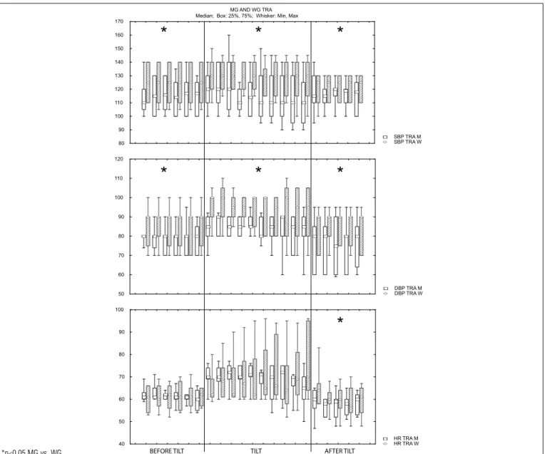
for the SBP and DBP in the MG are signii cantly lower, both in the pre and post-training conditions. h e values for HR, however, were signii cantly lower in the MG in the supine position, pre and post Tilt in the SED condition, and in post Tilt in the TRA condition.
h e behavior of the iRR in the MG versus the WG, in both SED and TRA conditions, can be seen in Figures 3 and 4.
h e MG shows an increased iRR when compared to the WG
throughout most of the Tilt protocol both before and after training, indicating a greater bradycardia.
Analysis of the cardiorespiratory variables on the
cycle ergometer
Table 2 displays the behavior of the cardiorespiratory variables obtained during the continuous cycle ergometer protocol, before and after the APT, for the MG and the WG. It is possible to verify that men had higher values at the aerobic threshold (AT) and at peak of exercise (PEAK) in both SED and TRA condition (*p<0.05) compared to women. It is important to highlight that only the HR (AT and PEAK) was not statistically dif erent between the two groups in the moments of collection.
Discussion
h e present study had as its main focus the verii cation of possible dif erences between the behaviors of cardiovascular and respiratory variables of middle-aged men and women, in dif erent training conditions. h e occurrence of bradycardia at rest is evident in both groups after the aerobic training sessions (Figures 1 and 2), which could indicate a greater parasympathetic participation and/or a lower sympathetic stimulation.
During the postural change, quick cardiovascular adjustments must take place to maintain cardiac output as gravity works against venous return, which could cause a reduction in systolic volume. Both the MG and the WG showed an increase in HR as a response to the maneuver, agreeing with
Montano et al.25, who described the need for HR increase in the
tilted position during the Tilt Test caused by the sympathetic stimulation associated with the vagal withdrawal over the sinus node.
Nonetheless, it should be noted that, in the SED moment, the MG showed greater HR values than the WG, both in the supine position and at inclination. Similar HR results in the SED condition are also found in the study carried out by Shoemaker et al.26 during an inclination protocol at 60º. Although the study
was conducted with a younger population (mean age 30±11 years for men and 26±6 years for women), both HR and MBP were higher in the female group.
With regard to BP, our protocol demonstrated that women have higher values than men of the same age group during the entire Tilt Test protocol. h is corroborates the i ndings of Laitinen et al.22, who carried out a Tilt Test protocol at 70º
with individuals of dif erent age groups (23 to 77 years old) for only i ve minutes. Although the MG showed a higher SBP (ns) after APT in the supine stages of the protocol, these values are still lower than those shown by the WG, despite the latter’s signii cant SBP reduction in the TRA condition.
Bigger-Jr et al.27 reported that the passive postural
maneuver causes a reduction in the high frequency component (parasympathetic indicator) and a substantial increase in the sympathovagal balance, obtained through the spectral analysis of the iRR. h is would indicate that during the maneuver there is vagal inhibition and an increase of the sympathetic stimulation. Although the present study did not analyze the values of the spectral variables, the iRR obtained in time series during the i rst minute rel ect the autonomic modulation that takes place in both conditions.
Montano et al.25 reported that iRR is lower during the
active postural maneuver as this kind of maneuver also allows greater involvement by the central nervous system, as well as a more ef ective inl uence of muscular and respiratory activity as a rel ective action of stress control. However, our protocol was deliberately carried out with a passive postural maneuver to eliminate possible external inl uences over cardiovascular neural control. It was possible to observe that the individuals of both genders in the TRA condition had changes in the behavior
HR (bpm) E(L/min) O2(L/min) CO2(L/min) O2 (mL/kg/min) POW (W)
SED TRA SED TRA SED TRA SED TRA SED TRA SED TRA
AT MGWG 115105 118108 33.318.9* 42.721.6* 1.210.63* 0.68*1.44 0.56*0.99 0.60*1.36 13.99.6* 10.4*17.3 33*82 10545*
PEAK MG 160 155 93.5 103 2.39 2.41 2.17 2.86 29.0 30.4 168 190
WG 152 157 46* 59.2* 1.16* 1.25* 1.40* 1.58* 16.8* 19.5* 78* 97*
Table 2. Median values for cardiorespiratory variables at anaerobic threshold (AT) and at peak of exercise (PEAK) of both groups, in SED and TRA conditions.
Figure 2. Minute to minute changes (boxplot) of SBP(A), DBP(B) and HR(C) at each stage of TILT, in TRA condition, for MG and WG groups.
80 90 100 110 120 130 140 150 160 170
DBP TRA M DBP TRA W 50
60 70 80 90 100 110 120
Median; Box: 25%, 75%; Whisker: Min, Max MG AND WG TRA
SBP TRA M SBP TRA W
HR TRA M HR TRA W 40
50 60 70 80 90 100
BEFORE TILT TILT AFTER TILT
*
*
*
*
*
*
*
*p<0.05 MG vs. WG.
Figure 3. Changes in iRR (boxplot) during the 1st minute at each stage of TILT, in SED condition, for MG and WG groups.
iRR TILT TEST MG AND WG SED Median; Box: 25%, 75%; Whisker: Min, Max
iRR iSED M iRR iSED W
10 30 50 p10 p30 p50 10po 30po 50po
seg 400
500 600 700 800 900 1000
BEFORE TILT TILT AFTER TILT
-*
* * * *
*
* * * * *
*
*
*
*p<0.05 MG vs. WG.
of the iRR during the Tilt Test. In the test, both the MG and the WG reduced the iRR compared to the SED condition, although the MG maintained higher values compared to the WG in both conditions.
Changes in the cardiorespiratory system after aerobic training are very well documented in the literature as physically i t individuals tend to have a lower HR at rest due to intrinsic modulations, greater parasympathetic activity, or reduction in sympathetic activity21. Nevertheless, it is not possible to
conclude that these changes are direct consequences of the physical training because there is a number of physiological changes that result from better aerobic conditioning and involve dif erent systems at various levels28.
It is also known that individuals with better aerobic conditioning have a more efficient autonomic control than those who are sedentary28, both young and old29. Less active
lifestyles also seem to play a critical role on the appearance and progression of cardiovascular (or related) diseases.
Interestingly, Rennie et al.30 compared genders and found
that men have a lower HR at rest in response to moderate and/or intense training. Women however only showed a reduction in HR at rest after intense training. In the present study, we observed a reduction in HR at rest in the WG (p<0.05) after the 12 weeks of aerobic training at moderate intensity (70 to 85%).
Overall, the present study showed improvements in the post-training condition for both men and women in all the
analyzed cardiorespiratory variables at the AT and at PEAK. Only the peak HR showed a non-significant reduction for both groups.
Conclusions
Through the analysis of the results, it can be concluded that the MG in the sedentary condition showed a greater parasympathetic influence on HR control, made evident by the greater iRR during the entire Tilt Test protocol (supine pre-tilt, tilt, supine post-tilt). In the TRA condition, the iRR values of both groups were similar. The women had greater iRR in the supine position, while the men had greater iRR during the stress of inclination of the Tilt Test. As for the BP values, both in the SED and in the TRA conditions, the women showed higher values than the men. The HR values were similar for both groups in the same condition during the inclination of the Tilt Test protocol, unlike the smaller supine-iRR values in the MG. This may confirm a lower autonomic responsiveness to stress for men in this age group. With regard to the cardiorespiratory capacity, the men had a similar behavioral pattern to the women after training, but higher values for all other variables except the absolute HR, both at AT and PEAK. It was also observed that after 12 weeks of APT there was a substantial reduction in the BP values in the WG, even though they were greater
Figure 4. Changes in iRR (boxplot) during the 1st minute at each stage of TILT, in TRA condition, for MG and WG groups.
Median; Box: 25%, 75%; Whisker: Min, Max
iRR TRA W 10 30 50 p10 p30 p50 10po 30po 50po
seg 400
500 600 700 800 900 1000 1100
Pré-Tilt Tilt Pós-Tilt
iRR TILT TEST MG AND WG TRA
iRR TRA M 10 30 50 p10 p30 p50 10po 30po 50po
seg 400
500 600 700 800 900 1000 1100
BEFORE TILT TILT AFTER TILT
*
* * * * *
* * * * * *
* * * *
1. Pollock ML, Dawson GA. Physiologic responses of men 49 to 65 years of age to endurance training. J Am Geriatr Soc. 1976;24(3):97-104.
2. Haddock BL, Marshak HPH, Mason JJ, Blix G. The effect of hormone replacement therapy and exercise on cardiovascular disease risk factors in postmenopausal women. Sports Med. 2000;29(1):39-49.
3. Liu CC, Kuo TB, Yang CC. Effects of estrogen on gender-related autonomic differences in humans. Am J Physiol Heart Circ Physiol. 2003;285(5):H2188-93.
4. Rosano GM, Vitale C, Fini M. Hormone replacement therapy and cardioprotection: what is good and what is bad for the cardiovascular system? Ann. N.Y. Acad Sci. 2006;1092:341-8.
5. Ghorayeb N, Baptista CA, Dioguardi GS, Reginatto LE. Atividade física na mulher. Rev Soc Cardiol Est São Paulo, SOCESP.1996;6:540-2.
6. Kannel WB, Hjortland MC, McNamara PM, Gordon T. Menopause and risk of cardiovascular disease: the Framingham study. Ann Intern Med. 1976;85(4):447-52.
7. Tank J. Does aging cause women to be more sympathetic than men? Hypertension. 2005;45(4):489-90.
8. Kuttenn F, Gerson M. Hormone replacement therapy of menopause, heart and blood vessels. Arch Mal Coeur Vaiss. 2001;94(7):685-9.
9. Vanoli E, De Ferrari GM, Stramba-Badiale M, Hull SS Jr, Foreman RD, Schwartz PJ. Vagal stimulation and prevention of sudden death in conscious dogs with a healed myocardial infarction. Circ Res. 1991;68(5):1471-81.
10. Smith JJ, Kampine JP. Regulation of arterial blood pressure. In Smith JJ, Kampine JP, editores. Circulatory physiology – the essentials. 3ªed. Baltimore: Williams & Wilkins; 1990.
11. Kuo TB, Lin T, Yang CC, Li CL, Chen CF, Chou P. Effect of aging on gender differences in neural control of heart rate. Am J Physiol.1999;277(6 Pt 2):H2233-9.
12. Evans JM, Ziegler MG, Patwardhan AR, Ott JB, Kim CS, Leonelli FM et al. Gender differences in autonomic cardiovascular regulation: spectral, hormonal, and hemodynamic indexes. J Appl Physiol. 2001;91(6):2611-8.
13. Pikkujämsä SM, Mäkikallio TH, Airaksinen KE, Huikuri HV. Determinants and interindividual variation of R-R interval dynamics in healthy middle-aged subjects. Am J Physiol Heart Circ Physiol. 2001;280(3):H1400-6.
14. Neves VF, Silva de Sá MF, Gallo L Jr, Catai AM, Martins LE, Crescêncio JC et al. Autonomic modulation of heart rate of young and postmenopausal women undergoing estrogen therapy. Braz J Med Biol Res. 2007;40(4):491-9
15. American College of Sports Medicine. ACSM’s guidelines for exercise testing and prescription /ACSM. Philadelphia: Lippincott Williams & Wilkins; 2005.
16. Maciel BC, Gallo Júnior L, Marin Neto JA, Lima Filho EC, Terra Filho J, Manço JC. Parasympathetic contribution to bradycardia induced by endurance training in man. Cardiovasc Res. 1985;19(10):642-8.
17. Martinelli FS. Respostas da freqüência cardíaca e da pressão arterial sistêmica às manobras postural passiva e de valsalva, em indivíduos sedentários e atletas corredores de longa distância [dissertação]. Campinas: Unicamp; 1996.
18. Goldsmith RL, Bigger JT Jr, Steinman RC, Fleiss JL. Comparison of 24-hour parasympathetic activity in endurance-trained and untrained young men. J Am Coll. Cardiol. 1992;20(3):552-8.
19. Shin K, Minamitani H, Onishi S, Yamazaki H, Lee M. Autonomic differences between athletes and nonathletes: spectral analysis approach. Med Sci Sports Exerc. 1997;29(11):1482-90.
20. Seals DR, Taylor JA, Ng AV, Esler MD. Exercise and aging: autonomic control of the circulation. Med Sci Sports Exerc.1994;26(5):568-76.
21. Chacon-Mikahil MPT. Estudo da variabilidade da freqüência cardíaca nos domínios do tempo e da freqüência antes e após o treinamento aeróbio em homens de meia idade [tese], Campinas: Unicamp; 1998.
22. Laitinen T, Niskanen L, Geelen G, Länsimies E, Hartikainen J. Age dependency of cardiovascular autonomic responses to head-up tilt test in healthy subjects. J Appl Physiol. 2004;96(6):2333-40.
23. Gordon CC, Chumlea WC, Roche AF. Stature, Recumbent Length, Weight. In: Lohman TG et al., editores. Anthropometric standardizing reference manual. Champaign, Illinois: Human Kinetics Books; 1988, p.3-8.
24. Wasserman K, Whipp BJ, Koyl SN, Beaver WL. Anaerobic threshold and respiratory gas exchange during exercise. J Appl Physiol. 1973;35(2): 236-43.
25. Montano N, Ruscone TG, Porta A, Lombardi F, Pagani M, Malliani A. Power spectrum analysis of heart rate variability to assess the changes in sympathovagal balance during graded orthostatic tilt. Circulation.1994;90(4):1826-31.
26. Shoemaker JK, Hogeman CS, Khan M, Kimmerly DS, Sinoway LI. Gender affects sympathetic and hemodynamic response to postural stress. Am J Physiol Heart Circ Physiol. 2001;281(5):H2028-35.
References
than those of the MG in both conditions. Finally, the analysis of the Tilt Test protocol revealed a decrease in the iRR and
a change in the BP and in the instantaneous HR values of both groups.
27. Bigger JT Jr, Fleiss JL, Steinman RC, Rolnitzky LM, Kleiger RE, Rottman JN. Correlations among time and frequency domain measures of heart period variability two weeks after acute myocardial infarction. Am J Cardiol. 1992;69(9):891-8.
28. Almeida MBE, Araújo CGS. Effects of aerobic training on heart rate. Rev Bras Med Esporte. 2003;9(2):113-20.
29. Yataco AR, Fleisher LA, Katzel LI. Heart rate variability and cardiovascular fi tness in senior athletes. Am J Cardiol. 1997;80(10):1389-91.