118
POWERLIFTING SESSIONS PROMOTE SIGNIFICANT
POST-EXERCISE HYPOTENSION
SESSÕES DE POWERLIFTING PROMOVEM HIPOTENSÃO SIGNIFICATIVA PÓS-EXERCÍCIO
SESIONES DE POWERLIFTING PROMUEVEN HIPOTENSIÓN SIGNIFICATIVA POST-EJERCICIO
Gustavo Allegretti João1,2
(Profissional de Educação Física) Danilo Sales Bocalini1
(Profissional de Educação Física) Daniel Rodriguez1
(Profissional de Educação Física) Mario Augusto Charro2
(Profissional de Educação Física) Fábio Ceschini1
(Profissional de Educação Física) Antônio Martins1
(Profissional de Educação Física) Aylton Figueira Junior1
(Profissional de Educação Física)
1. Universidade São Judas Tadeu, Departamento de Educação Física e Ciências do Envelhecimento, Laboratório de Fisiologia Translacional, São Paulo, SP, Brasil. 2. Faculdades Metropolitanas Unidas – FMU, Laboratório de Fisiologia do exercício e desempenho humano, São Paulo, SP, Brasil.
Correspondência: Rua Taquari, 546, Mooca, São Paulo, SP, Brazil. 03166-000. [email protected]
ORIGINAL ARTICLE
ARTIGO ORIGINAL
ARTÍCULO ORIGINAL
ABSTRACT
Introduction: Powerlifting (PWL) is a worldwide method, frequently used in resistance training programs. However, the relationship between cardiovascular responses and PWL is still unclear in the literature. Objective: To evaluate acute cardiovascular overload and post-exercise hypotension (PEH) after acute powerlifting exercise session in subjects with experience in the modality. Methods: Nine powerlifting athletes (34 ± 5 years) participated voluntarily in this study. The following exercises were used in the session: squat, bench press and deadlift (95% of 1 RM, 2 to 5 repetitions). The anthropometric parameters and blood pressure (systolic, diastolic and mean) were evaluated immediately, 5’, 10’, 30’, 60’ and 24 hours after the exercise session with a non-invasive automatic pres-sure monitor. Results: Significant differences (p<0.05) were found between rest and immediately after exercise on systolic (135 ± 6 vs. 153 ± 10 mmHg) and mean (102 ± 3 vs. 108 ± 3 mmHg) blood pressures, but no difference was found at diastolic (85 ± 3 vs. 85 ± 4 mmHg) blood pressure. Additionally, the increase in systolic pressure did not reach values considered as a risk of cardiovascular overload. Significant PEH was found after 60 minutes (systolic: -12 ± 12%, diastolic: -5 ± 6% and mean: -7 ± 5%) and 24 hours after PWL session (systolic: -5 ± 4%, diastolic: -8 ± 4% and mean: -7 ± 3%). Conclusion: Our data demonstrated that a PWL session does not increase systolic blood pressure up to the risk range and promotes PEH after 60 minutes of exercise and that this cardiovascular response persisted after 24 hours post-exertion in powerlifting athletes.
Keywords: hypotension; resistance training; exercise.
RESUMO
Introdução: O levantamento de peso básico ou powerlifting (PWL) é um método frequentemente utilizados em pro-gramas de treinamento resistido em todo o mundo. Contudo, a relação das respostas cardiovasculares e PWL ainda não está clara na literatura. Objetivo: Avaliar a sobrecarga cardiovascular aguda e a hipotensão pós-exercício (HPE) depois de uma sessão aguda de exercícios de PWL em indivíduos com experiência na modalidade. Métodos: Nove atletas de PWL (34 ± 5 anos) participaram voluntariamente deste estudo. Os seguintes exercícios foram realizados na sessão: agacha-mento, supino e levantamento terra (95% de 1 RM, 2 a 5 repetições). Os parâmetros antropométricos e a pressão arterial (sistólica, diastólica e média) foram avaliados imediatamente, 5’, 10’, 30 ‘, 60’ e 24 horas após a sessão de exercício com um monitor automático de pressão não invasivo. Resultados: Foram encontradas diferenças significativas (p < 0,05) de pressão sistólica (135 ± 6 vs. 153 ± 10 mmHg) entre o repouso e imediatamente após o exercício e na média (102 ± 3 vs. 108 ± 3 mmHg), porém, não se verificou nenhuma diferença na pressão diastólica (85 ± 3 vs. 85 ± 4 mmHg). Além disso, o aumento da pressão sistólica não atingiu valores considerados risco de sobrecarga cardiovascular. Constatou-se HPE significativa 60 minutos (sistólica: -12 ± 12%, diastólica: -5 ± 6% e média: -7 ± 5%) e 24 horas (sistólica: -5 ± 4%, diastólica: -8 ± 4 % e média: -7 ± 3%) depois da sessão de PWL. Conclusão: Nossos dados demonstraram que uma sessão de PWL não aumenta a pressão sistólica até a faixa de risco e promove HPE após 60 minutos de exercício e que essa resposta cardiovascular persistiu 24 horas pós-esforço nos atletas de powerlifting.
Descritores: hipotensão; treinamento de resistência; exercício.
RESUMEN
119
Article received on 07/15/2016 accepted on 10/19/2016 DOI: http://dx.doi.org/10.1590/1517-869220172302166667
sistólica hasta el rango de riesgo y promueve HPE después de 60 minutos de ejercicio y que la respuesta cardiovascular se mantuvo 24 horas después del ejercicio en atletas de powertlifting.
Descriptores: hipotensión; entrenamiento de resistencia; ejercicio.
INTRODUCTION
Scientific evidences suggest that regular resistance exercise (RE) is an important strategy to control systemic blood pressure, in both nor-motensive and hypertensive subjects1,2. Among the effects of physical
activity on the cardiovascular system, post-exercise hypotension (PEH) has been studied in hypertensive subjects with clinically relevant impli-cations3-5. There are several studies utilizing RE to promote BP reduction
at acute4,5 and chronic6 approaches. The mechanisms involved at PEH
have been attributed to reduced peripheral vascular resistance, reduced sympathetic activity, and diminished systolic volume and changes in the sensitivity of adrenergic cardiac and endothelial factors7-9.
The American College of Sports and Medicine1 and the American
Heart Association2 stated that RE in association with an aerobic based
exercise program are efficient to prevent, treat and control the arterial hy-pertensive, however, the RE and PEH effect still unclear4. In 2015 our group5
demonstrated that PHE occur independently of exercise intensity without expressed cardiovascular overload during the session, however, to the best of our knowledge, there are few study5,10-12 on literature evaluating different
manipulation of volume and intensity on RE and the magnitude of PEH. In this perspective, powerlifting method consists of practice by ex-ercises considered basic, such squat, bench press and deadlift, utilizing load near of maximal repetition test (1RM)13,14. Haslam et al.15 shown
that higher loadings lead to larger increases in blood pressure and heart rate, additionally, the exercises utilized on powerlifting exercise session is composed of movements can lead to high values of BP and HR16-19.
According to ACSM1 recommendations high intensity of RE (over
80% of 1RM) had been used by athletes, recreational exercisers and fit-ness center practitioners such strategy to increase of muscular strength, however, there are a gap on literature about influence exercise intensities using load near of 100% of 1RM. In this way, the purpose of this study was evaluated the acute cardiovascular overload and PEH after acute powerlifting exercise session on subjects with experience PWL modality.
METHODS
After approval by the Ethics Committee for Human Research of São Judas Tadeu University (n° 90801), nine subject with experience on power-lifting modality participated voluntarily in this study. All study participants signed the Informed Consent Form. The following exclusion criteria was considered: recent hospitalization, symptomatic cardiorespiratory disease or cardiac alterations, metabolic syndrome, renal or hepatic disease, cognitive impairment, progressive or debilitating condition, marked obesity with inability to exercise, involvement by muscle or tendon injury on the last month, being under treatment of infectious disease, using any type of medication that alters the cardiovascular, hormonal and / or metabolic responses, being submitted to a weight loss diet and/or any other medical contraindication to physical exercise. The inclusion criteria were having at least 1 year of experience in weightlifting training.
The exercise program was consisted at one single powerlifting exercise session, utilizing three exercises: squat, bench press and deadlift. For the training session athletes used 95% of 1RM, with 5 of 2 repetitions in each exercise with 5 minutes of rest. The execution of the exercises was standardized according to the competitive rules of the International Powerlifting Federation. To determine external load all subject performed
one repetition maximum (1RM) testing by repeating the methods previously published by our group20. All tests were performed with the same examiner
present and on the same equipment. The participants were instructed not to perform any other exercise during the period in which the experiment occurred. In each test session and between each exercise, 10 repetitions of the specific exercise were performed to warm-up specific muscles using 50% of the estimated load (approximating 20% and 40% of 1RM). Following a rest of 2–3 minutes, the test began. Four attempts were offered to reach 1RM, a widely accepted indicator of voluntary strength. In the first test set, the subjects were instructed to complete two repetitions. The second test set was performed after a 5-minute rest, with a greater or smaller load than that was applied in the previous test set. If the attempt was successful, weight was increased in the next set. If the attempt was unsuccessful, weight was reduced in the next set. This procedure was repeated during the third and fourth attempts to clearly identify the load corresponding to 1RM. The load corresponding to 1RM was defined as the weight with which the individual could only complete one correct repetition in a set. No more than five attempts were necessary to reach 1RM with any subject.
The anthropometric evaluation were conducted as previously study of our group13. Briefly, the height was measured on a Cardiomed (WCS
model) stadiometer, with an accuracy of 115/220 cm. The measurement was performed with the cursor at an angle of 90° with respect to scale, with the patient in the standing position with feet together, and while the subject made contact with the measuring instrument with the posterior surfaces of the heels, occipital bone, and scapula. The subjects were instructed to stay in inspiratory apnea with the top of the head parallel to the ground. Body mass was measured on a Filizola electronic scale (Personal Line Model 150) with a resolution of 100 g and a maximum capacity of 150 kg. Body composition was determined through skinfold thickness using the Lange Skinfold Caliper (Laffayete Instruments, USA). Fat mass (FM) was determined by equation (FM = %Fat x weigth ÷ 100) and the lean mass was estimated by the subtraction of fat mass from total body mass.
The systolic blood pressure (SBP), diastolic blood pressure (DBP), mean arterial blood pressure (MBP) (MBP = DBP + [SBP – DBP]/3) and HR were measured before, during, and immediately after each training session using an automated non-invasive BP monitor (Microlife 3AC1-1PC, Microlife, Widnau, Switzerland). Heart pressure product (HPP) was evaluated according to the following equation: RPP = HR*SBP. According to previously publication of our group5, the measurement was performed
120
Table 1. Sample caracteristics.
Mean ± DP
Age (years) 34 ± 5
Bodyweight (kg) 92 ± 14
Stature (m) 1.75 ± 7
Fat body (%) 14 ± 3
Fat mass (kg) 13 ± 4
Leanmass (kg) 79 ± 10
Experience (years) 7 ± 3
PAS rest (mmHg) 135 ± 6
PAD rest (mmHg) 84 ± 3
Test 1RM squat (kg) 184 ± 22
Test 1RM bench press (kg) 153 ± 13
Test 1RM deadlift (kg) 202 ± 30
values expressed in mean ± standard deviation.
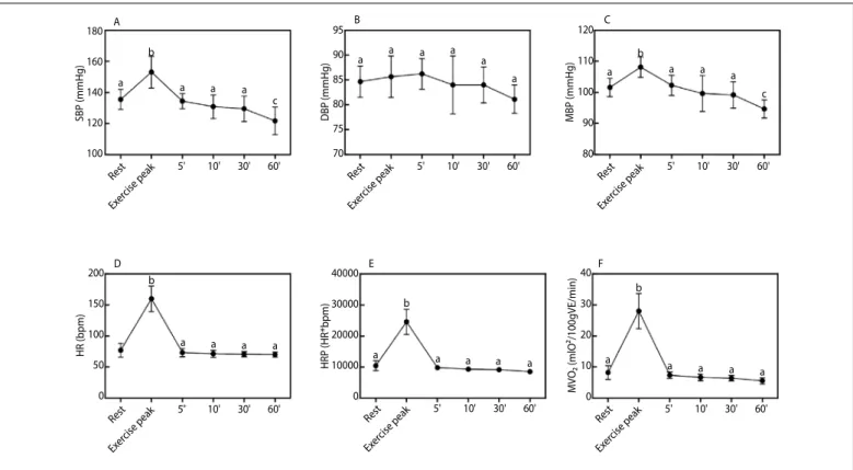
Figure 1. Values expressed as the mean ± standard error deviation of PHE to Panel A: systolic blood pressure (SBP), Panel B: diastolic blood pressure (DBP), Panel C: mean blood pressure (MBP), Panel D: heart rate (HR), Panel E: rate pressure product (RPP) and Panel E: myocardium oxygen volume (MVO₂)after powerlifting training session. Different letters indicate statistical differences (p<0.05) to ANOVA repeated measurements.
A
D E F
B C
Rest
Rest Rest Rest
Rest Rest
Exer cise peak
Exer cise peak
Exer cise peak
Exer cise peak Exer
cise peak
Exer cise peak
SBP (mmHg)
HR (bpm)
HRP (HR*bpm)
MVO₂ (mlO²/100gVE/min)
DBP (mmHg) MBP (mmHg) 180
160
140
120
100
200
150
100
50
0
40000
30000
20000
10000
0 95
90
85
80
75
70
120
110
100
90
80
40
30
20
10
0 10'
10' 10' 10'
10' 10'
5'
5' 5' 5'
5' 5'
a
a a a b
a
a a a a
a a
b
a a a a
b
a a a a a
a a a a a
c b
a b
a a
c
30'
30' 30' 30'
30' 30'
60'
60' 60' 60'
60' 60'
and MVO2. The following equation expressed in mlO₂/100gVE/min were used: MVO2= (DP x 0.0014) - 6.37.
Statistical analyses
All statistical analyses were performed using SPSS software (v 15.0; IBM, Armonk, NY, USA). Analysis of comparisons between groups over the periods was performed with one-way analysis of variance with repeated measures, followed by Kruskal–Wallis or Bonferroni’s post-hoc test when appropriate. The D’Agostino–Pearson test was applied to Gaussian distri-bution analysis. Statistical significance was established at p<0.05. Data is expressed as mean ± standard deviation. Additionally, the magnitude of the training effect was calculated using the mathematical formula that considers the final average the final average value subtracted from the initial average value in relation to the variation of initial mean: [Pre-Post ES = Posttest mean – Pretest mean / Pretest SD]21.
RESULTS
The anthropometric parameters are described on Table 1. The hemodynamic analysis of powerlifting exercise session at rest, exercise peak and recovery are presented at Figure 1. Significant increase (p<0.01) was observed from rest to exercise peak in SBP (Panel 1A: rest 135 ± 6 mmHg; exercise peak: 153 ± 10 mmHg; 11 ± 4 %:), MBP (Panel 1C: rest 102 ± 3 mmHg; exercise peak: 108 ± 3 mmHg; 6 ± 3 %), HR (Panel 1D: rest 77 ± 11; exercise peak: 160 ± 20 bpm; 50 ± 10 %), RPP (Panel 1E: rest: 10442 ± 1583 bpm*mmHg; exercise peak 24561 ± 4040 bpm*mmHg; 56 ± 10 %) and MVO₂ (Panel 1F: rest: 8.24 ± 0,73 mlO₂/100gVE/min; exercise peak 28.01 ± 1.88 mlO₂/100gVE/min; 69 ± 11%) but not to DBP (Panel 1B: 85 ± 3 mmHg; exercise peak 85 ± 4 mmHg; 0.99 ± 5 %). Additionally, the effect size in relationship rest versus exercise peak was large to SBP (2.65), MBP (2.18), HR (7.5), RPP (8.92), MVO₂ (8.92), but small to DBP (0.32).
During recovery, differences were found only on SBP (122 ± 9 mmHg) and MBP (95 ± 3 mmHg) at 60 min in relation at rest with trivial effect
size to both parameters (-2.11 and -1.07) respectively. No differences were presented on others parameters.
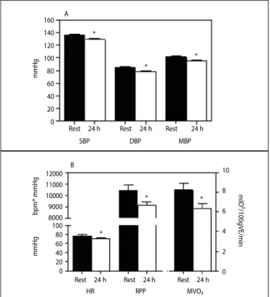
The 24h analyses recovery could be observed at Figure 2. Statistical reduc-tions (p<0.001) were found in all parameters 24h after powerlifting exercise ses-sion, at SBP (rest 135 ± 6 mmHg; 24h 128 ± 6 mmHg; -5 ± 3 %), DBP (rest 84 ± 3; 24h 78 ± 3 mmHg; -8 ± 4 %), MBP (rest 102 ± 3 mmHg; 24h 95 ± 3,2 mmHg; -7 ± 3 %) on Panel A, and HR (Panel B: rest 77 ± 3bpm; 24h 70 ± 2 bpm; -8 ± 9 %), RPP (Panel B: rest 10442 ± 1583 bpm*mmHg; 24h 9115 ± 1013 bpm*mmHg; -14 ± 12 %) and MVO₂ (rest: 8.24 ± 2.27 mlO₂/100gVE/min; 24h: 6.39 ± 1.42 mlO₂/100gVE/min; -29 ± 24 %) on Panel B. In all parameters the effect size of 24h post exercise to SBP (-1,05), DBP (-1,97), MBP (-2,16), HR (-0,56), RPP (-0,84), MVO₂ (-0,84.7). SBP (-1,05), MBP (2.18), HR (7.5), RPP (8.92) and MVO₂ (8.92) were trivial.
DISCUSSION
121
Figure 2. Values expressed as the mean ± standard error deviation of PHE to Panel A: systolic blood pressure (SBP), diastolic blood pressure (DBP) and mean blood pressure (MBP), Panel B: heart rate (HR), rate pressure product (RPP) and myocardium oxygen volume (MVO₂) after powerlifting training session. *p< 0.01 indicate statistically significant differences to rest.
A
B SBP
HR RPP MVO₂
DBP MBP
Rest
Rest Rest Rest
Rest Rest 24 h
24 h 24 h 24 h
24 h 24 h
m
lO
²/1
00
g
VE
/m
in
m
mHg
bpm* mmHg
mmHg
160 140 120 100 80 60 40 20
0
12000 11000 10000 9000 8000 100 80 60 40 20 0
10
8
6
4
2
0
*
*
* *
*
*
SBP after 60 min of exercise session, and 3) the PHE persisted after 24 hours after powerlifting training sessions.
Important information should be address in this study, although had been used very high training intensities (>90% of 1RM), the ex-ercise session does not promote hemodynamic overload during the practice. Some important consideration should be pointed about this fact, although the powerlift practice is usually done with high intensity, the values of systolic Blood pressure was not high then (160 mmHg).
Traditionally, powerlifting training sessions often use small volumes and high intensity could reach 100% of 1RM13,14, which could lead to a sharp
adjustment that provide unique and differentiated hemodynamic responses. There are few studies5,10-12 on literature that evaluated the impact of
alter-ations of external load (intensity) on PEH, demonstrating11,12 or absence22
PHE after load modification. In relationship to training volume, previous studies22-25 does not found effect on PHE magnitude after its modification.
In 2015 Cavalcante et al.5 demonstrated that PHE occur independently
of exercise intensity without expressed cardiovascular overload during the session, however, there are few study5,10-12 on literature evaluating different
manipulation of volume and intensity on RE and the magnitude of PEH. In this study the PHE were found after 60 min and 24 hour after PWL exercise session just in SBP. Duncan et al.3 and Hardy and Tucker26 found
a reduction in SBP that persisted for at least 1 hour after a RT session and Queiroz et al.4 found reduction of SBP after 24 hours after aerobic exercise.
Considering the influence of load at PHE, Fisher27studied normotensive
and hypertensive women after exercises at 50% of 1RM found significant SBP reduction for 60 minutes. Moraes et al.28also analyzed the hypotensive
effect of resistance exercise in hypertensive middle-aged men with loads of 60% of 1RM significantly reduced SBP, DBP, and MBP, respectively, by an average of 16, 12, and 13 mmHg to pre-hypertensive values. To our knowledge, there is only one study Brandão Rondonet al.9 that showed
a reduction in SBP, DBP, and MBP for 60 minutes simultaneously. Though the PHE had been much studied, the mechanisms
responsible for decreases in post resistance exercise BP are outside the scope for this study, however, theses have not been completely elucidated in the scientific literature29. Previous studies5,6,10, suggest that
several mechanisms can influence the cardiovascular and PHE behavior. So, overall the hypotensive effect may be associated with factors such as occlusion of the vessels and arteries, cardiac output, stroke volume, autonomic modulation of heart rate through the sympathetic and parasympathetic nerves, in addition to peripheral vascular resistance that can be changed during training30-32.
In relation to autonomic modulation evidences indicate that at higher intensities (above 80%) appears to be a reduction in cardiac output medi-ated decrease in stroke volume (SV)11. The drop in SV would be offset by
an increase in HR caused by increased sympathetic activity and parasym-pathetic reduction in heart. Additionally, the vasodilation mediated by the action of kallikrein system / kinin which also have higher concentrations after RE28 can be involved on this effect. Simultaneously, other peripheral
vasodilators such as nitric oxide, prostaglandins, adenosine, and potassium may also appear to influence the PHE33,34. It is speculated also that the
blood lactate concentration interferes with the blood pressure response after exercise35. The lactate accumulation could cause reduction in vascular
resistance with consequent PHE. In this case, exercises performed with higher intensities would cause increased release of lactate36.
According to Negrão et al.30 the aspects of training load should be
associated with hemodynamic response we should note the concern about the workload, and the percentage of 1 repetition maximum (% of 1RM), the number of series and number of repetitions, rest interval between series, factors that are associated with pressure changes during and after exercise session. Figueira Júnior et al.6 and Fagard36 considered
the volume of strength training as number of series, repetition number and amount of exercise, are the variables that can promote increases on blood pressure during and post exercise. Another factor important consideration to PHE and its influence should be address to muscle mass involved in exercise and training session. Polito and Farinatti37 was
not found changes PHE after leg extension exercise in different training models. Similar responses were found by D’Assunção et al.31 using
exer-cises with small muscle mass compared large mass.
Therefore, muscle group or larger amount of muscle mass involved during exercise, cannot be the main reasons for changing the hypotensive effect. In this case the powerlifting modality the PHE may be due to the involvement of a larger number of joints in the same movement increase need for blood in the active region, increasing vasodilation, reduce periph-eral resistance and favoring PHE6. Studies31,37,38 had showed differences on
mono and multi-joint exercises. In this case, the exercise performed in this study by powerlifting athlete has multi-joints characteristics associated with large muscle groups, which may have enhanced the hypotensive effect13.
Although few studies have evaluated the effects of strength training with high intensity in the cardiovascular response, we noted that the elevation of systolic and diastolic blood pressure increase progressively, reaching higher values in the last repetition, and early fatigue. In general, it can be seen that low intensity and high reps are more elevated blood pressure6,17. Muscle mass exerts influence on cardiovascular response,
122
The hypothesis for PHE on strength training with high intensity for powerlifting practitioner may be related to decreased cardiac output caused by a reduction in stroke volume. The increase in stroke volume is related to an increase in cardiac contractility, which led to gradual decrease in end systolic volume, in other words, greater volume of ventricular filling during diastole. The results obtained in this study may provide relevant information about PHE, the variables possibly related to hypotension mechanisms as sympathetic activity, blood flow, cardiac output and production of nitric oxide have not been evaluated. However, this experience becomes relevant, since it is the first to directly investigate the influence of the intensity of strength training on multi-joints exercises, involving the greatest amount of muscle mass and low number of repetitions, and the response of the PEH effect.
Our study does have some limitations that should be mentioned. First, the mechanisms of hypotension were not investigated in the present study. We did not assess the exercise effects on peripheral vascular resistance as well as sympathetic activity, systolic volume, beta-adrenergic receptors, or endothelial factors. Second, the auscul-tation method used for assessing blood pressure. This method, while universally used, has limitations compared to invasive methods, such as intra-arterial catheterization; however, all safeguards were taken to ensure that these measures were obtained in a consistent, reliable, and accurate manner. Third, the number of subjects on sample and a lack of control group (not athletes) may be useful to comparative effects.
Thus, it appears that the type of exercise, muscle mass involved, intensity and volume of strength training can be an important factor on cardiovascular response, especially on SBP and DBP post effort. More studies are needed to clarify the influence of RE variables on PEH in addition to the elucidation of the mechanisms involved in this phenomenon mainly in intense strength training in normotensive and hypertensive subjects.
CONCLUSION
In summary, the data presented in this study show that PEH effect in powerlifting practitioners after 60 min and 24 hours after exercise session without promote hemodynamic overload during an exercise session in PWL athletes.
ACKNOWLEDGMENTS
The authors thanks CAPES (Coordenação de Aperfeiçoamento de Pessoal de Nível Superior) fellowships addressed to João G.A., Rodriguez D. and Ceschini F. The fund providers had no role in decision to publish and preparation of the paper.
All authors have declared there is not any potential conflict of interests concerning this article.
REFERENCES
1. American College of Sports Medicine. American College of Sports Medicine position stand. Progression models in resistance training for healthy adults. Med Sci Sports Exerc. 2009;41(3):687-708. 2. Williams MA, Haskell WL, Ades PA, Amsterdam EA, Bittner V, Franklin BA, et al. Resistance exercise in
individuals with and without cardiovascular disease: 2007 update: a scientific statement from the American Heart Association Council on Clinical Cardiology and Council on Nutrition, Physical Activity, and Metabolism. Circulation. 2007;116(5):572-84.
3. Duncan MJ, Birch SL, Oxford SW. The effect of exercise intensity on postresistance exercise hypotension in trained men. J Strength Cond Res. 2014;28(6):1706-13.
4. Queiroz AC, Gagliardi JF, Forjaz CL, Rezk CC. Clinic and ambulatory blood pressure responses after resistance exercise. J Strength Cond Res. 2009;23(2):571-8.
5. Cavalcante PA, Rica RL, Evangelista AL, Serra AJ, Figueira A Jr, Pontes FL Jr, et al. Effects of exercise intensity on postexercise hypotension after resistance training session in overweight hypertensive patients. Clin Interv Aging. 2015;10:1487-95.
6. Figueira Júnior AJ, Rodriguez D, Gama EF, Pontes Jr FL, Bocallini DS. Exercise influence on morphological changes in cardiovascular control centers: Integration and adaptation. Rev Bras Ci e Mov. 2013;21(1):166-73. 7. Eysmann SB, Gervino E, Vatner DE, Katz SE, Decker L, Douglas PS. Prolonged exercise alters beta-adrenergic
responsiveness in healthy sedentary humans. J Appl Physiol (1985). 1996;80(2):616-22.
8. Kulics JM, Collins HL, DiCarlo SE. Postexercise hypotension is mediated by reductions in sympathetic nerve activity. Am J Physiol. 1999;276(1 Pt 2):H27-32.
9. Brandão Rondon MU, Alves MJ, Braga AM, Teixeira OT, Barretto AC, Krieger EM, et al. Postexercise blood pressure reduction in elderly hypertensive patients. J Am Coll Cardiol. 2002;39(4):676-82. 10. Bentes CM, Costa PB, Neto GR, Costa e Silva GV, de Salles BF, Miranda HL, et al. Hypotensive effects and
performance responses between different resistance training intensities and exercise orders in apparently health women. Clin Physiol Funct Imaging. 2015;35(3):185-90.
11. Rezk CC, Marrache RC, Tinucci T, Mion D Jr, Forjaz CL. Post-resistance exercise hypotension, hemodynamics, and heart rate variability: influence of exercise intensity. Eur J Appl Physiol. 2006;98(1):105-12. 12. Lizardo, JHF e Simões, HG. Efeitos de diferentes sessões de exercícios resistidos sobre a hipotensão
pós-exercício. Rev Bras Fisioter. 2005;9(3): 289-95.
13. João GA, Evangelista AL, Gomes JH, Charro MA, Bocalini DS, Cardozo D, et al. Effect of 16 weeks of periodized resistance training on strength gains of powerlifting athletes JEPonline 2014;17(3):102-9. 14. Pritchard HJ, Morton Rh. Powerlifting: success and failure at the 2012 Oceania and 2013 classic world
championships. J Australian Strength Cond 2015;23(6):67-70.
15. Haslam DRS, McCartney N, McKelvie RS, MacDougall JD.Direct measurements of arterial blood pressure during formal weightlifting in cardiac patients. J Cardiopulm Rehabil. 1988;8:213-25.
16. McCartney N. Acute responses to resistance training and safety. Med Sci Sports Exerc. 1999;31(1):31-7. 17. Boroujerdi SS, Rahimi R, Noori SR. Effect of high-versus low-intensity resistance training on post exercise
hypotension in male athletes Int Sport Med J 2009;10(2):95-100.
18. MacDougall JD, Tuxen D, Sale DG, Moroz JR, Sutton JR. Arterial blood pressure response to heavy resistance exercise. J Appl Physiol (1985). 1985;58(3):785-90.
19. MacDonald JR. Potential causes, mechanisms, and implications of post exercise hypotension. J Hum Hypertens. 2002;16(4):225-36.
20. Bocalini DS, Portes LA, Ribeiro KJ, Tonicelo R, Rica RL, Pontes Jr FL, et al. Insight for learning and stability
of one repetition maximum test in subjects with or without experience on resistance training. Gazz Med Ital Arch per le Sci Med. 2013;172(11):845-51.
21. Rhea MR. Determining the magnitude of treatment effects in strength training research through the use of the effect size. J Strength Cond Res. 2004;18(4):918-20.
22. Faraji H, Bab L, Ardeshiri H. Effects of resistance exercise intensity and volume on post exercise hypotensive responses. Braz J Biomotr 2010;4(1):65-73.
23. Simão R, Fleck SJ, Polito M, Monteiro W, Farinatti P. Effects of resistance training intensity, volume, and session format on the postexercise hypotensive response. J Strength Cond Res. 2005;19(4):853-8. 24. Jones H, George K, Edwards B, Atkinson G. Is the magnitude of acute post-exercise hypotension mediated
by exercise intensity or total work done? Eur J Appl Physiol. 2007;102(1):33-40.
25. Keese F, Farinatti P, Pescatello L, Monteiro W. A comparison of the immediate effects of resistance, aerobic, and concurrent exercise on postexercise hypotension. J Strength Cond Res. 2011;25(5):1429-36. 26. Hardy DO, Tucker LA. The effects of a single bout of strength training on ambulatory blood pressure
levels in 24 mildly hypertensive men. Am J Health Promot. 1998;13(2):69-72.
27. Fisher MM. The effect of resistance exercise on recovery blood pressure in normotensive and borderline hypertensive women. J Strength Cond Res. 2001;15(2):210-6.
28. Moraes MR, Bacurau RF, Ramalho JD, Reis FC, Casarini DE, Chagas JR, et al. Increase in kinins on post--exercise hypotension in normotensive and hypertensive volunteers. Biol Chem. 2007;388(5):533-40. 29. Gomes Anunciação P, Doederlein Polito M. A review on post-exercise hypotension in hypertensive
individuals. Arq Bras Cardiol. 2011;96(5):e100-9.
30. Negrão CE, Rodon, MUPB. Exercício físico hipertensão e controle barorreflexo da pressão arterial. Rev Bras Hipertens. 2001;8(1):89-95.
31. D’Assunção W, Daltro M, Simão R, Polito MD, Monteiro W. Resposta cardiovasculares agudas no treinamento de força conduzido em exercícios para grandes e pequenos grupos musculares. Rev Bras Med Esporte. 2007;13(2):118-22.
32. Rocha AC, Moraes-Silva IC, Quinteiro HRG, Sartori M, De Angelis K. Ajustes agudos, subagudos e crônicos da pressão arterial ao exercício resistido. Con Scien Saúde. 2012;11(4):685-90.
33. Goto C, Higashi Y, Kimura M, Noma K, Hara K, Nakagawa K, et al. Effect of different intensities of exercise on endothelium-dependent vasodilation in humans: role of endothelium-dependent nitric oxide and oxidative stress. Circulation. 2003;108(5):530-5.
34. Pescatello LS, Franklin BA, Fagard R, Farquhar WB, Kelley GA, Ray CA; American College of Sports Medicine. American College of Sports Medicine position stand. Exercise and hypertension. Med Sci Sports Exerc. 2004;36(3):533-53.
35. Simões GC, Moreira SR, Kushnick MR, Simões HG, Campbell CS. Postresistance exercise blood pressure reduction is influenced by exercise intensity in type-2 diabetic and nondiabetic individuals. J Strength Cond Res. 2010;24(5):1277-84.
36. Fagard RH. Exercise characteristics and the blood pressure response to dynamic physical training. Med Sci Sports Exerc. 2001;33(Suppl 6):S484-92.
37. Polito MD, Farinatti PT. The effects of muscle mass and number of sets during resistance exercise on postexercise hypotension. J Strength Cond Res. 2009;23(8):2351-7.
38. Goldring N, Wiles JD, Coleman DA. The effects of four weeks home-based isometric exercise training on resting blood pressure. Am Col Sports Med. 2012;44(5):S486-S612.