Role of keratinocytes in wound contraction: an impact
assessment using a model of collagen matrix populated
with ibroblasts
Papel do queratinócito na contração da ferida: avaliação de impacto usando um
modelo de matriz de colágeno povoada por ibroblastos
Study conducted at the Research
Laboratory of Cell Culture and
Wound Healing (LIM 04) of the Department of Plastic Surgery of Hospital das Clínicas of the School of Medicine of Universidade de São Paulo (HCFMUSP), São Paulo, SP, Brazil.
Submitted to SGP (Sistema de Gestão de Publicações/Manager Publications System) of RBCP
(Revista Brasileira de Cirurgia
Plástica/Brazilian Journal of
Plastic Surgery).
Received: June 7, 2011 Accepted: July 12, 2011
1. Doctor, head of the Research Laboratory of Cell Culture and Wound Healing (LIM 04) of the Department of Plastic Surgery of Hospital das Clínicas
of the School of Medicine of Universidade de São Paulo (HCFMUSP), São Paulo, SP, Brazil.
2. Doctor, head of the Tissue Bank of the Central Institute of HCFMUSP, São Paulo, SP, Brazil.
3. Practitioner Resident of Plastic Surgery at HCFMUSP, São Paulo, Brazil. 4. Doctor, assistant professor at Monash University, Australia
5. MSc, researcher at the Laboratory of Cell Culture of the Department of Plastic Surgery of HCFMUSP, São Paulo, SP, Brazil.
6. Post-doctoral degree, researcher at the Institute of Energy and Nuclear Research (IPEN), São Paulo, SP, Brazil.
7. Full Professor at the Discipline of Plastic Surgery of HCFMUSP, São Paulo, SP, Brazil.
CÉSAR ISAAC1
ANDRÉ OLIVEIRA PAGGIARO2
JOHNNY LEANDRO CONDUTA
BORDA ALDUNATE3
MARISA ROMA HERSON4
SILVANA CEREIJIDO ALTRAN5
MÔNICA BEATRIZ MATHOR6
MARCUS CASTRO FERREIRA7
ABSTRACT
Background: The possible participation of keratinocytes in wound remodeling has been
widely studied. This study investigated the impact of keratinocytes in wound contraction.
Methods: Murine type I collagen gels populated by human ibroblasts and seeded with human keratinocytes on the surface to form a dermo-epidermal equivalent were used as the study
group. Collagen gels populated by only ibroblasts were used as the control group. The criteria for the preparation and storage of gels were similar for both groups. Results: An evident and
statistically signiicant increase in gel contraction was observed in samples populated by kera -tinocytes compared to the control group. Conclusions: These results suggest that keratinocytes
not only modulate ibroblast proliferation but also play an active role in wound contraction per se. Further research on the mechanisms involved in the communication pathways between cells and between cells and the matrix shall be assessed from the perspective of keratinocyte
participation in wound healing and pathologic scarring.
Keywords: Keratinocytes. Wound healing. Cell culture techniques. Fibroblasts.
RESUMO
Introdução: A eventual participação de queratinócitos na remodelagem da ferida tem sido
estudada há muito tempo. Este trabalho investigou o impacto dos queratinócitos na contra
-ção da ferida. Método: Foi utilizado gel de colágeno tipo I murino povoado por ibroblastos
humanos com queratinócitos humanos semeado na superfície (grupo estudo), formando um equivalente dermoepidérmico. Géis de colágeno povoado apenas por ibroblastos foram uti
-lizados como grupo controle. Os critérios de confecção e armazenagem dos géis foram iguais para ambos os grupos. Resultados: Houve aumento evidente e estatisticamente signiicante
na contração de gel das amostras povoadas por queratinócitos, em comparação ao grupo con -trole. Conclusões: Esses resultados sugerem que os queratinócitos não só podem modular a
proliferação de ibroblastos, mas também, por si só, desempenhar papel ativo na contração da ferida. Novas investigações sobre mecanismos envolvidos nas vias de comunicação entre células e entre célula e matriz devem ser avaliadas sob o ponto de vista de participação dos queratinócitos na cicatrização de feridas e formação de cicatrizes patológicas.
INTRODUCTION
Tissue damage induces a sequence of biochemical and cell
reactions that are intended to repair the integrity of tissues.
The healing process involves a tuned “orchestra” of cells, in
which each participant interacts with others.
Many aspects of this intercellular communication
remain unclear, despite the unquestionable fact that cells are responsible for growth factor release and other signaling
molecules that actively participate in the wound healing process, modulating the microenvironment and cellular responses1,2. Among the components of this orchestra,
fibroblasts perform many functions in wound healing. First, fibroblasts proliferate at the wound site and secrete collagen
in the extracellular matrix, filling the void and creating a new structural support for the epidermis to proliferate.
Second, fibroblasts contract the wound bed, an action that begins 4 to 5 days after injury and reaches its peak 12 to 15 days after injury. This feature can persist for longer periods,
especially if the wound is still open3-7.
Bioengineering has revolutionized research in the healing
area, enabling the establishment of in vitro models and cell
culture systems that mimic different stages of wound healing.
In particular, keratinocyte cultures on dermal substitutes provide an improved setting for observing these cells with
the underlying matrix.
In 1979, Bell et al.8 established the model of a murine
collagen gel populated with ibroblasts, whose contractile function determined by such cells could be assessed and modiied by the action of modulators. This became a classic
model for collagen remodeling assessment and wound contraction studies9,10.
The possible involvement of keratinocytes in wound remodeling was suggested by Souren et al.11 in 1989. These authors reported that collagen gels could be rearranged if
keratinocytes were seeded on their surfaces, an effect that
was not observed if the cells were seeded inside a gel. Research on dermo-epidermal substitutes by Ralston et
al.12 using a collagen gel populated with ibroblasts as a bed for cultured keratinocytes revealed that keratinocytes parti-cipate in the contraction of the extracellular matrix.
During the preparation of dermo-epidermal composites
in the research laboratory where this study was conducted, it was observed that the epidermis had an area larger than its dermal substrate when it was released from the dermis, as if the keratinocyte layer had been released from the force of contraction and adhesion. It was also observed that dermal
surfaces populated with keratinocytes suffered progressive contraction as the cells proliferated and reorganized in a
conluent and multi-layered epithelium13. These intriguing observations reinforce the suggestion that keratinocytes play
an active role in wound contraction.
This study aimed to assess and quantify the potential
role of keratinocytes in wound contraction using an in vitro and preset model of collagen gel contraction populated with
ibroblasts.
METHODS
Primary Culture of Keratinocytes and Fibroblasts
Human keratinocytes and fibroblasts were isolated from surgical remains of skin donated for research by five healthy women ranging in age from 18 to 42 years.
Keratinocytes were isolated by chemical enzymatic dissociation, as described by Rheinwald & Green14. Cells obtained between the second and fifth passages were
counted and resuspended in culture medium containing 60%
Dulbecco’s Modified Eagle’s Medium (DMEM) (GIBCO – Life Technologies, Baltimore, USA), 30% Ham 12% (GIBCO – Life Technologies, Baltimore, USA), 10% fetal calf serum (GIBCO – Life Technologies, Baltimore, USA), 4 mM glutamine (GIBCO – Life Technologies, Baltimore, USA), 0.18 mM adenine (Sigma Chemical – St. Louis, USA), 5 μg/ml insulin (Sigma Chemical - St. Louis, MO, USA), 0.4 μg/ml hydrocortisone (Sigma Chemical - St. Louis, USA), 0.1 mM cholera toxin (Sigma Chemical - St. Louis, USA), 2 mM triiodothyronine (Sigma Chemical - St. Louis, USA), and an antibiotic solution (penicillin 100 UI/ ml/streptomycin 100 μg/ml) (Sigma Chemical - St. Louis,
USA).
Primary keratinocyte cultures were established by seeding cells (5 × 10⁶) suspended in 5 ml of the culture
medium described above in 25-cm² cell culture flasks (Falcon). Cells were maintained in an incubator (Fisher)
with an atmosphere of 5% CO2 at 37 °C. After 24 hours, 10
μg/ml of epidermal growth factor was added to the culture medium (Sigma Chemical - Louis, USA). The medium was changed every 48 hours until the cells became subconfluent,
after which they were trypsinized and seeded on collagen
gels previously populated by fibroblasts.
Human fibroblasts were obtained by the explantation technique described by Carrel & Burrows15: skin fragments of partial thickness and devoid of subcutaneous tissue were attached to 25-cm² surface culture plates and bathed in DMEM culture medium, 10% fetal calf serum, and 1% antibiotic solution (100 IU/ml of penicillin/streptomycin 100 μg/ml). The culture medium was changed every three days, and cells were kept in an incubator with an atmosphere
of 5% CO2 at 37 °C. Fibroblasts migrated to the exterior of the skin fragments after a few days, and the fragments
Preparation of a Collagen Matrix Populated with Fibroblasts
The collagen solution was prepared according to the protocol proposed by Bell et al.8, in which the tail tendons
of Wistar-Furth type rats were aseptically removed and placed in a container with diluted acetic acid at a ratio
of 1:1000 (250 ml of acid solution/tail). The mixture was stored at 4 °C. After two weeks, a viscous solution was obtained that split into the following two phases after
centrifugation at 500 rpm for one hour: a supernatant
fraction that mainly contained solubilized type I collagen, which was removed and stored at 4 °C (soluble acid), and a residual fraction that represented acid-insoluble collagen,
which was discarded.
The protocol proposed by Collagen Biomaterials16 was used to produce type I collagen gels. The gels consisted of a mixture of 100 µl of PBS (10×) with 100 µl of sodium hydroxide (NaOH) (0.1 M), 400 µl of the fibroblast suspen
-sion (5 × 10⁴) in PBS (1×), and 400 µl of collagen solution.
Two-thirds of the prepared gels were used for kerati -nocyte seeding (hereafter called co-cultures, i.e.,
kerati-nocytes in a collagen gel populated with fibroblasts), and
a third of these were used as control samples (collagen gels containing only keratinocytes).
Keratinocyte Seeding on Collagen Gels Containing Fibroblasts
After collagen gels containing ibroblasts had been poly
-merized for 12 hours, keratinocytes (1.2 × 10⁵) were seeded
on the surface of each gel. Both co-cultures and controls were
embedded in the culture medium and kept in an incubator at 37 °C and 5% of CO2 for another 48 hours. This procedure was repeated in duplicate for each of the ive different kera -tinocyte cell donors.
Collagen Gel Contraction
The area of the collagen gel populated with fibroblasts
was photographed using a digital camera1 (SONY
Cyber-shot, 3.2 megapixels) at 0, 12, 24, 36, and 48 hours after detachment from the well matrices. Gel area measurement was performed using the software UTHSCSA Image Tool
for Windows version 3.017. The calculation was performed in triplicate and the resulting value represented the
arith-metic mean of the triplicates, which was obtained in pixels
and converted to square centimeters.
Statistical Analysis
The areas of the gels (co-culture and control) at the
measured intervals were assessed using the nonparametric statistical test of ANOVA.
RESULTS
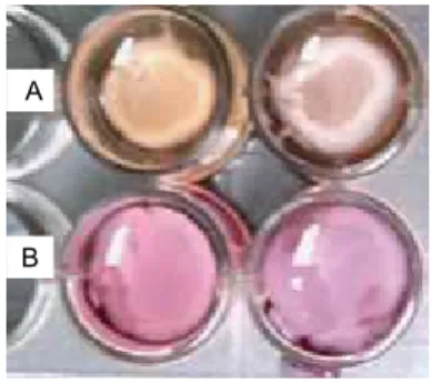
Progressive contraction of matrices occurred within a range of 48 hours after the gels were released from the walls
of the wells. Macroscopically detectable contraction was not observed after this period (Figure 1).
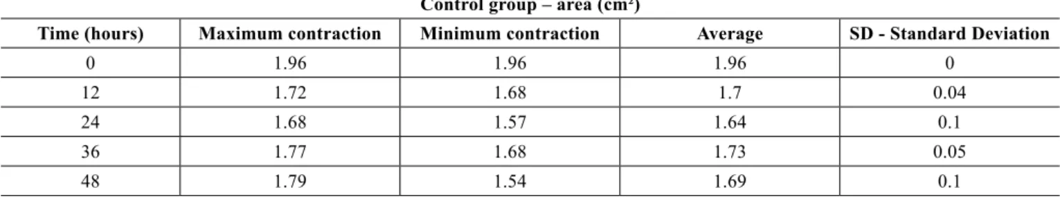
Table 1 shows the average area of control gels (ibroblast/ collagen gel) during different intervals (initial - time 0 = 1.96 cm² area). The average areas of co-culture gels (keratinocytes + ibroblasts/collagen gel) measured at different intervals are shown in Table 2 (initial - time 0 = 1.96 cm² area).
The contraction of control gels was signiicantly smaller (approximately 0.27 cm² area reduction) compared to the contraction of co-culture gels (mean reduction of 1.53 cm²). Figure 2 shows the different contractions of collagen gels populated only by ibroblasts (controls) and kerati
-nocytes seeded on collagen gels populated with ibroblasts
(co-cultures) for the period of the experiment.
The gels seeded with keratinocytes (co-cultures) showed signiicantly greater contraction than the control group (P ≤ 0.0001 and α = 0.05).
DISCUSSION
This experimental study assessed changes in the contrac
-tion of collagen gels populated with ibroblasts in the
presence of keratinocytes.
The experimental model of extracellular matrix contrac
-tion has been used in wound healing studies since its deve
-lopment by Bell et al.8. Scattered ibroblasts in the collagen
matrix behave more similarly to ibroblasts in vivo than those
grown in a monolayer. Likewise, proliferation is slower in cells scattered in the extracellular matrix.
Generally speaking, three-dimensional models are best used in the study of relationships and interferences between
components of the analyzed system, whereas monolayer
cultures better assess behavior and cellular activity. However,
Figure 1 – Matrix contraction 48 hours after gels were released from the walls of the wells. A, a collagen gel populated with
ibroblasts and keratinocytes (study group). B, a collagen matrix
populated only with ibroblasts (control group).
some authors observed that the initial construction of the
model may modify the results of gel contraction measure-ment18,19.
Bell et al.8 showed that after 48 hours of the onset of
contraction, the matrix becomes stable without signiicant signs of collagen gel contraction. This information was used to establish the timetable proposed in this paper.
The literature indicates that gels attached to well walls exhibit reduced thickness without changes in the diameter.
However, the mechanical release of gels from the walls
results in a three-dimensional contraction of these gels, with
thickness and diameter changes due to the random distribu
-tion of collagen ibers and ibroblast ac-tion over such ibers.
Another factor that may modify model contraction is
the number of ibroblasts seeded in the collagen solution: in theory, the observed contraction increases as the number of cells increases. However, after a certain number of ibroblasts
is reached per volume unit of collagen solution, no apparent
signiicant increase or measurable change is observed in the
matrix contraction19. The literature indicates that quantities
between 3 × 10⁵ and 5 × 10⁵ ibroblasts/well of 1.96 cm² of
area are suficient to promote gel contraction consistent with
what occurs in vivo20.
A previous studies conducted in the same laboratory as the work described herein determined that less than 5 × 10⁴
ibroblasts seeded in 1.96 cm² wells do not generate contrac
-tion (unpublished data). Therefore, this number of cells was selected for the present study so that there was less inluence of ibroblasts in the contraction of these gels, thus allowing better characterization of the participation of keratinocytes
in this phenomenon.
These results clearly demonstrate that the collagen gels populated with ibroblasts that received keratinocytes had signiicantly higher rates of contraction compared to controls. The inal area of gels seeded with keratinocytes was up to ive times smaller than the area of control gels. Given that
the systems were similar and varied only in the action of
Table 2 – Co-culture gel: maximum, minimum, average, and the standard deviation of the areas (cm²) at different experimental times. Study group – area (cm²)
Time (hours) Maximum contraction Minimum contraction Average SD - Standard Deviation
0 1.96 1.96 1.96 0
12 1.18 0.51 0.85 0.34
24 1.05 0.46 0.67 0.3
36 1 0.4 0.6 0.27
48 0.51 0.21 0.43 0.15
Table 1 – Control gel: areas (cm²) at different times of the experiment - maximum, minimum, average, and standard deviation. Control group – area (cm²)
Time (hours) Maximum contraction Minimum contraction Average SD - Standard Deviation
0 1.96 1.96 1.96 0
12 1.72 1.68 1.7 0.04
24 1.68 1.57 1.64 0.1
36 1.77 1.68 1.73 0.05
48 1.79 1.54 1.69 0.1
Figure 2 – Model of a collagen matrix populated with cells (cm²) in
relation to the experimental time frame (P ≤ 0.0001).
Influence of the keratinocytes on the collagen gel contraction
Area (cm
2)
time (hours)
keratinocytes, it is clear that keratinocytes have the ability to interfere with the function of collagen substrate reorgani
-zation of ibroblasts.
Data from this study suggest that several concepts regar-ding the contractile and proliferative phases of wound
healing should be revised. Although most experts believe that ibroblasts are solely responsible for wound contraction and
collagen rearrangement, the present study suggests a clear
collaboration of keratinocytes in these processes. However, the exact mechanisms underlying this participation must be better understood.
CONCLUSION
Keratinocytes play a role in extracellular matrix
reorga-nization. The experimental model of contraction used in this
paper may allow further investigation of the communication
pathways between interacting cells and modulators, provi -ding new insight into the role of keratinocytes in wound healing and pathologic scarring.
REFERENCES
1. Peacock EE, Cohen IK. Wound healing. In: McCarthy J, ed. Plastic surgery. Philadelphia: WB Saunders; 1990. p. 161-85.
2. Grinnel F. Fibroblasts, myoibroblasts, and wound contraction. J Cell Biol. 1994;124(4):401-4.
3. Finesmith TH, Broadley KN, Davidson JM. Fibroblasts from wounds of different stages of repair vary in their ability to contract a collagen gel in response to growth factors. J Cell Physiol. 1990;144(1):99-107. 4. Coulomb B, Lebreton C, Dubertret L. Inluence of human dermal
ibroblasts on epidermalization. J Invest Dermatol. 1989;92(1):122-5. 5. Gabbiani G, Ryan GB, Majne G. Presence of modiied ibroblasts in
granulation tissue and their possible role in wound contraction. Expe
-rientia. 1971;27(5):549-50.
6. Darby I, Skalli O, Gabbiani G. Alpha-smooth muscle actin is transiently expressed by myoibroblasts during experimental wound healing. Lab Invest. 1990;63(1):21-9.
7. Ross R. The ibroblast and wound repair. Biol Rev Camb Philos Soc. 1968;43(1):51-96.
8. Bell E, Ivarsson B, Merrill C. Production of tissue-like structure by contraction of collagen lattices by human ibroblasts of different proli
-ferative potential in vitro. Proc Natl Acad Sci USA. 1979;76(3):1274-8. 9. Kamamoto F, Paggiaro AO, Rodas A, Herson MR, Mathor MB, Ferreira
MC. A wound contraction experimental model for studying keloids and
wound-healing modulators. Artif Organs. 2003;27(8):701-5. 10. Isaac C, Mathor MB, Bariani G, Paggiaro AO, Herson MR,
Goldenstein-Schainberg C, et al. Pentoxifylline modiies three-dimensional collagen lattice model contraction and expression of collagen types I and III by human ibroblasts derived from post-burn hypertrophic scars and from normal skin. Burns. 2009;35(5):701-6.
11. Souren JM, Ponec M, van Wijk R. Contraction of collagen by human ibroblasts and keratinocytes. In Vitro Cell Dev Biol.
1989;25(11):1039-45.
12. Ralston DR, Layton C, Dalley AJ, Boyce SG, Freedlander E, MacNeil S.
Keratinocytes contract human dermal extracellular matrix and reduce
soluble ibronectin production by ibroblasts in a skin composite model. Br J Plast Surg. 1997;50(6):408-15.
13. Herson MR, Mathor MB, Altran S, Capellozzi VL, Ferreira MC. In vitro construction of a potential skin substitute through direct human
keratinocyte plating onto decellularized glycerol-preserved allodermis.
Artif Organs. 2001;25(11):901-6.
14. Rheinwald JG, Green H. Serial cultivation of strains of human epidermal
keratinocytes: the formation of keratinizing colonies from single cells.
Cell. 1975;6(3):331-43.
15. Carrel A, Burrows MT. Cultivation of adult tissues and organs outside the body. JAMA. 1910;55:1379-81.
16. Collagen biomaterials. Available at: http://www.vasoseal.com/cb/cbbio
-materials.html. Access on: 5/2/2011.
17. Wilcox CD, Dove SB, McDavid WD, Greer DB. UTHSCSA ImageTool, Version 3.0 Final. Available at: http://ddsdx.uthscsa.edu/dig/itdesc.
html
18. Rompré P, Auger FA, Germain L, Bouvard V, López Valle CA, Thibault J, et al. Inluence of initial collagen and cellular concentrations on the inal surface area of dermal and skin equivalents: a Box-Behnken analysis. In Vitro Cell Dev Biol. 1990;26(10):983-90.
19. Zhu YK, Umino T, Liu XD, Wang HJ, Romberger DJ, Spurzem JR, et al. Contraction of ibroblast-containing gels: initial collagen concentration
regulates the degree of contraction and cell survival. In Vitro Cell Dev
Biol Anim. 2001;37(1):10-6.
20. Dallon JC, Ehrlich HP. A review of ibroblast-populated collagen lattices. Wound Repair Regen. 2008;16(4):472-9.
Correspondence to: César Isaac