Ana Célia Caetano, Raquel Gonçalves, Carla Rolanda
Eosinophilic esophagitis-endoscopic distinguishing indings
Ana Célia Caetano, Raquel Gonçalves, Carla Rolanda, De-partment of Gastroenterology, Braga Hospital, 471243 Braga, Portugal
Ana Célia Caetano, Carla Rolanda, Life and Health Sciences Research Institute, School of Health Sciences, University of Minho, 47157 Braga, Portugal
Ana Célia Caetano, Carla Rolanda, Life and Health Sciences Research Institute/3Bs-PT Government Associate Laboratory, 47157 Braga, Portugal
Author contributions: Caetano AC contributed to the analysis, interpretation of data and bibliographic review; Gonçalves R and Rolanda C contributed to the critical revision and inal approval of the version to be published.
Correspondence to: Ana Célia Caetano, MD, Department of Gastroenterology, Braga Hospital, Sete Fontes-São Victor, 471243 Braga, Portugal. [email protected] Telephone: +35-191-53319 Fax: +35-125-327999 Received: February 22, 212 Revised: May 2, 212 Accepted: May 26, 212
Published online: August 21, 212
Abstract
Eosinophilic esophagitis (EE) is the most frequent con-dition found in a group of gastrointestinal disorders called eosinophilic gastrointestinal diseases. The hypo-thetical pathophysiological mechanism is related to a hypersensitivity reaction. Gastroesophageal relux dis-ease-like complaints not ameliorated by acid blockade or occasional symptoms of dysphagia or food impac-tion are likely presentaimpac-tions of EE. Due to its unclear pathogenesis and unspeciic symptoms, it is dificult to diagnose EE without a strong suspicion. Although histo-logical criteria are necessary to diagnosis EE, there are some characteristic endoscopic features. We present the case of a healthy 55-year-old woman with dyspha-gia and several episodes of esophageal food impaction over the last six months. This case report stresses the most distinguishing endoscopic indings-mucosa rings, white exudative plaques and linear furrows-that can help in the prompt recognition of this condition.
© 2012 Baishideng. All rights reserved.
Key words: Distinguishing indings; Dysphagia; Eosino-philic esophagitis; Gastro esophageal reflux disease; Histological criteria
Peer reviewer: Dr. Xiaoyun Liao, Department of Medical On-cology, Dana-Farber Cancer Institute, 45 Brookline Avenue, Room JF-28E, Boston, MA 2215, United States
Caetano AC, Gonçalves R, Rolanda C. Eosinophilic esophagitis-endoscopic distinguishing indings. World J Gastroenterol 212; 18(31): 4221-4223 Available from: URL: http://www.wjgnet. com/17-9327/full/v18/i31/4221.htm DOI: http://dx.doi. org/1.3748/wjg.v18.i31.4221
INTRODUCTION
Eosinophilic gastrointestinal diseases (EGD) are rare conditions of growing interest due to their increasing diagnostic frequency in well developed countries[1]. Eo-sinophilic esophagitis (EE) is the most common EGD, and its clinical presentation varies extensively making the diagnosis difficult and clinical suspicion fundamen-tal. Although not entirely clear, given that EE correlates with other atopic disorders and has a good response to corticoid treatment, it seems that its pathophysiological mechanism is related to a hypersensitivity reaction[1].
In this case report, through several expressive images, we highlight the set of endoscopic features which helped in the early recognition of EE.
CASE REPORT
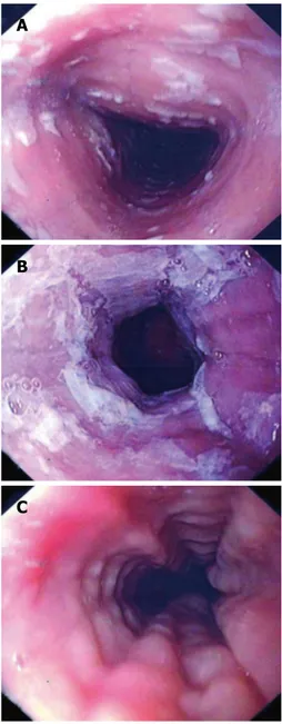
A 55-year-old woman with no previous medical his-tory presented with dysphagia and several episodes of esophageal food impaction over the last six months. Up-per gastrointestinal (GI) endoscopy revealed scattered white plaques in the proximal esophagus (Figure 1A), a whitish exudate coating the mucosa in the distal part of the esophagus (Figure 1B), and characteristic images of concentric transient rings and linear furrows (Figure 1C).
CASE REPORT
Online Submissions: http://www.wjgnet.com/esps/ [email protected]
doi:10.3748/wjg.v18.i31.4221
4221 August 21, 2012|Volume 18|Issue 31|
WJG|www.wjgnet.com
Biopsy specimens showed dense eosinophilic iniltrates,
> 20 eosinophils/high power field (HPF) and micro-abscesses (Figure 2A and B). Gastroesophageal reflux disease (GERD) was excluded when no improvement was observed following the administration of a proton pump inhibitor (PPI). The patient started treatment with a budesonide inhaler twice daily (instructions to swallow) and experienced symptom relief.
DISCUSSION
EE is part of a group of disease known as the eosino-philic gastrointestinal disorders. The pathogenesis of EE is not yet understood, although it appears to be related
to a hypersensitivity reaction. Some studies suggest that increased mucosa permeability allows contact with poten-tial allergenic digestion products leading to a consequent immunologic response[2]. EE tends to be a chronic dis-order with intermittent or persistent symptoms, usually GERD-like complaints which are not ameliorated by acid blockade with PPI. Additionally, patients may present with symptoms of dysphagia or food impaction. Due to its unspecific esophageal symptoms, clinical suspicion is critical in the diagnosis of EE. Although endoscopy may be normal in one third of cases, images of mucosal rings, white exudative plaques and esophageal strictures are characteristic findings of this pathology. Neverthe-less, multiple biopsies should be performed in differ-ent esophageal locations, as well as in the stomach and duodenum as the diagnosis of EE relies on histological criteria-one HPF must contain, at least, 15 intraepithelial eosinophils. Additional histological features include eo-sinophilic microabcesses[1,3].
To date, there are no large randomized controlled trials on EGD treatment. The majority of data are from smaller studies where corticosteroids play a role in the treatment of these disorders. Generally, oral or topical corticoid therapy is given to the patient for at least eight weeks followed by a gradual taper. The symptoms usu-ally recur, suggesting the need for continuous therapy. Some case reports show evidence of better symptom control following maintenance treatment with mast cell inhibitors or leukotriene receptor antagonists, however,
4222 August 21, 2012|Volume 18|Issue 31|
WJG|www.wjgnet.com
Caetano AC et al. How to promptly recognize eosinophilic esophagitis
Figure 1 A 55-year-old woman presented with dysphagia and several episodes of esophageal food impaction over the last six months. A: Scat-tered white plaques in the proximal esophagus; B: Whitish exudate coating the mucosa in the distal part of the esophagus; C: Concentric transient rings and linear furrows on esophagoscopy.
Figure 2 Histological indings in esophageal biopsy specimen. A: Dense
eosinophilic iniltrates; B: Microabscesses on esophageal microscopy.
C B A
larger trials are needed[2,3].
REFERENCES
1 Furuta GT, Liacouras CA, Collins MH, Gupta SK, Justinich C, Putnam PE, Bonis P, Hassall E, Straumann A, Rothenberg ME. Eosinophilic esophagitis in children and adults: a
sys-tematic review and consensus recommendations for diagno-sis and treatment. Gastroenterology 2007; 133: 1342-1363 2 Shiflet A, Forouhar F, Wu GY. Eosinophilic digestive
dis-eases: eosinophilic esophagitis, gastroenteritis, and colitis. J Formos Med Assoc 2009; 108: 834-843
3 Dellon ES. Diagnosis of eosinophilic esophagitis: current ap-proach and future directions. Curr Gastroenterol Rep 2011; 13: 240-246
S- Editor Gou SX L- Editor Webster JR E- Editor Zhang DN
4223 August 21, 2012|Volume 18|Issue 31|
WJG|www.wjgnet.com