Prevalence of fungemia in a tertiary hospital: Analysis of the
last decade
LUÍSA LIMA CASTRO1, MANUEL SCHÜTZE2, DANIEL HENRIQUE BÜCKER3, LEONARDODE SOUZA VASCONCELLOS4*
1MD from Faculdade de Medicina, Universidade Federal de Minas Gerais (UFMG), Belo Horizonte, MG, Brazil 2MD from Faculdade de Medicina, UFMG. MSc in Molecular Medicine from UFMG, Belo Horizonte, MG, Brazil
3Biologist, degree from Universidade Federal de Rondônia. MSc in Genetics from UFMG. Employee of the Laboratory Medicine Service at Hospital das Clínicas, UFMG, Belo Horizonte, MG, Brazil 4MSc and PhD in Medicine from UFMG. Adjunct Professor, Department of Complementary Propedeutics, Faculdade de Medicina, UFMG. MD, Clinical Pathologist, Belo Horizonte, MG, Brazil
S
UMMARYStudy conducted by the Grupo
de Pesquisa em Patologia Clínica/ Medicina Laboratorial (GPPCML) da
Universidade Federal de Minas Gerais (UFMG), Belo Horizonte, MG, Brazil
Article received: 10/29/2014
Accepted for publication: 11/3/2014
*Correspondence:
Dpto. de Propedêutica Complementar da
Faculdade de Medicina da UFMG Address: Av. Prof. Alfredo Balena, 190, sala
403, Santa Eigênia Belo Horizonte, MG – Brazil
Postal code: 30130-100 [email protected]
http://dx.doi.org/10.1590/1806-9282.62.04.315
Introduction: The prevalence of nosocomial fungemia has increased worldwide, and mortality caused by this disease is high.
Objective: To assess progress in the last decade, and the prevalence and profile of fungal agents isolated in blood cultures performed in a tertiary university hospital.
Method: All the results of blood cultures processed at Hospital das Clínicas, Universidade Federal de Minas Gerais (HC-UFMG), in the time intervals 2001-2003 and 2011-2013 were analyzed retrospectively. For each three-year period, the number of collected blood cultures, the overall positivity rate and the per-centage of fungemia were recorded. In addition, all identified fungal species were cataloged. All blood samples were incubated in the BacT/ALERT® (bioMérieux) automation system.
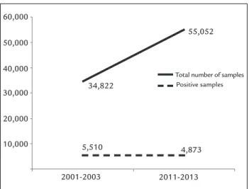
Results: In 2001-2003, 34,822 samples were evaluated, with 5,510 (15.8%) pos-itive results. In 2011-2013, the number of blood cultures processed increased to 55,052 samples, with 4,873 (8.9%) positive results. There was an increase in the number of positive cultures for fungi in the analyzed period (2001-2003: 4.16%; 2011-2013: 5.95%; p<0.001). Among the agents, candidemias were predominant, especially those caused by non-albicansCandida species (2001-2003: 57.64%; 2011-2013: 65.17%; p<0.05). There was also an increase in fungemia caused by other genera (2001-2003: 2.62%; 2011-2013: 4.48%; p<0.01).
Conclusion: There was an increase in the prevalence of fungemia in the last de-cade at HC-UFMG. Although candidemias have been responsible for most of the cases, there has been an increase in fungemias caused by other species.
Keywords:Candida, non-albicans Candida, fungemia, tertiary health care, prevalence.
INTRODUCTION
The term fungemia indicates the presence of viable fun-gi in the bloodstream, confirmed by laboratory tests. Cur-rently, over 80% of infections in the bloodstream, wheth-er caused by fungi or bactwheth-eria, are acquired in hospitals or other medical care centers.1
Mortality among patients with nosocomial funge-mia is high, reaching rates as high as 50 to 80%, and has been attributed mainly to absence or inadequacy of ini-tial antifungal therapy.2,3
The prevalence of hospital fungemia has increased in recent decades worldwide.3,4 Several studies have shown that the main risk factors for fungemias are prolonged steroid therapy, chemotherapy, malnutrition,
malignan-cy, previous fungal colonization, dialysis, abdominal sur-gery and immunosuppression.5,6 Other determining fac-tors for the increased occurrence of hospital fungemia are the growing use of broad-spectrum antibiotics and invasive technical procedures, such as central venous cath-eters, mechanical ventilation, parenteral nutrition, and the growing number of organ transplants.2,4,7
in a multicentric study carried out in Spain, C. albicans
caused 49.08% of the infections, followed by C. parapsilosis
(20.73%), C. glabrata (13.61%) and C. tropicalis (10.77%).10 The incidence of non-albicansCandida species has shown significant increase in recent decades.3 As susceptibility to antifungal drugs varies among species of Candida, it is im-portant to know the prevalence of each of them in hospi-tals.11 Early distinction between candidemia and blood in-fection by other fungi is essential for effective therapy.4
In Brazil, epidemiological surveys of fungemias in communities and hospitals have become increasingly fre-quent. Motta et al., in a study conducted at the Faculdade de Medicina, Universidade de São Paulo (FMUSP), Hos-pital das Clínicas, reported a 4% prevalence of fungemia in blood cultures performed in 2006, with approximate-ly 86% of cases related to species of the Candida genus. The species isolated most often were C. albicans (52.2%), C. parap-silosis (22.1%), C. tropicalis (14.7%), C. glabrata (6.6%).12
In addition to damage to the health of the population, fungemias also cause economic losses to public health sys-tems. Typically, antifungal treatments are long, which in-creases the length of hospital stay and the cost of drugs.13-16 In a recent article, Bloos et al. demonstrated the higher fi-nancial cost in patients hospitalized due to candidemia compared with sepse caused by other agents.15
Because of the inconvenience caused by this disease, each health care institution should know the profile of agents that cause fungemia in its population. This is vital not only for epidemiological purposes, but especially for care and therapy, serving also as parameter for implementation of preventive measures and hospital infection control.
Our study aims to compare prevalence progression and the profile of fungal species found in blood cultures of pa-tients admitted to the Universidade Federal de Minas Gerais (UFMG), Hospital das Clínicas, Brazil, in the last decade.
METHOD
All the results of blood cultures processed in the sector of Microbiology, Laboratory Medicine Service of Hospi-tal das Clínicas, Universidade Federal de Minas Gerais (HC-UFMG), in 2001-2003 and 2011-2013 were assessed. For each period, the number of samples collected, the overall positivity rate and the percentage of fungemia were recorded. In addition, all identified fungal species were cataloged.
In order to perform the blood cultures at the HC-UFMG, blood samples were taken after skin antisepsis with 0.5% alcoholic chlorhexidine. The number of sam-ples varied according to the physician’s discretion. Usu-ally, three samples were collected from different
anatom-ical sites in the case of adult patients, and a single sample for children. For adults, 16 mL to 20 mL of blood were collected from each sample, distributed into two bottles: Green lid (aerobic) and orange lid (anaerobic). For the children, the volume of blood varied from 1 mL to 5 mL, collected in a single jar with a yellow lid.
The blood culture bottles were transported to the Mi-crobiology sector and incubated in a BacT/ALERT® (bio-Mérieux) device. This is an automated system for incuba-tion and identificaincuba-tion of microbial growth, based on the colorimetric detection of CO2 through sensors positioned on the bottom of culture flasks. Growth of microorgan-isms increases CO2, showing positivity in the bottle.
The incubation time for antimicrobial growth was based on recommendations by the Clinical and Labora-tory Standards Institute (CLSI). Between 2001 and 2003, blood cultures lacking growth of microorganisms with-in seven days of with-incubation were considered negative. In case of suspected fungemia reported in the medical test request form, the incubation period would be increased to 30 days.16,17 From 2011 to 2013, in turn, according to the recommendations in force, incubation periods were reduced to 5 and 14 days, respectively.16,17
The positive samples were subjected to Gram stain-ing and morphological analysis usstain-ing optical microsco-py. After the identification of yeast-form or mycelial fila-ments using Gram staining, the samples were seeded on glass slides containing a Sabouraud medium and incu-bated at 37°C to determine the species. For yeast-form growth, the following tests were performed to identify
Candida: resistance to cycloheximide, germ tube forma-tion, microculture and physiological fermentation. When-ever Cryptococcus was suspected, urea test and inoculation in Niger culture medium were performed. Mycelial fun-gi were subjected to microculture in Sabouraud’s agar. Once the fungi were identified, the results were passed on to the Laboratory Informatics System and electroni-cally released for consultation by the requesting physi-cian and the patient.
For statistical analysis of the data, a two-sided chi-square test of the periods analyzed was adopted. P-val-ue<0.05 was considered significant.
RESULTS
From January 2001 to December 2003, and from January 2011 to December 2013, 89,874 blood samples were an-alyzed in the sector of Microbiology, Laboratory Medi-cine Service of HC-UFMG.
the latter, fungi were identified in 229 (16.4%) samples. In 2011-2013, 55,052 blood culture bottles were analyzed, with 4,873 (8.9%) positive samples, of which 290 (5.95%) contained fungi (Figure 1).
Among the isolated fungal species, prevalence of can-didemias was observed: 97.38% (2001-2003) and 95.52% (2011-2013). The main species isolated in both periods was
Candida albicans: 39.74% (2001-2003) and 30.34% (2011-2013). There was an increase in the number of positive cultures for fungi in the analyzed period (2001-2003: 4.16%; 2011-2013: 5.95%; p<0.001). There was also an increase in the number of non-albicansCandida species (2001-2003: 57.64%; 2011-2013: 65.17%; p<0.05) and in non-Candida genera (2001-2003: 2.62%; 2011-2013: 4.48%; p<0.01). The fungi isolated in each analyzed period are shown in Table 1.
TABLE 1 Fungal species isolated from blood cultures performed at the Laboratory Medicine Service of Hospital das Clínicas, Universidade Federal de Minas Gerais, Brazil, in 2001-2003 and 2011-2013.
Species 2001-2003 2011-2013 Total
N % N % N %
Candida albicans 91 39.7 88 30.3 179 34.5
Candida glabrata 1 0.4 7 2.4 8 1.5
Candida guilliermondii 3 1.3 4 1.4 7 1.3
Candida kefyr 0 0 2 0.7 2 0.4
Candida krusei 0 0 9 3.1 9 1.7
Candida parapsilosis 60 26.2 79 27.2 139 26.8
Candida spp. 29 12.7 24 8.3 53 10.2
Candida tropicalis 39 17 64 22.1 103 19.8
Cryptococcus neoformans 3 1.3 7 2.4 10 1.9
Cryptococcus spp. 3 1.3 4 1.4 7 1.3
Fusarium sp. 0 0 2 0.7 2 0.4
Total 229 100 290 100 519 100
N: Absolute number of cases; %: Percentage of cases.
DISCUSSION
Candida spp. is found in the gastrointestinal tract in 20 to 80% of the healthy adult population. These microor-ganisms can become pathogenic if there are changes in host defense mechanisms. Candidemia has a high inci-dence in tertiary hospitals, with overall mortality of 60%. Rapid detection of the etiological agent causing the in-fection is of fundamental importance to allow early ad-justment of treatment and hence, the reduction of hos-pital mortality by fungemia.4
The HC-UFMG is a teaching hospital that offers pub-lic and general care, health assistance, research services
and further training. It admits patients of the Unified Health System (SUS) only and is located in Belo Horizon-te, Minas Gerais, Brazil. The HC-UFMG is a reference hos-pital in the municipal and state healthcare spheres for me-dium and high-complexity procedures, mainly in oncohematologic, infectious and parasitic, endocrine-met-abolic, and mother and child diseases, as well as transplan-tation of organs and tissues. The HC-UFMG hospital com-plex has 511 beds; it includes one central building and seven other buildings for out- and in-patient care com-prising all specialties and sub-specialties covered by the SUS system. Each month in these facilities, about 2,300 emergency room visits, 1000 hospitalizations, 24,000 out-patient visits and 155,000 laboratory tests take place.18
In this study, the authors observed an increased in-cidence of fungemia in the last decade. This increase was consistent with epidemiological data from other nation-al and internationnation-al hospitnation-als.6,13,19-21 The increased prev-alence of fungemia in hospital services may be related to advances in health care, which allowed greater survival of immunosuppressed patients.19 The frequent use of inva-sive instruments and materials in hospital care, and the indiscriminate use of broad-spectrum antimicrobials also contributed to the increase in fungemia.2,19
The increase in the percentage of fungemia diagnosed in the last decade can be more significant, considering that false-negative results can occur if the minimum in-cubation time for growth of fungal species is not observed. At HC-UFMG, only samples reported as suspected funge-mia were subjected to longer incubation times, either from 7 to 30 days (2001-2003) or from 5 to 14 days
(2011-60,000
50,000
40,000
30,000
20,000
10,000
Total number of samples Positive samples
34,822
55,052
5,510 4,873
2001-2003 2011-2013
FIGURE 1 Number of total and positive samples of blood cultures,
2013).16 Unfortunately, the possibility of fungemia was not always informed on the request form for laboratory testing. In these cases, the samples were discarded early, probably hindering a proper diagnosis of fungemia.
We must also bear in mind that blood culture is a test that has intermediate sensitivity to detect fungal species, and may exhibit a sensitivity of only 50% for candidemias, for example.22 Therefore, the prevalence of fungemia de-tected in the HC-UFMG, and in other hospitals, can be falsely lower than the actual prevalence of this infection. The most frequent microorganism in this study was
Candida spp., which accounted for almost all of the iso-lated fungi, and Candida albicans, which was the most fre-quent species. These results are in agreement with other studies.2,3,8,22
Even though C. albicans was the most prevalent spe-cies, our results revealed an increase in the prevalence of other fungal species, especially non-albicansCandida spp. Other authors have demonstrated high rates of candi-demia caused by non-albicans species.3,23 The reasons for the emergence of other species have not been clarified. However, some risk factors are strongly associated to fungemia by certain species. C. parapsilosis has been linked to vascular catheter and parenteral nutrition.24,25 C.
tropi-calis was associated with cancer and neutropenia.26,27 C.
krusei has been associated with prior use of azoles, neu-tropenia and hematologic malignancies.28C. glabrata has been associated with prior use of azoles, and transplants of solid organ and hematopoietic cells.2,29-31
Some Candida spp. were isolated between 2011 and 2013: Candida kefyr, Candida krusei and Fusarium sp. Re-cent isolation of C. glabrata can represent a trend of in-creased incidence of this species in Brazil, already report-ed in São Paulo between 2006 and 2010,32 whose mortality seems higher than that of C. albicans.3
An interesting fact is the increased prevalence of gen-era other than Candida, whose mortality rate is higher than that found in candidemias.4 Previous colonization by these fungal species, collagen diseases and dialysis are risk factors associated with fungemia caused by non- Can-dida genera.4
Laboratory diagnosis of fungemia is extremely im-portant to establish proper treatment and to reduce pa-tient morbidity and mortality. Knowledge on the most prevalent species responsible for cases of fungemia with-in the service will allow the attendwith-ing physician to with- ini-tiate the most appropriate empiric therapy, as well as the analysis of a possible impact of new anti-fungal drugs introduced to the market on the profile of hospi-tal fungemias.
CONCLUSION
There was an increase in the prevalence of fungemia among patients treated at the HC-UFMG in the last decade. Can-didemias represented almost all cases, but there was an increase in fungemias caused by other genera.
RESUMO
Prevalência de fungemia em um hospital terciário: análi-se da última década
Introdução: a prevalência de fungemia hospitalar tem aumentado em todo o mundo e a mortalidade por essa afecção é elevada.
Objetivo: avaliar a evolução, na última década, da preva-lência e do perfil dos agentes fúngicos isolados em hemo-culturas realizadas em um hospital universitário terciário.
Método: foram analisados retrospectivamente todos os resultados de hemocultura processados no Hospital das Clínicas da Universidade Federal de Minas Gerais (HC--UFMG), entre os períodos de 2001-2003 e de 2011-2013. Para cada triênio foram registrados o número de hemo-culturas coletadas, o percentual de positividade geral e o percentual de fungemia. Também foram catalogadas to-das as espécies fúngicas identificato-das. Toto-das as amostras sanguíneas foram incubadas no sistema de automação BacT/ALERT® (bioMérieux).
Resultados: entre 2001-2003, foram avaliadas 34.822 amos-tras, sendo 5.510 (15,8%) positivas. Entre 2011-2013, o nú-mero de hemoculturas processadas aumentou para 55.052 amostras, sendo 4.873 (8,9%) positivas. Observou-se um aumento do número de culturas positivas para fungos no período analisado (2001-2003: 4,16%; 2011-2013: 5,95%; p<0,001). Dentre os agentes, as candidemias foram predo-minantes, principalmente por espécies de Candida não al-bicans (2001-2003: 57,64%; 2011-2013: 65,17%; p<0,05). Houve também aumento da fungemia por outros gêneros (2001-2003: 2,62%; 2011-2013: 4,48%; p<0,01).
Conclusão: houve aumento da prevalência de fungemia na última década no HC-UFMG. Embora as candidemias tenham sido responsáveis pela maioria dos casos, houve aumento de fungemias causadas por outras espécies.
Palavras-chave:Candida, Candida não albicans, fungemia, atenção terciária à saúde, prevalência.
REFERENCES
2. Anunnatsiri S, Chetchotisakd P, Mootsikapun P. Fungemia in non-HIV-infected patients: a five-year review. Int J Infect Dis. 2009; 13(1):90-6. 3. Colombo AL, Guimarães T, Silva LR, de Almeida Monfardini LP, Cunha AK,
Rady P, et al. Prospective observational study of candidemia in São Paulo, Brazil: incidence rate, epidemiology and predictors of mortality. Infect Control Hosp Epidemiol. 2007; 28(5):570-6.
4. Yamamoto M, Takakura S, Hotta G, Matsumura Y, Matsushima A, Nagao M, et al. Clinical characteristics and risk factors of non-Candida fungaemia. BMC Infect Dis. 2013; 13:247-53.
5. Dóczi I, Pető Z, Fodor E, Bereczki L, Nagy E, Hajdú E. Evaluation of fungaemia infections in a Hungarian university hospital between 1996 and 2009. Acta Microbiol Immunol Hung. 2012; 59(1):29-41.
6. Rosas RC, Salomão R, da Matta DA, Lopes HV, Pignatari AC, Colombo AL. Bloodstream infections in late-stage acquired immunodeficiency syndrome patients evaluated by a lysis centrifugation system. Mem Inst Oswaldo Cruz. 2003; 98(4):529-32.
7. Asmundsdóttir LR, Erlendsdóttir H, Gottfredsson M. Increasing incidence of candidemia: results from a 20-year nationwide study in Iceland. J Clin Microbiol. 2002; 40(9):3489-92.
8. Almirante B, Rodríguez D, Park BJ, Cuenca-Estrella M, Planes AM, Almela M, et al.; Barcelona Candidemia Project Study Group. Epidemiology and predictors of mortality in cases of Candida bloodstream infection: results from population-based surveillance, Barcelona, Spain, from 2002 to 2003. J Clin Microbiol. 2005; 43(4):1829-35.
9. Costa SF, Marinho I, Araújo EA, Manrique AE, Medeiros EA, Levin AS. Nosocomial fungaemia: a 2-year prospective study. J Hosp Infect. 2000; 45(1):69-72.
10. Cisterna R, Ezpeleta G, Telleria O; Spanish Candidemia Surveillance Group. Nationwide sentinel surveillance of bloodstream Candida infections in 40 tertiary care hospitals in Spain. J Clin Microbiol. 2010; 48(11):4200-6. 11. Lai CC, Gau SJ, Tan CK. Distribution of Candida species causing bloodstream
infections. J Microbiol Immunol Infect. 2012; 45(2):161-2.
12. Motta AL, Almeida GMD, Almeida Júnior JN, Burattini MN, Rossi F. Candidemia epidemiology and susceptibility profile in the largest Brazilian teaching hospital complex. Braz J Infect Dis. 2010; 14(5):441-8.
13. Sampaio Camargo TZ, Marra AR, Silva CV, Cardoso MF, Martino MD, Camargo LF, et al. Secular trends of candidemia in a tertiary care hospital. Am J Infect Control. 2010; 38(7):546-51.
14. Rosenthal VD, Guzman S, Migone O, Crnich CJ. The attributable cost, length of hospital stay, and mortality of central line-associated bloodstream infection in intensive care departments in Argentina: a prospective, matched analysis. Am J Infect Control. 2003; 31(8):475-80.
15. Bloos F, Bayer O, Sachse S, Straube E, Reinhart K, Kortgen A. Attributable costs of patients with candidemia and potential implications of polymerase chain reaction-based pathogen detection on antifungal therapy in patients with sepsis. J Crit Care. 2013; 28(1):2-8.
16. Clinical and Laboratory Standards Institute/National Committee for Clinical Laboratory Standards – CLSI. Principles and Procedures for Blood Cultures; Approved Guideline. CLSI document M47-A. Wayne: Clinical and Laboratory Standards Institute, 2007.
17. Clinical and Laboratory Standards Institute/National Committee for Clinical Laboratory Standards - CLSI. Reference method for broth dilution
susceptibility testing of yeasts: approved standard. 3.ed. Wayne: Clinical and Laboratory Standards Institute, 2008.
18. Nossa História – Hospital das Clínicas UFMG. [cited 2014 Oct 12]. Available from: http://www.hc.ufmg.br/institucional/historia.
19. Giolo MP, Svidzinski TIE. Fisiopatogenia, epidemiologia e diagnóstico laboratorial da candidemia. J Bras Patol Med Lab. 2010; 46(3):225-34. 20. Banerjee SN, Emori TG, Culver DH, Gaynes RP, Jarvis WR, Horan T, et al.
Secular trends in nosocomial primary bloodstream infections in the United States, 1980-1989. National Nosocomial Infections Surveillance System. Am J Med. 1991; 91(3B):86S-9S.
21. Velasco E, Thuler LCS, Martins CAS, Nucci M, Dias LMC, Gonçalves VMSC. Epidemiology of bloodstream infections at a cancer center. Sao Paulo Med J. 2000; 118(5):131-8.
22. Pfaller MA, Diekema DJ. Epidemiology of invasive candidiasis: a persistent public health problem. Clin Microbiol Rev. 2007; 20(1):133-63. 23. Colombo AL, Nucci M, Salomão R, Branchini ML, Richtmann R, Derossi
A, et al. High rate of non-albicans candidemia in Brazilian tertiary care hospitals. Diagn Microbiol Infect Dis. 1999; 34(4):281-6.
24. Clark TA, Slavinski SA, Morgan J, Lott T, Arthington-Skaggs BA, Brandt ME, et al. Epidemiologic and molecular characterization of an outbreak of Candida parapsilosis bloodstream infections in a community hospital. J Clin Microbiol. 2004; 42(10):4468-72.
25. Levy I, Rubin LG, Vasishtha S, Tucci V, Sood SK. Emergence of Candida parapsilosis as the predominant species causing candidemia in children. Clin Infect Dis. 1998; 26(5):1086-8.
26. Muñoz P, Giannella M, Fanciulli C, Guinea J, Valerio M, Rojas L, et al. Candida tropicalis fungaemia: incidence, risk factors and mortality in a general hospital. Clin Microbiol Infect. 2011; 17(10):1538-45.
27. Nucci M, Colombo AL. Candidemia due to Candida tropicalis: clinical, epidemiologic, and microbiologic characteristics of 188 episodes occurring in tertiary care hospitals. Diagn Microbiol Infect Dis. 2007; 58(1):77-82. 28. Marr KA, Seidel K, White TC, Bowden RA. Candidemia in allogeneic blood
and marrow transplant recipients: evolution of risk factors after the adoption of prophylactic fluconazole. J Infect Dis. 2000; 181(1):309-16.
29. Trick WE, Fridkin SK, Edwards JR, Hajjeh RA, Gaynes RP; National Nosocomial Infections Surveillance System Hospitals. Secular trend of hospital-acquired candidemia among intensive care unit patients in the United States during 1989-1999. Clin Infect Dis. 2002; 35(5):627-30. 30. Aquino VR, Lunardi LW, Goldani LZ, Barth AL. Prevalence, susceptibility
profile for fluconazole and risk factors for candidemia in a tertiary care hospital in southern Brazil. Braz J Infect Dis. 2005; 9(5):411-8.
31. Ortega M, Marco F, Soriano A, Almela M, Martínez JA, López J, et al. Candida species bloodstream infection: epidemiology and outcome in a single institution from 1991 to 2008. J Hosp Infect. 2011; 77(2):157-61. 32. Moretti ML, Trabasso P, Lyra L, Fagnani R, Resende MR, de Oliveira Cardoso