Extracorporeal shock wave lithotripsy in the treatment of renal
and ureteral stones
FÁBIO CÉSAR MIRANDA TORRICELLI1*, ALEXANDRE DANILOVIC1, FÁBIO CARVALHO VICENTINI1, GIOVANNI SCALA MARCHINI1, MIGUEL SROUGI2,
EDUARDO MAZZUCCHI3
1Assistant Physician to the Endourology Group at the University of São Paulo Medical School’s Hospital das Clínicas (HC-FMUSP), São Paulo, SP, Brazil 2Full Professor of Urology at the HC-FMUSP
3Post-doctoral Professor, Head of the Endourology and Lithiasis Group at the HC-FMUSP
S
UMMARYStuded conducted at Hospital das Clínicas, University of Sao Paulo Medical School
Article received: 2/24/2014
Approved for publication: 4/2/2014
*Correspondence:
Address: Avenida Dr. Enéas de Carvalho Aguiar, 255 7o Andar, Secretaria da Divisão de Urologia
Postal Code: 05403-010 [email protected]
http://dx.doi.org/10.1590/1806-9282.61.01.065
Conlict of interest: none
The use of certain technical principles and the selection of favorable cases can optimize the results of extracorporeal shock wave lithotripsy (ESWL). The aim of this study is to review how ESWL works, its indications and contraindications, predictive factors for success, and its complications. A search was conducted on the Pubmed® database between January 1984 and October 2013 using “shock
wave lithotripsy” and “stone” as key-words. Only articles with a high level of ev-idence, in English, and conducted in humans, such as clinical trials or review/ meta-analysis, were included. To optimize the search for the ESWL results, sev-eral technical factors including type of lithotripsy device, energy and frequency of pulses, coupling of the patient to the lithotriptor, location of the calculus, and type of anesthesia should be taken into consideration. Other factors related to the patient, stone size and density, skin to stone distance, anatomy of the excre-tory path, and kidney anomalies are also important. Antibiotic prophylaxis is not necessary, and routine double J stent placement before the procedure is not routinely recommended. Alpha-blockers, particularly tamsulosin, are useful for stones >10mm. Minor complications may occur following ESWL, which gener-ally respond well to clinical interventions. The relationship between ESWL and hypertension/diabetes is not well established.
Key-words: lithotripsy, renal colic, ureter, urinary calculi, kidney calculi.
I
NTRODUCTIONExtracorporeal shock wave lithotripsy (ESWL) was intro-duced into medical practice in the 1980s, and since then has become one of the main treatment options in patients with renal and/or ureteral calculi. However, the progress of endourology and minimally invasive surgeries with their high success rates has reduced its applicability. From then on, it has become necessary to search for the optimal tech-nical parameters and careful selection of candidates for ESWL in order to optimize its results and justify its indi-cation. The aim of this study is to review how ESWL works,
its indications and contraindications, predictive factors for success, and its early and late complications.
M
ETHODSA search was conducted on the Pubmed® database between
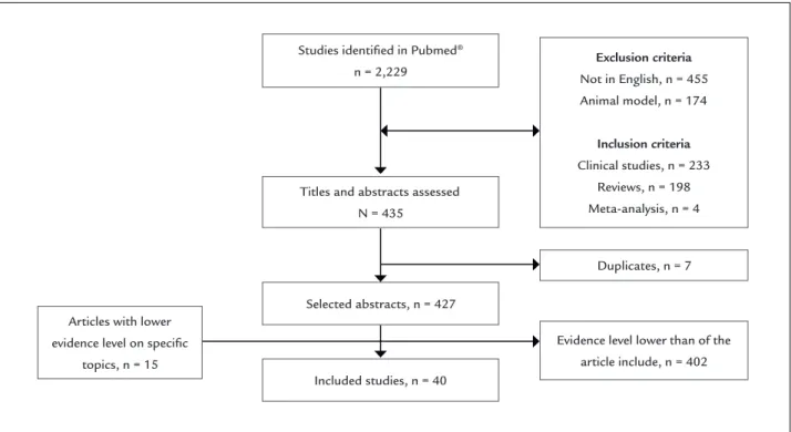
to-tal of 428 studies, excluding duplicates. The summaries of these articles were assessed by two independent authors in order to select the studies with the best level of evidence in each theme covered during the review. Articles on speciic and relevant topics without a level of evidence over II were also included (Figure 1).
T
ECHNICALPRINCIPLESESWL consists of the fragmentation of calculi by means of pulsed acoustic waves at high intensity and low fre-quency directed at the stone from an external power sour-ce to the patient, called a lithotriptor. To optimize the re-sults of ESWL, several technical factors must be taken into consideration, such as the type of device,1 the energy
level, the frequency of the pulses, the quality of coupling between patient and lithotripsy machine, the focal zone, the site of the calculus, and anesthetic technique.
The procedure should start with a low energy level (13-14KV) in each pulse, which is then gradually increa-sed.2 The successive shock waves result in direct shearing
forces, as well as in the formation of cavitation bubbles around the stone, which when ruptured generate energy that enhances the fragmentation of the calculus.3
Curren-tly, frequencies from 60-90 shocks/minute have been cho-sen, with a gradual increase in energy with the aim of in-creasing the fragmentation of the stone and reducing the
morbidity of the procedure.4,5 A recent meta-analysis
pro-ved the superior eficacy of ESWL with a frequency of 60 in relation to 120 shocks per minute.4 And as
demons-trated by Pace et al.,6 this beneit is clearest in calculi over
10 mm.
The correct coupling of patients to lithotripsy machi-nes increases the success of ESWL. The presence of air in the path of the shock waves is inversely proportional to its effectiveness.7-9 The focal zone has also been investigated
to optimize the delivery of shock waves, with current commendations for a larger focal zone (50 x 9 mm) for re-nal calculi and a smaller one (28 x 6 mm) for ureteral ones.3
The preparation of the patient consists of an appro-priate assessment before the procedure, using non-con-trast spiral computed tomography as the investigation of choice, as it provides fundamental information regar-ding indication and prognosis.10 Coagulation proiles and
urine cultures should be checked prior to the procedure. The patient usually remains in a supine position, but in cases of distal ureter calculi, horseshoe kidney or pelvic kidney, changing to a ventral position generates a better “window”, free from the iliac crest. According to the size, density and location of the calculus, its identiication is made either by luoroscopy or ultrasound. The latter me-thod has the advantages of not using ionizing radiation and a greater sensitivity in the characterization of low
FIGURE 1 Flowchart for the identiied, selected and/or excluded studies in the review.
Titles and abstracts assessed N = 435
Exclusion criteria
Not in English, n = 455 Animal model, n = 174
Inclusion criteria
Clinical studies, n = 233 Reviews, n = 198 Meta-analysis, n = 4
Duplicates, n = 7
Selected abstracts, n = 427 Articles with lower
evidence level on specific topics, n = 15
Included studies, n = 40
Evidence level lower than of the article include, n = 402 Studies identified in Pubmed®
density renal and ureteral stones. The exact location of the calculus is essential to the success of ESWL, so that the use of high frequency ventilation with low current volu-me to reduce breathing movevolu-ments is a valid alternati-ve.11, 12 Increasing the focal zone or the use of automated
tracking systems via luoroscopy also help the correct de-livery of shock waves on the calculus.3 After general
anes-thesia, which is preferred owing to better results,13 or
se-dation, the procedure can begin. In most services, about 3,000 pulses are applied, with a total procedure time of around 1 hour.
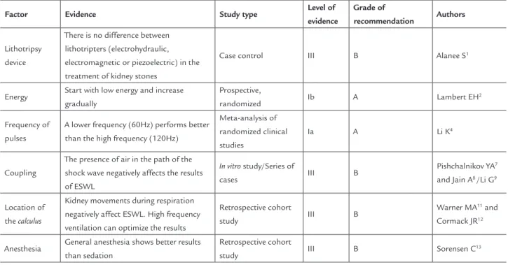
Table 1 summarizes the technical factors and their respective levels of evidence/grade of recommendation impacting the ESWL results.
Indications
Currently, ESWL is considered a irst-line treatment for renal calculi under 2.0 cm, with a success rate ranging from 33 to 91%.14 Some series have already reported the use of
ESWL for calculi over 2.0 cm, but the low success rates and the need for multiple sessions to optimize results are li-miting factors.14 Owing to its low invasiveness, ESWL is
also recommended in the use of ureteral calculi.15 A recent
meta-analysis demonstrated that emergency ESWL for ureteral calculi presents an overall stone-free rate of 78% (75-82%), with 79% (61-95%) in proximal ureter calculi,
78% (69-88%) in mid ureter calculi and 79% (74-84%) in distal ureter calculi.16
Contraindications
The formal contraindications for ESWL are: pregnancy, un-treated urinary tract infection/urosepsis, decompensated coagulopathy, uncontrolled arrhythmia, and abdominal aortic aneurysm >4.0 cm.17 In the presence of any of these
conditions, other treatment methods should be proposed.
Predictors of success
A series of factors can inluence the results of ESWL, espe-cially factors related to the calculus (size, location, compo-sition-density), factors related to renal anatomy (obstruc-tion/stasis, hydronephrosis, stenosis of the ureteropelvic junction, calyceal diverticula, horseshoe kidney, ectopic kid-ney/renal fusion) and patient-related factors (obesity, skin to stone distance, renal failure).
ESWL presents results inversely proportional to the size of the calculus. A meta-analysis published on 1994 by Lingeman et al.14 demonstrated that ESWL has a success
rate of 74, 56 and 33% for kidney stones up to 1.0 cm, 1.0 to 2.0 cm and greater than 2.0 cm, respectively.
Various articles have been published studying predic-tors of the success of ESWL. Al-Ansari et al.18 in a
retros-pective study of 427 patients with calculi up to 3.0 cm
un-TABLE 1 Technical factors impacting the extracorporeal shock wave lithotripsy results
Factor Evidence Study type Level of
evidence
Grade of
recommendation Authors
Lithotripsy device
There is no difference between lithotripters (electrohydraulic, electromagnetic or piezoelectric) in the treatment of kidney stones
Case control III B Alanee S1
Energy Start with low energy and increase gradually
Prospective,
randomized Ib A Lambert EH
2
Frequency of pulses
A lower frequency (60Hz) performs better than the high frequency (120Hz)
Meta-analysis of randomized clinical studies
Ia A Li K4
Coupling
The presence of air in the path of the shock wave negatively affects the results of ESWL
In vitro study/Series of
cases III B
Pishchalnikov YA7
and Jain A8 /Li G9
Location of the calculus
Kidney movements during respiration negatively affect ESWL. High frequency ventilation can optimize the results
Retrospective cohort
study III B
Warner MA11 and
Cormack JR12
Anesthesia General anesthesia shows better results than sedation
Retrospective cohort
study III B Sorensen C
of ESWL for ureteral calculi. In a similar study, Kanao et al.21 studied 435 patients with renal and ureteral calculi and
developed a nomogram considering size, location (renal pelvis renal vs. renal calyx vs. proximal ureter vs. distal ure-ter) and number of calculi as predictors of success. The highest success rate was obtained for single proximal ure-teral calculi less than 5 mm (93.8%) while the worst was for multiple calycine calculi larger than 21 mm (10.5%).
There are few prospective studies evaluating the pre-dictors of success of ESWL in renal calculi. A study on 120 patients with a single renal calculus between 0.5 and 2.5 cm undergoing ESWL showed good results on a three--month follow-up with computed tomography, with 87.5% of patients free of stones or with residual calculi less than 4 mm. In this study, only the body mass index (p = 0.04) and calculus densities greater than 1000 Hounsield units (p = 0.02) were predictors of success after the multivaria-te analysis.10
A topic still under debate regarding ESWL is its in-dication for lower calyx calculi, where renal anatomy, more precisely the infundibular calicinal angle, infun-dibular length, width and height can have a negative impact.22-24
Table 2 summarizes the main factors for poor prog-nosis in the success of ESWL, as well as the levels of evi-dence/grade of recommendation.
ESWL adjuvant factors
The use of antibiotics for ESWL in patients with sterile urine is not necessary.25 A meta-analysis involving nine
studies with a total of 1,364 patients showed that anti-biotic prophylaxis did not reduce the incidence of fever (RR 0,36 95% CI 0.07-2.36, p=0.31) and urinary infection (RR 0.54, 95% CI 0.29-1.01, p=0.05) (Level of evidence: 1a; Grade of recommendation A).
Routine double-J stenting before ESWL does not in-crease the stone-free rate and does not reduce complica-tions, and therefore should not be encouraged (Level of evidence: 1a; Grade of recommendation A). Even in pa-tients with a single kidney the procedure without urete-ral catheter is feasible, although it requires a careful se-lection of candidates.26 A systematic review,27 using
sources such as the PubMed®, Embase® and Cochrane
databases evaluated the results and complications of ESWL in the treatment of calculi of the upper urinary tract with or without double-J stenting before the proce-dure. Rate of calculus-free response, steinstrasse, lower urinary tract symptoms, hematuria, fever, infection, pain, dergoing ESWL reported a success rate (deined as patients
free of calculi or fragments under 4 mm) of 78% in three months, while 53.1% of these required more than one ses-sion and 8.4% had treatment complemented by another procedure (percutaneous nephrolithotomy, lexible ure-teroscopy or double-J stenting). In this study of cases, the size, location and number of calculi, as well as renal ana-tomy and congenital anomalies have an impact on the success rate. Calculi smaller than 10 mm had a rate of suc-cess of 90%, while those larger than 10 mm presented a rate of 70% (p <0.05). Calculi located in the renal pelvis and upper pole had a success rate of 87.3 and 88.5%, res-pectively, while for lower pole calculi this was 69.5% (p <0.05). Kidneys with a single calculus had a 78.3% success rate compared to 62.8% in multiple kidney calculi (p<0.01). Kidneys without dilation had an 83% success rate, while for kidneys with hydronephrosis success reached 76% (p<0.05). Kidneys without congenital anomalies had a success rate of 79% compared to 54% in kidneys with ano-malies (p<0.03). In a larger study of cases encompassing 2,954 patients with calculi less than 3.0 cm undergoing ESWL, monotherapy with ESWL presented a stone-free rate of 86.7% at a 3 month follow-up. A logistic regres-sion conirmed that size, location, number of calculi, re-nal anatomy and congenital disorders are predictors of success.
Some studies have analyzed the characteristics of the
calculus and the patient. In a retrospective study with 111 patients with calculi under 2.0 cm submitted to ESWL, Perks et al.19 reported a stone-free rate of 40% and complete
frag-mentation in 24%. The multivariate analysis including body mass index, size, location, attenuation, skin-to-stone dis-tance (SSD) and composition of the calculation showed that the latter three factors were signiicantly and indepen-dently associated with elimination or complete fragmen-tation outcomes. Patients with favorable parameters had signiicantly better results than patients with unfavorable parameters (odds ratio = 7.1, 95% conidence interval = 1.6 to 32, p=0.01).
In an attempt to create a clinical nomogram for pre-dicting the success of ESWL for renal and ureteral calculi, Wiesenthal et al.20 studied 422 patients with renal or
nausea and vomiting were analyzed, as well as the need for analgesia and auxiliary procedures for elimination of the calculi. Eight randomized studies were identiied, in-cluding a total of 876 patients divided into two groups: 453 with catheters and 423 without catheters. The result of the meta-analysis showed no signiicant difference between groups (RR 0.97, 95% CI 0.91-1.03, p=0.27). The incidence of steinstrasse in the catheter group was simi-lar to the group without catheters, except in one study. Nevertheless, the incidence of lower urinary tract symp-toms was much higher in patients with a catheter (RR 4.10, 95% CI 2.21-7.61, p<0.00001). There was no signii-cant difference in rates of hematuria, fever, infection, pain, nausea or vomiting, and need for analgesia or auxiliary procedures between the groups. It was concluded that double-J stenting prior to ESWL does not increase the rate of patients free from stones or reduce the need for auxiliary procedures, and boosts the presence of lower urinary tract symptoms.
In relation to care after ESWL, there is good eviden-ce that drug treatment with alpha-blockers, speciically tamsulosin, provides beneits (Level of evidence: 1a; Gra-de of recommendation A). A recent meta-analysis has been published assessing the real eficacy of tamsulosin in patients undergoing ESWL,28 verifying that the drug
increases the average calculus elimination rate by 16% (5 to 27%) and decreases calculus elimination time by 8 (3 to 20) days on average. Other medication such as
nifedipi-ne have also proved effective as an adjunct to ESWL treat-ment, but with a higher rate of side effects such as dizzi-ness and hypotension.29
Complications
A series of minor complications can occur after ESWL. Pain in the costovertebral angle and lank, the appea-rance of petechiae or subcutaneous bruising at the entry and exit point of the shock waves are common, requi-ring analgesics in up to 40% of cases.30 Microscopic
maturia occurs in virtually all cases, however gross he-maturia appears only in about one third of patient.31 A
prospective study of 3,241 patients with calculi larger than 4 mm undergoing ESWL (7,245 sessions) and mo-nitored for a period of three months reported 4,075 com-plications, including renal colic (40%), gross hematuria (32%), urinary obstruction (30.9%) and perirenal hema-toma or subclinical subcapsular hemahema-toma (4.6%) as the most common. Furthermore, symptomatic bacteriu-ria was diagnosed in 9.7% of cases.32 Patients with pain
are effectively treated with anti-spasmodic or anti-in-lammatory drugs without further intervention requi-red in most cases, such as repeated ESWL or ureteros-copy. Patients with gross hematuria present spontaneous improvement within 48 hours in 85% of cases, and in 10 days, in virtually 100% of cases.32 Patients with
uri-nary obstruction can be clinically treated with alpha--blockers or surgically through double-J stenting or ure-TABLE 2 Predictors of the success of extracorporeal shock wave lithotripsy
Factor Evidence Study type Level of
evidence
Grade of recommendation
Authors
Size of the
calculus
Size is inversely proportional to the ESWL result Meta-analysis of cohort studies
IIa B Lingeman JE14
calculus
density
High density calculi present worse results (>1000
UH)
Prospective cohort study
IIb B El-Nahas AR10
Location of the calculus
Lower pole renal calculi present worse results than
mid-pole and upper pole calculi. Calycine calculi
have worse outcomes compared to renal pelvic and ureteral stones.
Retrospective cohort study
III B Al Ansari
A18 and Kanao
K21
Skin-to-stone distance
Distances over 9 cm negatively affect ESWL Retrospective cohort study
III B Perks AE19 and
Wiesenthal JD20
Anatomy of the excretory path
Unfavorable anatomy (infundibulopelvic angle <90°, infundibular length > 3.0 cm and
infundibular width of <4-5 mm) negatively affect ESWL
Retrospective cohort study
III B Elbahnasy22
Kidney anomalies
Kidneys with congenital anomalies have lower elimination rates
Retrospective cohort study
teroscopy depending on the size, number and location of the calculi. Perirenal hematomas should be monito-red with imaging exams and control of hemoglobin and hematocrit when large in size. Rare cases of post-ESWL renal explosion have been reported and even then a con-servative treatment may be appropriate.33 In a study of
6,172 ESWL sessions, Razvi et al.34 evaluated the risk
fac-tors for the development of perirenal hematoma and identiied intraoperative hypertension (hazard ratio = 3.3, 95% CI 1.6-10.2, p = 0.03) and use of anticoagulant/ antiplatelet agents (hazard ratio = 4.2, 95% CI 1.1 -15.9, p = 0.03) as signiicant.
In relation to late complications, a series of articles has sought to demonstrate an association between ESWL and the development of hypertension and diabetes. In a retrospective study of 727 patients undergoing ESWL, Chew et al.35 did not ind a higher incidence of these
di-seases in the study group compared with the popula-tion mean. In a study of 4,782 patients with calculi and without hypertension monitored for an average of 8.7 years, Krambeck et al.36 also found no association between
ESWL and hypertension in both univariate and multi-variate analyses, including age, sex and obesity (hazard ratio = 1.03; 95% CI 0.8-1.2, p=0.77). However, in a study with the collection of prospective data via a question-naire sent to and answered by 2,041 patients undergoing ESWL, B arbosa et al.37 found a signiicant yet small
in-crease in the incidence of hypertension in these patients compared to controls matched for age, sex and body mass index (37.8 versus 32.5%, p = 0.0009). In relation to the development of diabetes, a study with an identical design to the one mentioned above and with 1,869 pa-tients submitted to ESWL did not ind an increased in-cidence of diabetes in such patients compared to con-trols matched for age, sex and body mass index (5.2
versus 5.8%; p=0.47).38 A study of 5,287 patients with
cal-culi and without diabetes monitored for an average of 8.7 years found no association between ESWL and the appearance of diabetes in both univariate and multiva-riate analyses, including age, sex and obesity (hazard ra-tio = 0.92; 95% CI 0.71-1.18).39 The association between
ESWL and the development of chronic diseases (hyper-tension and diabetes) is unclear and studies with higher levels of evidence are needed to conirm or rule out this association (Level of evidence 2b; Grade of recommen-dation B). Lastly, in relation to a possible worsening of renal function after ESWL, in a study of 156 patients with a single kidney undergoing ESWL and an average follow up of 3.8 years, El-Assmy et al.40 found no
chan-ges in creatinine levels, demonstrating the safety of this
method at least in the medium term (Level of evidence: 3; Grade of recommendation B).
C
ONCLUSIONESWL shows good results in the treatment of kidney sto-nes up to 2.0 cm and is an alternative to ureteroscopy in the management of ureteral calculi. Several technical tors are important for the optimization of results, and fac-tors related to the patient and the stone, such as its size, density, skin to stone distance, anatomy of the excretory system and renal anomalies, help predict the chances of success. Antibiotic prophylaxis and double J stent place-ment before the procedure are not required. Alpha-bloc-kers may increase success rates. Early and important com-plications are rare and late comcom-plications are not yet proven.
R
ESUMOLitotripsia extracorpórea no tratamento de cálculos re-nais e ureterais.
A utilização de certos princípios técnicos e a seleção de ca-sos favoráveis podem otimizar os resultados da litotripsia extracorpórea por ondas de choques (LECO). O objetivo deste trabalho é revisar os princípios de funcionamento da LECO, suas indicações e contraindicações, fatores prediti-vos de sucesso e suas complicações. Realizou-se uma pes-quisa na base de dados do Pubmed® entre janeiro/1984 e
outubro/2013 utilizando como palavras chaves shock wave lithotripsy e stone. Apenas artigos com bom nível de evidên-cia, de língua inglesa, em seres humanos, do tipo clinical trials ou de revisão/metanálise foram incluídos. Na busca pela otimização dos resultados da LECO, diversos fatores técnicos, como o tipo de aparelho de litotripsia, energia e frequência dos pulsos, acoplamento do paciente ao lito-tridor, localização do cálculo e tipo de anestesia, devem ser levados em consideração. Fatores relacionados ao doen-te e ao cálculo, como seu tamanho, densidade, distância pele-cálculo, anatomia da via excretora e anomalias renais, também são importantes. A proilaxia com antibiótico não é necessária, e a passagem de duplo J de rotina não é recomendada. A prescrição de alfabloqueadores, particu-larmente a tansulosina, é benéica em cálculos > 10 mm. Complicações menores podem ocorrer após LECO e ge-ralmente respondem bem a condutas clínicas. A relação entre LECO e o surgimento de hipertensão e diabetes não está comprovada.
R
EFERENCES1. Alanee S, Ugarte R, Monga M. The effectiveness of shock wave lithotripters: a case matched comparison. J Urol. 2010;184:2364-7.
2. Lambert EH, Walsh R, Moreno MW, Gupta M. Effect of escalating versus
ixed voltage treatment on stone comminution and renal injury during extracorporeal shock wave lithotripsy: a prospective randomized trial. J Urol. 2010;183:580-4.
3. Rassweiler JJ, Knoll T, Kohrmann KU, McAteer JA, Lingeman JE, Cleveland RO et al. Shock wave technology and application: an update. Eur Urol. 2011;59:784-96.
4. Li K, Lin T, Zhang C, Fan X, Xu K, Bi L, et al. Optimal frequency of shock wave lithotripsy in urolithiasis treatment: a systematic review and meta-analysis of randomized controlled trials. J Urol. 2013;190:1260-7. 5. Mazzucchi E, Brito AH, Danilovic A, Ebaid GX, Chedid Neto E, Azevedo JR,
et al. Comparison between two shock wave regimens using frequencies of 60 and 90 impulses per minute for urinary stones. Clinics. 2010;65:961-5. 6. Pace KT, Ghiculete D, Harju M, Honey RJ, University of Toronto. Lithotripsy
A. Shock wave lithotripsy at 60 or 120 shocks per minute: a randomized, double-blind trial. J Urol. 2005;174:595-9.
7. Pishchalnikov YA, Neucks JS, VonDerHaar RJ, Pishchalnikova IV, Williams JC Jr., McAteer JA. Air pockets trapped during routine coupling in dry head lithotripsy can signiicantly decrease the delivery of shock wave energy. J Urol. 2006;176:2706-10.
8. Jain A, Shah TK. Effect of air bubbles in the coupling medium on eficacy of extracorporeal shock wave lithotripsy. Eur Urol. 2007;51:1680-6. 9. Li G, Williams JC Jr, Pishchalnikov YA, Liu Z, McAteer JA. Size and location
of defects at the coupling interface affect lithotripter performance. BJU Int. 2012;110:E871-7.
10. El-Nahas AR, El-Assmy AM, Mansour O, Sheir KZ. A prospective multivariate analysis of factors predicting stone disintegration by extracorporeal shock wave lithotripsy: the value of high-resolution noncontrast computed tomography. Eur Urol. 2007;51:1688-93.
11. Warner MA, Warner ME, Buck CF, Segura JW. Clinical eficacy of high frequency jet ventilation during extracorporeal shock wave lithotripsy of renal and ureteral calculi: a comparison with conventional mechanical ventilation. J Urol. 1988; 139: 486-7.
12. Cormack JR, Hui R, Olive D, Said S. Comparison of two ventilation techniques during general anesthesia for extracorporeal shock wave lithotripsy: high-frequency jet ventilation versus spontaneous ventilation with a laryngeal mask airway. Urology. 2007;70:7-10.
13. Sorensen C, Chandhoke P, Moore M, Wolf C, Sarram A. Comparison of intravenous sedation versus general anesthesia on the eficacy of the Doli 50 lithotriptor. J Urol. 2002;168:35-7.
14. Lingeman JE, Siegel YI, Steele B, Nyhuis AW, Woods JR. Management of lower pole nephrolithiasis: a critical analysis. J Urol. 1994;151:663-7. 15. Lindqvist K, Holmberg G, Peeker R, Grenabo L. Extracorporeal shock-wave
lithotripsy or ureteroscopy as primary treatment for ureteric stones: a retrospective study comparing two different treatment strategies. Scand J Urol. 2006;40:113-8.
16. Picozzi SC, Ricci C, Gaeta M, Casellato S, Stubinski R, Ratti D, et al. Urgent shock wave lithotripsy as irst-line treatment for ureteral stones: a meta-analysis of 570 patients. Urol Res. 2012;40:725-31.
17. Streem SB. Contemporary clinical practice of shock wave lithotripsy: a reevaluation of contraindications. J Urol. 1997;157:1197-203.
18. Al-Ansari A, As-Sadiq K, Al-Said S, Younis N, Jaleel OA, Shokeir AA. Prognostic factors of success of extracorporeal shock wave lithotripsy (ESWL) in the treatment of renal stones. Int Urol Nephrol. 2006;38:63-7.
19. Perks AE, Schuler TD, Lee J, Ghiculete D, Chung DG, D’A Honey RJ, et al. Stone attenuation and skin-to-stone distance on computed tomography predicts for stone fragmentation by shock wave lithotripsy. Urology. 2008;72:765-9.
20. Wiesenthal JD, Ghiculete D, Ray AA, Honey RJ, Pace KT. A clinical nomogram to predict the successful shock wave lithotripsy of renal and ureteral calculi. J Urol. 2011;186:556-62.
21. Kanao K, Nakashima J, Nakagawa K, Asakura H, Miyajima A, Oya M, et al. Preoperative nomograms for predicting stone-free rate after extracorporeal shock wave lithotripsy. J Urol. 2006;176:1453-6.
22. Elbahnasy AM, Clayman RV, Shalhav AL, Hoenig DM, Chandhoke P, Lingeman JE, et al. Lower-pole caliceal stone clearance after shockwave lithotripsy, percutaneous nephrolithotomy, and lexible ureteroscopy: impact of radiographic spatial anatomy. J Endourol. 1998;12:113-9.
23. Tuckey J, Devasia A, Murthy L, Ramsden P, Thomas D. Is there a simpler method for predicting lower pole stone clearance after shockwave lithotripsy than measuring infundibulopelvic angle? J Endourol. 2000;14:475-8. 24. Sampaio FJ, DAnunciacao AL, Silva EC. Comparative follow-up of patients
with acute and obtuse infundibulum-pelvic angle submitted to extracorporeal shockwave lithotripsy for lower caliceal stones: preliminary report and proposed study design. J Endourol. 1997;11:157-61.
25. Lu Y, Tianyong F, Ping H, Liangren L, Haichao Y, Qiang W. Antibiotic prophylaxis for shock wave lithotripsy in patients with sterile urine before treatment may be unnecessary: a systematic review and meta-analysis. J Urol. 2012;188:441-8.
26. Kumar S, Sakthivel A, Chacko KN, Kekre NS, Ganesh G. Shock wave lithotripsy in solitary functioning kidneys: is prophylactic stenting necessary? Urol Int. 2006;77:179-81.
27. Shen P, Jiang M, Yang J, Li X, Li Y, Wei W, et al. Use of ureteral stent in extracorporeal shock wave lithotripsy for upper urinary calculi: a systematic review and meta-analysis. J Urol. 2011;186:1328-35.
28. Zhu Y, Duijvesz D, Rovers MM, Lock TM. Alpha-Blockers to assist stone clearance after extracorporeal shock wave lithotripsy: a meta-analysis. BJU Int. 2010;106:256-61.
29. Vicentini FC, Mazzucchi E, Brito AH, Chedid Neto EA, Danilovic A, Srougi M. Adjuvant tamsulosin or nifedipine after extracorporeal shock wave lithotripsy for renal stones: a double blind, randomized, placebo-controlled trial. Urology. 2011;78:1016-21.
30. Newman RC, Bezirdjian L, Steinbock G, Finlayson B. Complications of extracorporeal shock wave lithotripsy: prevention and treatment. Semin Urol. 1986;4:170-4.
31. Sofras F, Karayannis A, Kostakopoulos A, Delakas D, Kastriotis J, Dimopoulos C. Methodology, results and complications in 2000 extracorporeal shock wave lithotripsy procedures. BJU Int. 1988;61:9-13.
32. Salem S, Mehrsai A, Zartab H, Shahdadi N, Pourmand G. Complications and outcomes following extracorporeal shock wave lithotripsy: a prospective study of 3.241 patients. Urol Res. 2010;38:135-42.
33. Marchini GS, Lopes RI, Bruschini H, Torricelli F, Lopes RN. Conservative treatment of severe renal trauma after extracorporeal shockwave lithotripsy. Rev Col Bras Cir. 2011;38:447-9.
34. Razvi H, Fuller A, Nott L, Méndez-Probst CE, Leistner R, Foell K, et al. Risk factors for perinephric hematoma formation after shockwave lithotripsy: a matched case-control analysis. J Endourol. 2012;26:1478-82.
35. Chew BH, Zavaglia B, Sutton C, Masson RK, Chan SH, Hamidizadeh R, et al. Twenty-year prevalence of diabetes mellitus and hypertension in patients receiving shock-wave lithotripsy for urolithiasis. BJU Int. 2012; 109:444-9.
36. Krambeck AE, Rule AD, Li X, Bergstralh EJ, Gettman MT, Lieske JC. Shock wave lithotripsy is not predictive of hypertension among community stone formers at long-term followup. J Urol. 2011;185:164-9.
37. Barbosa PV, Makhlouf AA, Thorner D, Ugarte R, Monga M. Shock wave lithotripsy associated with greater prevalence of hypertension. Urology. 2011;78:22-5.
38. Makhlouf AA, Thorner D, Ugarte R, Monga M. Shock wave lithotripsy not associated with development of diabetes mellitus at 6 years of follow-up. Urology. 2009;73:4-8.
39. De Cógáin M, Krambeck AE, Rule AD, Li X, Bergstralh EJ, Gettman MT, et El. Shock wave lithotripsy and diabetes mellitus: a population-based cohort study. Urology. 2012;79:298-302.