SUMMARY
Although noroviruses (NoVs) were the irst viral agents linked to gastrointestinal disease, for a long time they have been considered secondary cause of gastroenteritis, second to rotaviruses as etiologic agents. he development of molecular techniques in diagnosing NoV provided a clearer insight into the epidemiological impact of these viruses, which are currently recognized not only as the leading cause of non-bacterial gastroenteritis outbreaks, but also as a major cause of sporadic gastroenteritis in both children and adults. his review focuses on the required knowledge to understand their morphology, genetics, transmission, pathogenesis, and control. Since no vaccine is available, preven-tion of NoV infecpreven-tion relies mainly on strict community and personal hygiene measures. Keywords: Norovirus; gastroenteritis; diarrhea.
Study conducted at Instituto Adolfo Lutz, Virology Center, Nucleus of Enteric Diseases, São Paulo, SP, Brazil
Submitted on: 01/18/2011 Approved on: 05/01/2011
Correspondence to: Maria do Carmo Sampaio Tavares
Timenetsky Av. Dr. Arnaldo, 355 CEP 01246-902 São Paulo, SP, Brazil Phone: 55 + 11 3068 2909 Fax: 55 + 11 3085 3505 [email protected]
Conlict of interest: None.
©2011 Elsevier Editora Ltda. All rights reserved.
Norovirus: an overview
SIMONE GUADAGNUCCI MORILLO1, MARIADO CARMO SAMPAIO TAVARES TIMENETSKY2
1 M.Sc. in Sciences, Laboratory Research Program in Public Health/Disease Control Coordination Ofice, State Secretary of Health, SP; Biologist, Nucleus of Enteric Diseases, Virology Center, Instituto Adolfo Lutz, State Secretary of Health, São Paulo, SP, Brazil
Figure 1 – Bovine calicivirus, viral particles extracted from stool sample (Stewart McNulty, Veterinary Sciences, Queen’s University, Belfast). Negative dye technique by direct electron microscopy. Magniication 33,000 X. Available at: http: //www.qub.ac.uk
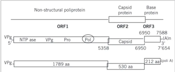
Figure 2 – Norovirus genomic organization. Localization of the three ORFs and the Pol region used for the design of the primer pool used in the RT-PCR for the identiication of genogroups and genotypes. From Atmar & Estes, 2001.29 INTRODUCTION
Acute gastroenteritis is one of the most common diseases in humans; in the United States, it is the second leading cause for reporting, followed by respiratory infections1.
A billion cases of acute diarrhea are estimated to occur yearly in children and adults worldwide2. Gastroenteritis
is usually expressed as a mild diarrhea, but it can be seen as a severe form with enhanced symptoms (nausea and vomiting), possibly leading to dehydration and death. he annual mortality associated with gastroenteritis has been estimated as four to six million people2.
he etiology of diarrheas can involve several agents, such as viruses, bacteria, and parasites. Bacterial agents are relatively more important in developing countries, where-as viral agents are more relevant in industrialized coun-tries. he importance of these agents is related to hygiene and sanitation conditions for the population1. In 1972, a
27-nm viral particle was discovered in an infectious iltrate of human fecal samples over a gastroenteritis outbreak in Norwalk, Ohio3. Since then, the number of viral agents
as-sociated with gastroenteritis has progressively increased, with rotaviruses4, astroviruses5, and Norwalk-like viruses6
being identiied.
Currently, most gastroenteritis in children are con-sidered to be caused by viruses included in four diferent families: Reoviridae (rotavirus), Caliciviridae (norovirus
and sapovirus), Astroviridae (astrovirus), and Adenoviri-dae (adenovirus)7.
VIRALPARTICLESTRUCTUREOF NOROVIRUSES
Virions consist of a capsid and a nucleic acid measuring about 27 to 30 nm in diameter. hey have no envelope. he nucleocapsid is rounded and exhibits an icosahedral sym-metry. he surface structure reveals a regular model with distinct features8. he capsomere arrangement is clearly
visible (Figure 1).
he virus genome consists of a linear molecule of sin-gle-strand RNA with a positive polarity. he genomes with these features serve as mRNA. As soon as they enter the target-cell, they are bound to cell ribosomes and protein translation occurs. he genome RNA serves as a template for a complementary negative strand being transcribed into genome RNA through the viral polymerase. he com-plete genome contains approximately 7.5 kb and consists of 45%-56% of cytosine + guanine (C + G). he genome 5’ end presents the VPg protein, having an essential role in virus infectivity and initial translation; in the 3’ end, the poli A tail addition occurs ater the gene synthesis and its function is giving stability to the molecule and helping translation8.
he three open read frames (ORF) of the virus ge-nome can be observed in Figure 2: the irst ORF encodes a 194-kDa polyprotein which is cleaved by the virus prote-ase 3C into six likely proteins, including RNA-dependent RNA polymerase. hus, the 5’-end in the genome encodes a precursor of non-structural proteins involved in the vi-rus transcription and replication9. he second ORF
en-codes a 60-kDa capsid protein (VP1), a structural protein with a major role in virus replication10. he third ORF is
considered the most variable region in the genome and en-codes the 23-kDa basic protein (VP2) interacting with the genome RNA when the virion formation occurs11.
NOMENCLATUREAND CLASSIFICATION
Because of low viral (NoV) load in feces and diicult spread in both cell culture and laboratory animals, the vi-rus classiication was deined only from 1990. Since then, some calicivirus genomes have been sequenced, allowing framing most of these viruses into the Caliciviridae
fam-ily12.
In 2005, a new classiication system was established and based on ORF2 phylogenetic analyses of 164 NoV sequences. NoVs can be subdivided into ive genogroups (GI, GII, GIII, GIV, GV), consisting of at least 31 genetic clusters or genotypes: 8 genotypes in GI genogroup, 17 in GII, 2 in GIII, 1 in GIV, and 1 in GV13. Across all of them,
Non-structural poliprotein
NTP ase VPg
5’
5358 6950
212 aa 530 aa
1789 aa
7588 6950
7’654 VPg
VPg
Capsid
Pro Pol
Capsid protein
Base protein
ORF1 ORF2 ORF3
(A)n
(poli A)
GI and GII are the genogroups presenting the largest ge-netic diversity; six new genotypes were new genotypes de-ined and described: GI/8 in serogroup GI and GII/13-17 in genogroup GII14. NoVs with genogroups GI, GII, and
GIV are found in humans, except for the sample of NV S11/GII found in swine; genogroups GIII and GV are found in cattle and mice, respectively15. Recently,
molecu-lar epidemiology studies have demonstrated 70% of NoV outbreaks are caused by the variant genotype GII.416,17.
PATHOGENESISANDREPLICATION
Human caliciviruses cause infection predominantly by the oral route. Virions are stable in acid and they can survive ater passing through the stomach. NoVs are highly infec-tious due to the combination of low infecting dose (DI 50 < 20 virus particles), high virus excretion level (108 to
1010 copies of RNA per gram of feces) and extended
ex-cretion ater clinical recovery18,19. he virus is replicated in
the enterocyte cytoplasm, where the positive polarity RNA acts as mRNA. Studies showed the virus elimination can continue for over 2 weeks ater the symptomatic phase of the disease, as well as cases of asymptomatic infection 20
with an implication on outbreaks caused by food transmis-sion diseases21. here is little evidence that NoVs can cause
a chronic infection in a normal host; however, a study per-formed in immunocompromised children and adolescents by Levett et al.22 reported NoV GII shedding over at least
8 months.
TRANSMISSION
NoVs are the main cause of acute nonbacterial human gastroenteritis, being transmitted from food or from per-son to perper-son via a fecal-oral route, afecting adults and children all over the world23. Indirect evidence in
epide-miological studies suggests the virus transmission can be airborne, such as in explosive vomiting occurred during the disease24. Transmission can also occur via water
reser-voir when groundwater is contaminated25. hey are highly
contagious, possibly occurring in either sporadic cases or in great acute diarrhea outbreaks in wards, hospitals, schools, universities, camping places, cruises, hotels, and restaurants26. Food quality control is oten based on
bacte-rial contamination; thus, virus contamination may not be reported oten27. Virus transmission in hospital wards is
diicult to identify26. Filtering animals living in
contami-nated waters and which are eaten raw, such as oysters, are major transmission routes28.
Clinical features are characterized by nausea, abdomi-nal pain, vomiting, mild, self-limited, and non-bloody diarrhea. However, some patients can have severe forms, with symptoms linked to nausea and vomiting, followed by copious diarrhea, which can result in dehydration and occasionally death. Incubation period is 24 to 48 hours, with the symptoms lasting 12 to 60 hours29. Low fever and
abdominal pain can also be associated with a virus infec-tion, with the term “stomach lu” being used to describe the disease, although no biological association with in-luenza virus can be found. Approximately 10% of people with NoV require a medical visit, including hospitalization and dehydration treatment. Deaths caused by NoV are more frequently reported in elderly people. Around 30% of infections from NoV are asymptomatic; however, these individuals can transmit the virus, although in lower levels than symptomatic individuals30.
IMMUNITY
Because of a lack of animal model and resources to grow this virus in cell cultures, in vitro neutralization tests are
not feasible, and data of immunity development ater NoV infection is obtained from human studies with vol-unteers31. Studies indicate approximately 50% of people
exposed to the virus acquired short-term homologous im-munity, which is correlated with the serum antibody lev-el32. However, people with preexisting high antibody levels
to NoV may become ill if exposed to the virus26,32. A
candi-date vaccine has been developed, although it is not known whether the vaccine induces homotypic or heterotypic im-mune protection31,33. As no suitable prevention and
con-trol method is available, the development of a vaccine to NoV could be the best solution for this infectious disease34.
Studies suggest there is a short-term immunity ater infec-tion, and some individuals are susceptible to symptomatic infection, whereas others never develop symptoms, even ater a direct contact32.
PREVENTIONANDTREATMENT
Stopping transmission is the irst strategy for prevention, especially in hospitals and day-care centers. A number of precautions, such as hand washing with water and soap before and ater contacting the patient or objects used by him/her, must be taken when caring for a patient diag-nosed with an acute gastroenteritis. It is also required to clean all surfaces with 2% hypochlorite35, as NoV persist
in dry inanimate surfaces over eight hours to seven days36.
To avoid secondary transmissions, prevention of food contaminations during the preparation by a continuous hand washing is required. hose who handle food must wear plastic gloves when preparing raw food37. Afected
workers must not prepare food for a minimum period of three days ater the disease to avoid gastroenteritis out-breaks38.
In 2006, Rossignol40 analyzed a new drug, the nitazoxanide,
indicated to treat diarrhea caused by virus gastroenteritis. In this study, the drug eicacy in several patients with symptoms and positive diagnosis for rotavirus, enteric ad-enovirus, norovirus, and astrovirus was observed. Howev-er, higher drug efectiveness was found against rotavirus, compared with other viral pathogens.
LABORATORYDIAGNOSIS
he classic diagnosis method is electron microscopy (EM), detecting virus particles with 27 to 30 nm in diameter, the so-called SRSV. his method is used in public health labo-ratories in many countries; however, it requires a highly qualiied microscopist and very expensive equipment, making epidemiological or clinical studies impracticable41.
he immunoenzymatic method (ELISA) to detect the virus antigen uses norovirus capsid proteins expressed on baculovirus as a reactant in immunoenzymatic tests29. his
method has been recently made commercially available to diagnose NoV directly from feces (Dako Cytomation, Ely, UK 2001; Denka Seiken, Tokyo, Japan, 2002; R-Biopharm AG, Germany 2004). hese kits have low diagnostic sen-sitivity, as reported by Bull et al.42. However, the
develop-ment of new kit generations, such as RIDASCREEN 3rd
Generation kit (R-Biopharm AG, Darmstadt, Germany), which is more sensitive and speciic, entailed beneits for NoV quick diagnosis, mainly targeting outbreaks43.
he RT-PCR molecular technique, developed to iden-tify NoV, is sensitive and speciic, enabling epidemiologi-cal studies to identify gastroenteritis outbreaks44.
Interna-tional collaborative studies45 demonstrated that, among
several primer pools developed for regions ORFs 1, 2, and 3, those showing the best results were primers in POL re-gion of ORF 1 (preserved rere-gion). Phylogenetic analysis of 145 nucleotides in the POL gene region was used as a pat-tern to identify genotypes. NoV sequencing has assisted in epidemiological investigations relating clinical cases to determine a common source and to diferentiate outbreaks that could be wrongly related45.
REAL-TIME TaqMan46,47 RT-PCR and SYBR Green48
techniques quantify speciic DNA or RNA sequences in clinical samples and the gene expression from emit-ted luorescence detection since the irst ampliication cycle. hese methods have advantages over regular PCR, such as higher speciicity, sensitivity, and reproducibil-ity, in addition to allowing real-time monitoring; quicker cycling; lower RNA amount in RT-PCR reactions; and elimination of post-PCR product handling, thus reduc-ing contamination45.
EPIDEMIOLOGY
NoVs are considered the most common viral etiologic agents in outbreaks of virus gastroenteritis transmitted by water and food49. he epidemiology of diarrheal diseases
transmitted by water and food is quickly changed from the human behavior changes concerning global economy, the industry, and microbiological adaptations. he high-est disease incidence is among children under ive years of age50. However, the highest economic impact is among
elderly residing in nursing homes51. Although diarrhea
outbreaks can occur all over the year, some seasonality patterns have been observed. hese patterns are difer-ent in Northern and Southern hemispheres. In Northern hemisphere, gastroenteritis caused by NoV is more com-monly seen in winter and early spring22. In the Southern
hemisphere, outbreaks are more frequent over the spring and summer52.
From 1999 to 2002, 170 NoV outbreaks occurred in Spain. By employing EM, RT-PCR and sequencing tech-niques, the genogroup GII was observed as predominat-ing53. In England, Lopman et al.51, by analyzing samples
from outbreaks occurred between 1995 and 2002, ob-served NoV had a peak infection over the summer, in contrast with the reports from that time, describing higher occurrence in the winter.
Chapin et al.54 found NoV transmission from food
contamination crosses borders; the virus from the United States reached Guatemala and Mexico, and the samples were analyzed by RT-PCR and sequencing, with NoV genogroup GI being identiied in 65% of positive cases.
In the state of São Paulo, during the summer of 1995, a gastroenteritis outbreak occurred, afecting around 3,500 people (CVE data). EM analysis of fecal samples from this outbreak detected virus particles with SRSV morphology identiied as Norwalk-like by immunoelectron micros-copy (IEM)55. hese samples were further analyzed by
RT-PCR and sequencing and characterized as calicivirus SMA (Snow Mountain Agent) type, currently termed GII genogroup NoV56.
From 2005 to 2008, a surveillance NoV study was con-ducted in the state of Rio de Janeiro. A total 1,087 fecal samples were analyzed and about 35% were positive for NoV, with a 96% prevalence of GII genogroup and 80% GII.4; the requirement of implanting NoV diagnosis in the surveillance laboratories was described57.
In the state of São Paulo, also in 2005, several gastroen-teritis outbreaks occurred in children and adolescents, and the sample analysis demonstrated the circulation of difer-ent viruses around the same region. NoVs were detected in 21.4% of samples, followed by rotavirus, with 14.5%, astro-virus with 13.2%, and adenoastro-virus with 2.1%58.
CONCLUSION
implementation for NoV detection was initiated to detect NoV in IAL59. In 2009, with the enhancement of
conduct-ing, 15 identiied NoV outbreaks were detected in Catan-duva, Mongaguá, Piracicaba, Pitangueiras, Praia Grande, Ribeirão Preto, Sabino, São Paulo, Taubaté, Votuporanga60.
In January 2010, Guarujá, in Baixada Santista, faced a severe diarrhea outbreak with NoV (28%) and rotavirus (20%) (data from IAL). he easiness with which norovi-ruses are transmitted and the low infecting dose required to set an infection results in extensive outbreaks. he accu-rate diagnosis of virus gastroenteritis is essential to reduce the impact of the disease on the society.
REFERENCES
1. Parashar UD, Gibson CJ, Breese JS, Glass RI. Rotavirus and severe childhood diarrhea. Emerg Infect Dis 2006;12:304-6.
2. World Health Organization. WHO. State of the art of new vac-cines: research and development. 2005. p.1-13: Diarrhoeal diseases. Available from: http://www.who.int/vaccine_research/documents/ Dip%20814.pdf. [cited 2011 mar 15]
3. Kapikian AZ, Wyatt RG, Dolin R, hornhill TS, Kalica AR, Chanock RM. Visualization by immune electron microscopy of a 27 nm par-ticle associated with acute infectious nonbacterial gastroenteritis. J Virol 1972;10:1075-81.
4. Bishop RF, Davidson GP, Holmes IH, Ruck BJ. Virus particles in epithelial cells of duodenal mucosa from children with acute non-bacterial gastroenteritis. Lancet 1973;2:1281-3.
5. Madeley CR, Cosgrove BP. Letter: 28 nm particles in faeces in infan-tile gastroenteritis. 1975; Lancet 1975;2:451-2.
6. Kaplan JE, Feldman R, Campbell DS, Lookabaugh C, Gary GW. Frequency of a Norwalk-like pattern of illness in outbreaks of acute gastroenteritis. Am J Public Health 1982;72:1329-32.
7. Yamashita T, Ito M, TsuzukI H, Sakae K. Identiication of Auchi virus infection by measurement of immunosorbent assay. J Clin Microbiol 2001;39:4178-80.
8. Cubitt D, Bradley DW, Carter MJ, Chiba S, Estes MK, Saif LJ et al. Caliciviridae. ICTVdB Index of Viruses. 2003. [cited 15 Oct 2004]. Available at: http://www.ncbi.nlm.nih.gov/ICTVdb.
9. Belliot G, Sosnovtsev SV, Mitra T, Hammer C, Garield M, Green KY. In vitro proteolytic processing of the MD 145 norovírus ORF 1 non-structural polyprotein yields stable precursors and products similar to those detected in calicivirus-infected cells. J Virol 2003;77:10957-74. 10. Green SM, Dingle KE, Lambden PR, Caul EO, Ashley CR, Clarke IN.
Human enteric Caliciviridae: a new prevalent SRSV group deined by
RNA-dependent RNA polymerase and capsid diversity. J Gen Virol 1994;75:1883-8.
11. Green KY, Chanock RM, Kapikian AZ. In: Knipe DM, Howley PM,
editors. Human caliciviruses: ields virology. Philadelphia: Lippin-cott Williams and Wilkins; 2001. v. 1, p.841-74.
12. Mayo MA. A summary of taxonomic changes recently approved by ICTV. Arch Virol 2002;147:1655-63.
13. Zheng DP, Ando T, Fankhauser RL, Beard RS, Glass RI, Monroe SS. Norovirus classiication and proposed strain nomenclature. Virology 2006;346:312-23.
14. Vinjé J, Hamidjaja RA, Sobsey MD. Development and application of a capsid VP1 (region D) based reverse transcription PCR assay for genotyping of genogroup I and II Noroviruses. J Virol Methods 2004;116:109-17.
15. Green J, Vinje J, Gallimore CI, Koopmans M, Hale A, Brown DWG. Capsid protein diversity among Norwalk-like viruses. Virus Genes 2000;20:227-36.
16. Gallimore CI, Green J, Lewis D, Richards AF, Lopman BA, Eglin R et al. Diversity of noroviruses cocirculating in the north of England from 1998 a 2001. J Clin Microbiol 2004;42:1396-401.
17. Tu ET, Nguyen T, Lee P, Bull RA, Musto J, Musto J et al. Norovi-rus GII.4 strains and outbreaks, Australia. Emerg Infect Dis 2007;13:1128-30.
18. Lee N, Chan MC, Wong B, Chor KW, Sin W, Choi KW et al. Fecal viral concentration and diarrhea in norovirus gastroenteritis. Emerg Infect Dis 2007;13:1399-401.
19. Tu ET, Bull RA, Kim MJ, Mc Ivir CJ, Heron I, Mc Iver CJ et al. Noro-virus excretion in an aged- care setting. J Clin Microbiol 2008; 46: 2119-21.
20. Okhuysen PC, Jiang X, Ye L, Johnson PC, Estes MK. Viral shedding and fecal IgA response ater Norwalk virus infection. J Infect Dis 1995;171:566-9.
21. Parashar UD, Dow L, Fankhauser RL, Miller J, Ando J, Williams KS et al. An outbreak of viral gastroenteritis associated with consump-tion of sandwiches: implicaconsump-tions for the control of transmission by food handlers. Epidemiol Infect 1998;121:615-21.
22. Levett PN, Gu M, Luan B, Fearson M, Stubber J, Jamieson F et al. Longitudinal study of molecular epidemiology of small round-struc-tured viruses in a pediatric population. J Clin Microbiol 1996; 34: 1497-501.
23. Kirkwood CD, Bishop RF. Molecular detection of human calicivirus in young children hospitalized with acute gastroenteritis in Mel-bourne, Australia, during 1999. J Clin Microbiol 2001;39:2722-4. 24. Caul EO. Small round structured viruses: Airborne transmission and
hospital control. Lancet 1994;343:1240-2.
25. Leclerc H, Schwartzbroad L, Dei-Cas E. Microbial agents as-sociated with waterborne contaminants. Crit Rev Microbiol 2002;28:371-409.
26. Fankhauser RL, Noel JS, Monroe SS, Ando T, Glass RI. Molecular Epidemiology of “Norwalk - like viruses” in outbreaks of gastroen-teritis in the United States. J Infect Dis 1998;178:1571-8.
27. Haramoto E, Katayama H, Ohgaki S. Detection of noroviruses in tap water in Japan by means of a new method for concentrating en-teric viruses in large volumes of freshwater. Appl Environ Micronbiol 2004;70:2154-60.
28. Nishida T, Kimura H, Saitoh M, Shhinohara M, Kato M, Fukuda S et al. Detection, quantiication, and phylogenetic analysis of norovirus in Japanese oysters. Appl Environ Microbiol 2003;69:5782-6. 29. Atmar RL, Estes MK. Diagnosis of no cultivatable gastroenteritis
vi-ruses, the human Caliciviruses. Clin Microbiol Rev 2001;14:15-37. 30. Hall AJ, Vinjé J, Lopman B, Park GW, Yen C, Gregoricus N et al.
Centers of Disease Control and Prevention MMWR Morb Mortal Wkly Rep 2011;60:1-16.
31. Wyatt RG, Dolin R, Blacklow NR, Du Pont HL, Buscho RF, horn-hill TS et al. Comparison of three agents of acute infectious non-bacterial gastroenteritis by cross-challenge in volunteers. J Infect Dis1974;129:709-14.
32. Johnson PC, Mathewson JJ, Dupont HL, Greenberg HB. Multiple challenge study of host susceptibility to Norwalk gastroenteritis in US adults. J Infect Dis 1990;161:18-21.
33. Madore HP, Treanor JJ, Buja R, Dolin R. Antigenic relatedness among the Norwalk-like agents by serum antibody rises. J Med Virol 1990;32:96-101.
34. Estes MK, Ball JM, Crawford SE, O´Neal C, Opekun AA, Graham AA et al. Virus like particle vaccines for mucosal immunization. Adv Exp Med Biol 1997;412:387-95.
35. Wilhelmi I, Roman E, Sanchez - Fauquier A. Viruses causing gastro-enteritis. Clin Microbiol Infect 2003;9:247-62.
36. Clay S, Maherchandani S, Malik YS, Goyal SM. Survival on uncom-mon fomites of feline calicivirus, a surrogate of noroviruses. Am J Infect Dis 2006;34:41-3.
37. hornton AC, Jennings-Conklin KS, McCormick MI. Noroviruses. Agents in outbreaks of acute gastroenteritis. Disaster Manag Re-sponse 2004;2:4-9.
38. Parashar UD, Quiraz ES, Mounts AW, Monroe SS, Fankhauser AL, Ando T et al. “Norwalk-like viruses”: Public health consequenc-es and outbreak management. MMWR Morb Mortal Wkly Rep 2001;50:1-17.
39. Treanor JJ, Dolin R. Norwalk virus and other caliciviruses. In:
Man-dell GL, Bennett JE. Principles and practice of infectious diseases. 5th
ed. Philadelphia: Churchill Livingstone; 2000. v.2, p.1949-56. 40. Rossignol JF, El-Gohary YM. Nitazoxanide in the treatment of viral
41. Wright PJ, Gunesekere IC, Doultree JC, Marshall JA. Small round-structured (Norwalk-like) viruses and classical human caliciviruses in Southeastern Australia, 1980-1996. J Med Virol 1998;55:312-20. 42. Bull RA, Tu ETV, McIver CJ, Rawlinson WD, White PA. Emergence
of a New Norovirus Genotype II.4 Variant Associated with Global Outbreaks of Gastroenteritis. J Clin Microbiol 2006;44:327-33. 43. Castriciano S, Luinstra K, Petrich A, Smieja M, Lee C, Jang D et al.
Comparison of the RIDASCREEN® Norovirus enzyme immunoas-say to IDEIA NLV GI/GII by testing stool also asimmunoas-sayed by RT-PCR and electron microscopy. J Virol Methods 2007;141:216-9.
44. Schwab KJ, Estes MK, Neill FH, Atmar RL. Use of heat release and internal RNA standard control in reverse transcription - PCR de-tection of Norwalk virus from stool samples. J Clin Microbiol 1997;35:511-4.
45. Vinjé J, Vennema H, Maunula L, Bonsdorf CHV, Hoehne M, Sch-reier E. International Collaborative Study to Compare Reverse Tran-scriptase PCR Assays for Detection and Genotyping of Noroviruses. J Clin Microbiol 2003;41:1423-33.
46. Trujillo AA, McCaustland KA, Zheng DP, Hadley LA, Vaughn G, Adams SM et al. Use of TaqMan Real-Time reverse transcription-PCR for rapid detection, quantiication, and typing of norovirus. J Clin Microbiol 2006;44:1405-12.
47. Utagawa ET, Hara M, Takahashi K, Watanabe M, Wakita T. Develop-ment of a rapid high-throughput method for high-resolution melt-ing analysis for routine detection and genotypmelt-ing of noroviruses. J Clin Microbiol 2009;47:435-40.
48. Jor E, Myrmel M, Jonassen CM. SyBr green based real-time RT-PCR assay for detection and genotype prediction of bovine noroviruses and assessment of clinical signiicance in Norway. J Virol Methods 2010;169(1):1-7.
49. Jiang X, Wang M, Wang K, Estes MK. Sequence and genomic organi-zation of Norwalk virus. Virology 1993;195: 51-61.
50. Glass RI, Bresee J, Jiang B, Gentsch J, Ando T, Fankhauser RL et al. Gastroenteritis viruses: an overview. Novartis Found Symp 2001;238:5-19, discussion 25.
51. Lopman BA, Reacher M, Gallimore C, Adak GK, Gray JJ, Brown DWG. A summertime peck of “winter vomiting disease”: Surveil-lance of noroviruses in England and Wales, 1995 to 2002. BMC Pub-lic Health 2003;13:1-4.
52. Marshal IA, Hellard ME, Sinclair MI, Fairly CK, Cox BJ, Catton MG et al. Incidence and characteristics of endemic Norwalk - like virus - associated gastroenteritis. J Med Virol 2003;69:568-78.
53. García R, Hernández-Pezzi G, Ordóñez P, Varela MC. Boletin epi-demiológico semanal. Red Nacional de Vigilância Epidemiológica 2004;12:1-12.
54. Chapin AR, Carpenter CM, Dudley WC, Gibson LC, Pratdesaba R, Torres O et al. Prevalence of Norovirus among visitors from United States to Mexico and Guatemala who experience traveler’s diarrhea. J Clin Microbiol 2005;43:1112-7.
55. Okada S, Sekine S, Ando T, Hayashi Y, Murao M, Yabuuchi K et al. Antigenic characterization of small, round-structured viruses by im-mune electron microscopy. J Clin Microbiol 1990;28:1244-8. 56. Timenetsky MCST, Kisielius JJ, Grisi SJFE, Escobar AMU, Ueda
M, Tanaka H. Rotavírus, adenovírus, astrovírus, calicivírus e small round virus particles em fezes de crianças, com e sem diarréia aguda, no período de 1987 a 1988, na Grande São Paulo. Rev Inst Med Trop São Paulo 1993;53:275-80.
57. Ferreira MSR, Victoria M, Carvalho-Costa FA, Vieira CB, Xavier MPTP, Fioretti JM et al. Surveillance of norovirus infections in the state of Rio de Janeiro, Brazil 2005-2008. J Med Virol 2010;82:1442-8. 58. Castilho JG, Munford V, Resque HR, Fagundes-Neto U, Vinjé J, Rácz
ML. Genetic diversity of norovírus among children with gastroen-teritis in São Paulo state, Brazil. J Clin Microbiol 2006;44:3947-53. 59. Morillo SG. Identiicação e caracterização molecular de norovírus
em surtos de gastroenterites no Estado de São Paulo [dissertação]. São Paulo: Coordenadoria de Controle de Doenças, Secretaria de Es-tado da Saúde de São Paulo; 2007.