Matos asB etal.
318
Rev Assoc Med Bras 2010; 56(3): 318-21I
ntroductIonLesions that project from the gallbladder wall into the gall-bladder interior are called gallgall-bladder polyps (GPs).1 Diagnosis
of GP has increased greatly due to widespread use of abdominal ultrasound,1 and is now a common motive for surgery
consul-tations. In the majority of patients, diagnosis is an incidental inding of a routine abdominal ultrasound, or of one performed because of another pathology. Gallbladder polyps are diagnosed in around 5% of patients in the general population.3
Gallbladder polyps are classiied as benign or malignant.1-4
Benign GPs are subdivided into: pseudotumors (cholesterol polyps, inlammatory polyps; cholesterolosis and hyperplasia); epithelial tumors (adenomas) and mesenchymatous tumors (ibroma, lipoma, hemangioma).2,3,5
Malignant GPs are gallbladdercarcinomas.2
Recent studies have shown that the majority of GPs are benign and that 90% of them are cholesterol polyps. These emerge because of cholesterin decompensation,1 with accumulation of
cholesterol in histiocytes covered with columnar epithelium.5
They are normally attached to the vesicular mucosa by a deli-cate pedicle, are characteristically found in multiples (>3) and are small and the gallbladder mucosa is intact.3 They are often
associated with vesicular cholesterolosis and have no malignant potential whatsoever.
Vesicular cholesterolosis is characterized by diffuse deposi-tion of cholesterol and lipid esters in macrophages of the lamina propria, creating a surface of yellow papules, with a diameter of around 1 mm.2
Inlammatory polyps are uncommon. They are local epithelial proliferation inlammatory reactions with iniltration of inlamma -tory cells and are often associated with chronic cholecystitis.2
Although adenomas are benign polyps they can exhibit premalignant behavior.2,3 These lesions are habitually
pedun-culated single lesions and may be associated with gallstones or chronic cholecystitis. Some authors claim that many gall-bladder carcinomas emerge from dysplastic epithelium, in a
*Correspondence: Praceta Mota Pinto, 3000 Coimbra, Portugal
AbstrACt
objectIve. The objective of this study was to determine the correct therapeutic management for patients
with gallbladder polyps (GPs), what type of surveillance should be employed and how to differentiate between benign and malignant polyps in addition to also to providing reassurance in cases of “cance-rophobia”. Study design: This was a 5-years retrospective study. Location: The study was conducted at a Surgery Department at the Hospitais da Universidade de Coimbra. Population: We analyzed all patients operated on at the Surgery Service II from January 2003 to December 2007 who had had a preoperative diagnosis of GP.
Methods. Clinicopathological correlations were traced for all patients. The following were analyzed:
demographic data, clinical presentation, principal symptoms, associated pathologies, supplementary tests and diagnoses.
results. We studied 93 patients, 91 of whom had benign polyps and two of whom had malignant
polyps. Of the 91 benign polyps, 73 (78.5%) were cholesterol polyps, 14 (15%) were hyperplastic and two (2.2%) were adenomas. Two (2.2%) patients had malignant polyps, both adenogallbladder carcinomas. The mean diameter of benign polyps was 6 mm and 40 (43%) patients had multiple lesions. The mean diameter of malignant and premalignant polyps taken together was 18.8 mm, all were single polyps and the mean age of this patient subset was 57.7 years.
conclusIon. It was concluded that the surgical option for GPs is cholecystectomy and that this should
only be undertaken in cases where there are clinical signs of GP; polyps with diameters greater than 10 mm; fast-growing polyps; sessile polyps or wide-based polyps; polyps with long pedicles; patient aged over 50; concurrent gallstones; polyps of the gallbladder infundibulum or abnormal gallbladder wall ultrasound.
Keywords: Polyps. Diseases of the gallbladder. Gallbladder.
gallbladder
polyps
:
how
should
they
be
treated
and
when
?
ana sofIa bentode Matos1*, haMIlton neves baptIsta2, carlos pInheIro3, fernando MartInho4
Study conducted at the Hospitais da Universidade de Coimbra, Coimbra, Portugal
1. Especialista – Médica no Hospitais da Universidade Coimbra, Coimbra, Portugal 2. Assistente Graduado – Cirurgião no Hospitais da Universidade Coimbra, Coimbra, Portugal 3. Médico - Residente Cirurgia Geral no Hospitais da Universidade Coimbra, Coimbra, Portugal 4. Chefe de Serviço – Cirurgião no Hospitais da Universidade Coimbra, Coimbra, Portugal
GallBladderpolyps: howshouldtheyBetreatedandwhen?
319
Rev Assoc Med Bras 2010; 56(3): 318-21progression from adenoma to adenocarcinoma that is already known.4,5
The poor prognosis of gallbladder carcinoma patients means it is important to differentiate between benign polyps and malig-nant or premaligmalig-nant polyps, in order to choose the appropriate treatment.
The objective of this review was to determine the correct therapeutic approach for these patients and what type of surveillance is needed and also to provide reassurance in cases of “cancerophobia”.
M
ethodsThis paper describes the results of a review of the inter-national literature on the subject and a retrospective study of patients operated at the Surgery Service II at the Hospitais da Universidade de Coimbra (H.U.C).
A retrospective study was undertaken of all patients operated at the Surgery Service II from January 2003 to December 2007 and clinicopathological correlations with preoperative GP diag-noses were analyzed. Patients with GP who were not referred for surgical treatment were excluded. These patients’ clinical iles were reviewed and data was collated on demographics, clinical presentation, principle symptoms, associated patholo-gies, supplementary tests and diagnoses. Surgery, postopera-tive complications and 1-year postoperapostopera-tive follow-up were all analyzed.
The imaging exam chosen to investigate these patients was abdominal ultrasound, which has sensitivity and speciicity greater than 90% for diagnosis of GPs, even where lesions have small dimensions.
r
esultsDuring the study period, 95 patients with a preoperative diagnosis of GP were treated, but two patients were excluded from the study because clinical iles were missing.
The patients studied had a mean age of 48.3 years (ages ranging from 21 to 69 years) and there were 31 men and 62 women at a ratio of 1:2.
The symptoms presented were as follows: 46 patients had dyspepsia, nine patients had epigastralgia, ive patients had recurrent pain in the right hypochondrium, one had acute cholecystitis and 32 patients had not presented symptoms and GP had been diagnosed as a result of ultrasound indings. Thirty-three patients had an associated pathology.
All patients had abdominal ultrasound, 11 patients had serial ultrasound and three had abdominal CT (Computerized Axial Tomo-graphy). Patients who underwent abdominal CT did so for suspected gallbladder carcinoma. All patients had gallbladder polyps conirmed by anatomopathological study. Abdominal ultrasound achieved 100% sensitivity for diagnosis of gallbladder polyps in our patients. Twelve patients also had concomitant gallstones, which were only diagnosed with preoperative ultrasound in 10 cases
Laparoscopic cholecystectomy was performed on 86 patients and classical cholecystectomy on six patients, while one patient underwent classical cholecystectomy with segmentectomy IV and Va with lymphadenectomy of the hepatic hilum.
All surgical specimens were sent for anatomopathological study and it was found that 91 patients had benign polyps and two patients had malignant polyps. Of the 91 benign polyps, 73 (78.5%) were cholesterol polyps, 14 (15%) were hyperplasia and two (2.2%) were adenomas. Two (2.2%) patients had malignant polyps, which were gallbladder adenocarcinomas. The mean diameter of the benign polyps was 6 mm, the mean age of these patients was 48.2 years and 40 (43%) patients had multiple lesions. The mean diameter of the malignant polyps was 21.5 mm, all were single lesions and the mean age of these patients was 58.5 years.
Analyzing the malignant and premalignant (adenomas) polyps together, the mean diameter was 18.8 mm, all were single lesions and mean age was 57.7 years, (Tables I and II).
In this study none of the malignant or premalignant lesions were smaller than 10 mm and the only patient who was not over 50 was 49 years old.
Postoperative morbidity was 4.3%; two patients (2.2%) had supericial infections of the surgical wound and two patients (2.2%) had diarrhea. Mortality was zero.
When the patients’ preoperative clinical status was compared with their postoperative status, during 1-year follow-up, it was found that 78 patients (83.9%) still had the same complaints and had obtained no clinical beneit from surgery.
d
IscussIonGallbladder polyps are nowadays the most common surgical vesicular pathology and are found during routine abdominal ultrasound with ever-growing frequency. Many studies have been conducted into the prevalence in the general population, with igures ranging from 4.6% to 6.9%.5,6
In this study, prevalence was greater among women than among men, in contrast with the majority of publications in which the ratio is the inverse, being more prevalent in men.5
The majority of patients are asymptomatic; in our series 34.4% did not present any clinical sign and 49.5% of the patients only presented dyspepsia. Clinically, GPs may cause some patients to suffer nausea, vomiting and occa-sional pain in the right hypochondrium, due to intermittent obstructions caused by small fragments of cholesterol that become detached from the gallbladder mucosa. There are descriptions of polyps that protrude greatly obstructing the cystic canal or the primary biliary ducts, causing acute cholecystitis or obstructive jaundice, but these are very rare complications.5
<10 mm >10 mm
benign polyps 79 10
Malignant and premalignant polyps 0 4
<50 Years >50 Years
benign polyps 48 40
Matos asB etal.
320
Rev Assoc Med Bras 2010; 56(3): 318-21Abdominal ultrasound is the ideal exam for diagnosing GPs, not only because of its accessibility and low cost, but also because of its elevated sensitivity and speciicity, respectively 93% and 95.8%.1,3,4,5 The polyps can be located, counted and
measured with ultrasound, and the three layers of the gallbla-dder wall and any abnormalities can be viewed. The sensitivity of abdominal ultrasound for diagnosis of GP is superior to both oral cholecystography and CT. Experienced imaging specialists can use ultrasound to distinguish a cholesterol polyp from an adenoma or an adenocarcinoma. A cholesterol polyp shows as a mass with similar echogenicity to the gallbladder wall and with no shadow cone.4,5,7,8,9 However, the distinction is dificult to
make, and the status of polyps as benign or malignant cannot be determined with abdominal ultrasound alone. Intravenous cholecystography is a safe technique, but gallbladder polyps do not become suficiently opaque.7 Abdominal CT is incapable of
detecting low density lesions and its sensitivity for diagnosis of GPs is between 44% and 77%. It is useful for studying gall-bladder carcinoma, anatomic correlations and for investigating metastases of the ganglia.1,7 Retrograde endoscopic
cholangio-graphy and percutaneous transhepatic cholangiocholangio-graphy allow the biliary anatomy to be observed and its pathologies identiied, but they are both invasive procedures associated with complications. All of the studies published in the international literature are designed to identify discriminatory factors that allow benign polyps to be differentiated from malignant ones; namely the size of the polyp, growth rate and pedicle, in addition to gallbladder wallabnormalities.2
The relationship between adenoma and adenocarcinoma, which has already been described and conirmed by several authors, is proportional to the size and growth of the lesion in question; adenomas are smaller than 12 mm, in situ adenocarci-nomas are from 12 mm to 30 mm and invasive adenocarciadenocarci-nomas are larger than 30 mm.5
The majority of polyps are benign, are composed of cholesterol and have no potential for malignancy whatsoever.1 Comparative
studies have concluded that 94% of benign lesions are smaller than 10 mm and 88% of malignant lesions are larger than 10 mm.5,6
The treatment for gallbladder polyps is planned cholecystec-tomy , which, despite its low morbidity and mortality at specia-lized centers, is always an invasive approach and should only be performed when there is a demonstrable beneit for the patient.
The poor prognosis of patients with gallbladder carcinomas, with mean life expectancy of less than 6 months for those whose lesions cannot be completely excised, means that a GP with a diameter greater than 10 mm, gallbladder wall abnormalities or a rapidly growing GP are indications for cholecystectomy.1-5
Longitudinal follow-up studies show that small lesions (<10 mm) that are monitored with imaging technology have a low incidence of carcinoma3 and over a 5-year follow-up period no
morphological abnormalities, in terms of the size of polyps, were observed in around 88% of patients.5,8,10,11
Malignant GPs are signiicantly more common in patients aged over 50 and are single lesions of a sessile nature with a diameter of more than 10 mm.1,2,3,6 This is borne-out by the
results from our patient series, where the odds ratio was 3.6. The small size of our patient sample meant that Fisher’s exact test did not detect signiicant differences.
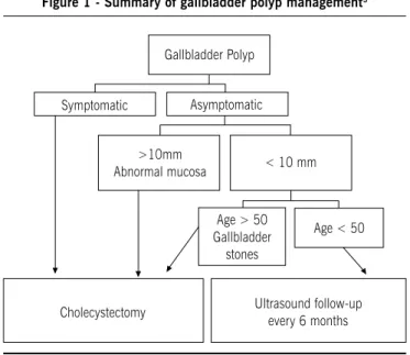
It can be concluded that the surgical treatment option for GPs is cholecystectomy and that this should only be undertaken when the following conditions are true: clinical signs of GP; polyps with diameters greater than 10 mm; fast-growing polyps; sessile polyps or wide-based polyps; polyps with long pedicles; patient aged over 50; concurrent gallstones; polyps of the gallbladder infundibulum or abnormal gallbladder wall ultrasound indings. (Figure 1).
Figure 1 - summary of gallbladder polyp management3
Young patients with polyps smaller than 10 mm, who are asymptomatic or have dyspeptic complaints only, do not need any treatment other than clinical follow-up with ultrasound every six months.
No conlicts of interest declared concerning the publication of this article.
r
eferences1. Sun XJ, Shi JS, Han Y, Wang JS, Ren H. Diagnosis and treatment of polypoid lesions of the gallbladder: report of 194 cases. Hepatobiliary Pancreat Dis Int. 2004;3:591-4.
2. Ljubičić N, Zovak M, Doko M, Vrkljan M, Vide L. Management of gallbladder
polyps: an optimal strategy proposed. Acta Clin Croat. 2001;40:57-60. 3. Josef E, Fischer MD. Mastery of surgery. 5th ed. Philadelphia: Lippincott
Willimas & Wilkins; 2006. p.1025;
4. Sugiyama M, Xiao-Yan Xie, Yutaka Atomi Y, Saito M. Differential diagnosis of small polypoid lesions of the gallbladder. the value of endoscopic ultrasonog-raphy. Ann Surg. 1999;229:498-504.
5. Csendes A, Burgos AM, Csendes P, Smok G, Rojas J. Late follow-up of polypoid lesions of the gallbladder smaller than 10 mm. Ann Surg. 2001;234:657-60.
6. Chattopadhyay D, Lochan R, Balupuri S, Gopinath BR, Wynne KS. Outcome of gallbladder polypoidal lesions detected by transabdominal ultrasound scanning: a nine year experience. World J Gastroenterol. 2006;11:2171-3.
7. Furukawa, H., Kosuge, T., Shimada, K., Yamamoto, J.,Kanai, Y., Mukai, K., Iwata, R. and Ushio, K. Small polypoid lesions of the gallbladder. Differential diagnosis and surgical indications by helical computed tomography. Arch Surg. 1998;133:735-9.
Gallbladder Polyp
Symptomatic Asymptomatic >10mm
Abnormal mucosa < 10 mm Age > 50 Gallbladder
stones
Age < 50
GallBladderpolyps: howshouldtheyBetreatedandwhen?
321
Rev Assoc Med Bras 2010; 56(3): 318-218. Kimura K, Fujita N, Noda Y, Kobayashi G, Ito K. Differential diagnosis of
large-sized pedunculated polypoid lesions of the gallbladder by endoscopic ultrasonography: a prospective study. J Gastroenterol. 2001;36:619-22. 9. Sugiyama M, Atomi Y, Yamato Y. Endoscopic ultrasonography for differential
diagnosis of polypoid gall bladder lesions: analysis in surgical and follow up series. Gut. 2000;46:250-4.
10. Escalona AP, León FG, Bellolio FR, Pimentel FM, Guajardo MB, Gennero R, et al.
Pólipos vesiculares: correlación entre hallazgos ecográicos e histopatológicos.
Rev Méd Chile. 2006;134:1237-42.
11. Kratzer W, Haenle MM, Voegtle A, Mason RA, Akinli AS, Hirschbuehl K, et al. and the Roemerstein study group. Ultrasonographically detected gallbladder polyps: a reason for concern? A seven-year follow-up study. BMC Gastroen-terology.2008;8:41.