Microscopic analysis of opacification in
Ioflex
®
hydrophilic acrylic intraocular lenses
A
nálise microscópica da opacificação de
lentes intraoculares acrílicas hidrofílicas Ioflex
®Bruna Vieira Ventura1, Marcelo Ventura1,2, Wagner Lira1, Camila Vieira Ventura1, Marcony Rodrigues
Santhiago3, Liliana Werner4
A
BSTRACTFive cases of intraocular lens (IOL) opacification in patients implanted with the Mediphacos Ioflex® IOL are described. Clinical data in each case was obtained from the patient’s medical record. The five explanted IOLs underwent gross and light microscopic analysis. Selected lenses were processed for further evaluation and multiple sagittal cuts were stained with the alizarin red and von Kossa methods. Light microscope analysis confirmed the presence of deposits on and within the lens, which stained positive for calcium. The reason why calcification has occurred in these cases remains unclear, but surgeons should be aware of this potential late postoperative complication.
Keywords: Lenses, intraocular; Lens implantation, intraocular; Reoperation; Device removal; Microscopy; Case reports
R
ESUMOCinco casos de opacificação de lente intraocular (LIO) em pacientes implantados com a LIO Ioflex® da Mediphacos são descritos. Os dados clínicos dos pacientes foram obtidos a partir dos prontuários médicos. As cinco LIOs explantadas foram enviadas para análise macroscópica e sob microscopia óptica. Algumas lentes foram processa-das para análise adicional e múltiplos cortes sagitais foram corados pelos métodos de vermelho de alizarina e von Kossa. A análise sob microscopia óptica confirmou a presença de depósitos na superfície e no corpo da lente, que coraram positivamente para cálcio. A razão pela qual a calcificação ocorreu nestes casos permanece obscura. Entretanto, cirurgiões devem estar atentos para esta potencial complicação tardia.
Descritores: Lentes intraoculares; Implante de lenteintraocular; Reoperação; Remoção de dispositivo; Microscopia; Relatos de casos
1Fundação Altino Ventura – FAV – Recife (PE), Brazil;
2Hospital de Olhos de Pernambuco – HOPE – Recife (PE), Brazil;
3Department of Refractive Surgery, Cole Eye Institute, Cleveland Clinic, Cleveland (OH), USA;
4Intermountain Ocular Research Center, John A. Moran Eye Center, University of Utah, Salt Lake City (UT), USA.
No author has a financial or proprietary interest in any material or method mentioned.
Financial support: none.
I
NTRODUCTIONP
ostoperative optic opacification is the leading cause of explantation of hydrophilic acrylic intraocular lens (IOL)(1). The Mediphacos Ioflex®(Belo Horizonte, Minas Gerais, Brazil) IOL is a foldable hydrophilic acrylic single-piece IOL with an optic diam-eter of 5.7 mm and an overall diamdiam-eter of 12.5 mm. The material used for the lens manufacture is a copolymer of poly-hydroxyethylmethacrylate with 25% water content. It was first introduced in the market in 2001. No case of IOL opacification has been reported in patients with this lens in articles published in scientific journals indexed in PubMed and SciELO (keywords searched: Ioflex, lens opacification, lens calcification). Although few hydrophilic acrylic IOL designs are approved by the Food and Drug Administration (FDA) to be used in the United States(2),
they are largely commercialized in other countries. The purpose of this study was to report the micro-scopic analysis of five cases of Mediphacos Ioflex® IOL
opacification. All IOLs were explanted due to signifi-cant visual impairment.
This study was approved by the Altino Ventura Foundation Ethics Committee and conducted at the Altino Ventura Foundation.
Case reports
Case 1
A 72-year-old female had an uneventful phacoemulsification with implantation of Mediphacos Ioflex® IOLs in the right eye (Lot G0718092) on august
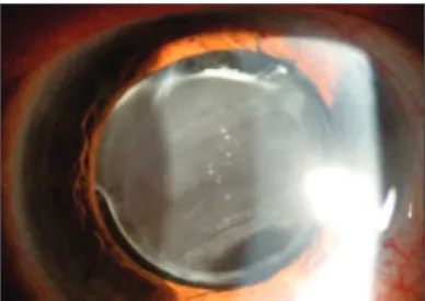
2007 and in the left eye (Lot H0731101) on november 2007. The patient had a medical history of hypertension. She presented 29 months after the surgery on the right eye with a decrease in best-corrected visual acuity (BCVA) in OD from 20/25 on the fifteenth postoperative day evalu-ation to 20/100. Examinevalu-ation revealed haziness of the IOL (Figure 1). The anterior chamber was quiet, with no signs of inflammation. Her fundus exam was normal. Neodymium:yag (Nd:YAG) laser treatment was ineffec-tive in removing the opacification from the lens. The IOL was explanted 31 months postoperatively and a hydro-phobic acrylic Type 7B (Alcon, Inc.) IOL was implanted in the capsular bag. There were no intraoperative or post-operative complications. The patient’s final BCVA on the right eye was 20/30. The BCVA of OS after phacoemulsification was always 20/20.
Case 2
An 86-year-old male with vascular hypertension was submitted to an uneventful phacoemulsification with im-plantation of Mediphacos Ioflex® IOLs in the capsular bag of the right eye (Lot C0715102) in august 2007 and of the left eye (Lot J0729351) in december 2007. On evaluation on the thirtieth postoperative day of the surgery in the left eye, his BCVA in OD was 20/30 and in OS was 20/25.
Thirty-three months later he returned referring a progressive decrease in the left eye’s visual acuity. He pre-sented with a 20/200 BCVA in this eye and 20/30 in OD. On examination IOL’s optic and posterior capsule opacifica-tion was noted. The eye had no inflammatory signs. A poste-rior capsulotomy was done with Nd:YAG laser, without vi-sion improvement. The IOL was explanted 35 months post-operatively. A dense fibrous tissue was connecting the lens haptics to the bag. The haptics were cut to facilitate IOL’s removal, the optic was explanted successfully and the haptics were left in the eye. An anterior vitrectomy was done and a poly(methyl methacrylate) (PMMA) OP-72 (Mediphacos, Belo Horizonte, Brazil) IOL was implanted in the sulcus. There were no intraoperative complications. On the first postoperative day, the eye had mild inflamma-tory signs and the IOL was centered.
Postoperative outcome evolved with secondary glaucoma treated medically, later requiring trabeculectomy. One month after this procedure, BCVA in the left eye was 20/400 and the IOP was 10 mm Hg.
Case 3
In september 2007 a 53-year-old female with se-vere non-proliferative diabetic and hypertensive retin-opathy was submitted to phacoemulsification with un-eventful implantation of a Mediphacos Ioflex® IOL in the right eye (Lot H0731101). There were no intraop-erative complications. At the moment of the surgery, the patient had hard exudates in the macula and few microaneurysms in the right eye. In the left eye she had mild nucleosclerosis. Pan-retinal photocoagulation and an intravitreal injection of triamcinolone acetonide were done in the right eye three and two years prior to cata-ract surgery, respectively.
One month after phacoemulsification she pre-sented with hard exudates in the macula and clinically significant macular edema. She was submitted to grid
tocoagulation. Her uncorrected vision after the laser was 20/60. Two months later a mild fibroglial proliferation was observed superiorly to the optic nerve and there were hard exudates temporally to the macula. Superior retinal photocoagulation and focal laser were done in OR.
Twenty-two months after surgery, her uncorrected visual acuity in the right eye was 20/30. She had no macu-lar edema or macumacu-lar exudates. However, thirty months postoperatively she presented with an uncorrected vi-sion on the right eye of 20/150. On biomicroscopy IOL and subcapsular opacification was noted. The patient was submitted to a Nd:YAG laser posterior capsulotomy, but her uncorrected visual acuity did not improve and one month later had decreased to 20/400.
The IOL was explanted seven months later. A dense fibrous tissue was connecting the lens haptics to the bag. The haptics were cut, the optic was removed successfully and the haptics were left in the eye. A
poste-rior vitrectomy was done. A hydrophobic acrylic Type 7B (Alcon, Inc.) IOL was implanted in the ciliary sulcus. Two months after IOL exchange, the uncorrected and corrected visual acuity in the right eye was 20/60. During fundus exam, macular edema was noted.
Case 4
A 79-year-old female had an uneventful phacoemulsification with implantation of a Mediphacos Ioflex® IOL in the capsular bag of the left eye (Lot
H0731101) on december 2007. The patient had a medi-cal history of hypertension and diabetes, and an ocular history of glaucoma (cup-to-disc ratio of 0.9 OU), con-trolled with timolol maleate 0.5% and travoprost 0.004%. She presented 33 months later with a decrease in BCVA from 20/30 on the thirtieth postoperative day evalu-ation to counting fingers at 4 meters. Biomicroscopy re-vealed IOL opacification. The eye had no signs of
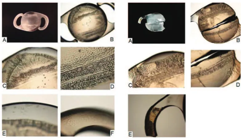
inflam-Figure 2: Explanted Ioflex® IOL of case 1. A: Gross photograph showing IOL opacification on the optic and haptics; B: Light photomicrograph. Dense deposits can be observed mostly on the anterior surface of the optic component, highlighting the marks caused by forceps during the folding process. Pits, corresponding to Nd:YAG laser application, can also be seen in the center of the optic component; C and D: Light photomicrographs showing confluent deposits arranged in a convoluted, “cerebriform” pattern. E: Light photomicrograph, small granular deposits are observed in areas outside of the confluent deposits; F: Light photomicrograph; multiple, small, granular deposits are seen within the haptics of the lens, close to the surface; B: unstained, original magnification X20; C: unstained, original magnification X40; D: unstained, origi-nal magnification X100; E, unstained, origiorigi-nal magnification X10; F, unstained, original magnification X100
mation. The IOL was explanted 39 months postoperatively and a hydrophobic acrylic Type 7B (Alcon, Inc.) IOL was implanted in the capsular bag. There were no intraopera-tive or postoperaintraopera-tive complications. The patient’s BCVA 1 month after IOL exchange on the left eye was 20/80.
Case 5
A 78-year-old male was submitted to a phacoemulsification with uneventful implantation of a Mediphacos Ioflex® IOL in the right eye (Lot J0726349)
on september 2007 and in the left eye (Lot G0726242) on october 2007. The patient had a medical history of hypertension and diabetes. There were no intraopera-tive or immediate postoperaintraopera-tive complications. His fun-dus exam was normal.
He presented 30 months after the surgery in the left eye with a decrease in this eye’s BCVA from 20/20 on the thirtieth postoperative day evaluation to 20/150. BCVA in the right eye was preserved. Ocular examina-tion only revealed haziness on the left IOL’s surfaces. Vision did not improve after Nd:YAG laser posterior capsulotomy. The IOL in the left eye was explanted 41 months postoperatively and a PMMA OP-72 (Mediphacos, Belo Horizonte, Brazil) IOL was implanted in the sulcus. This eye’s final BCVA was 20/30.
Laboratory findings
The explanted lenses were sent to the Intermoun-tain Ocular Research Center (John A. Moran Eye Cen-ter, University of Utah, USA) in the dry state. Each lens
underwent gross examination and light microscopy. Gross analyses were performed and photographs were taken for documentation using a Cyber-shot DSC-F707 (Sony, CA, USA). Light microscopy was performed us-ing a BX40 light microscope (Olympus, Japan) and pho-tomicrographs were taken with a DP20 digital camera (Olympus, Japan) attached to the light microscope.
Gross examination of the explanted IOLs showed a whitish discoloration of the specimens. Microscopic examination showed dense deposits forming an almost continuous crust, mostly on the anterior surface of the optic component. Confluent deposits were arranged in convoluted irregular areas forming a “cerebriform” pat-tern. Small granular deposits were observed in areas outside of the crust. The deposits were most confluent along linear areas. Multiple, small, granular deposits were also generally observed within the optic and haptics of the lenses, close to the surface. Some peripheral areas of the optic were relatively clear of surface and substance deposits/granules (Figures 2, 3 and 4).
In case 1, pits, corresponding to Nd:YAG laser application, were observed in the optic component. In case 3, there were brown deposits on the lens surface consistent with iris pigments.
Selected lenses underwent further examination. Two sagittal cuts were performed at the optic component of the lenses in cases 1 and 4 to obtain an optical cylinder. In case 1, the cylinder was processed and multiple sagittal cuts were stained with the von Kossa method for calcium. In case 4, the cylinder and the remainder of the lens were
Figure 4: Explanted Ioflex® IOL of case 3; A and B: Light
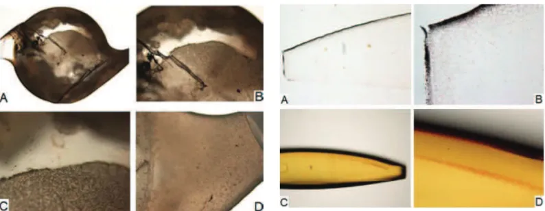
photomicrographs showing dense deposits mostly on the anterior surface of the optic component. Brown deposits, consistent with iris pigments, are seen on the lens surface; C: Light photomicrograph. Confluent deposits are arranged in a convoluted, “cerebriform” pattern. D: Light photomicrograph showing multiple, small, gra-nular deposits within the haptics of the lens, close to the surface. Part of the haptic was cut to facilitate IOL explantation. A: unstained, original magnification X20; B: unstained, original magnification X40; C: unstained, original magnification X100; D: unstained, original magnification X100
directly stained with alizarin red for calcium.
Analysis of the sections obtained from the lens in case 1 under the light microscope confirmed the presence of calcium deposits on and within the lens, which stained dark brown with the von Kossa method. Analysis of the optical cylinder of the lens in case 4 showed the calcium deposits stained in red by the alizarin red. They were present on the surface of the lens and close to the surface, at different depths within the optic component (Figure 5).
D
ISCUSSIONIOL calcification is a sight-threatening complica-tion of lens implantacomplica-tion. Nd:YAG laser treatment is in-effective in removing the calcified deposits from the lenses(1,3-5), as seen in most of our cases. The only
effec-tive treatment to restore vision is explantation and ex-change of the calcified IOL.
Calcification on the surface and in the substance of the lens in hydrophilic acrylic IOLs has been well documented(6-10). However, to the best of our knowledge
this is the first peer-reviewed report on opacification of the Mediphacos Ioflex® IOL. A previous study
analyz-ing an opacified hydrophilic acrylic AcquaSense®
(Oph-thalmic Innovative International, USA) IOL described the presence of calcium deposits on the surface and within the substance of the IOL optic and haptics(10). Similarly
to this and other studies regarding hydrophilic lenses, microscopic examination of the five Ioflex® IOLs
re-vealed that the opacification was due to calcium granu-lar deposits on the surface and within the optic and haptics of the lenses(7,8,10). Two histochemical methods for
cal-cium detection were used in these cases, and both of them yielded positive results, confirming the calcified nature of the deposits. The deposits were most confluent along linear areas, probably corresponding to marks caused by forceps during the folding process.
Calcification of hydrophilic acrylic lenses seems to have a multifactorial origin. Factors related to IOL manu-facture and packaging, surgical techniques, adjuvants, as well as patient metabolic and ocular conditions, may be involved(11). The formation of calcium deposits seems to
depend both on the material of the IOL and on the local chemical microenvironment of the aqueous humor(3). Groh
et al. described a possible association between IOL calci-fication and the metabolic disturbances in diabetes(12). The
level of phosphorus in the aqueous humor of diabetic pa-tients, particularly those with proliferative diabetic retin-opathy, is significantly higher than normal individuals, which may lead to opacification of hydrophilic acrylic IOLs(13). A previous study reported bilateral hydrophilic
IOL opacification in a diabetic patient(9). In the present
study, case 3 had severe non-proliferative diabetic retin-opathy and cases 4 and 5 also had diabetes. Interestingly enough, in case 5, only 1 of the lenses exhibited calcifica-tion. Both surgical implantations were performed within 1 month by the same surgeon, using the same solutions. This may suggest that local conditions of supersaturation,
ei-ther in the vicinity of the surface of the IOLs or within their substance, may promote salts development by diffu-sion of calcium/phosphate ions, as suggested in the study by Gartaganis et al.(3).
Additionally, all cases had arterial hypertension. Other studies have described IOL calcium deposits in patients with hypertension(3,5,8,14). However, not all
pa-tients with IOL calcification have underlying systemic diseases(3,5,8,14) and not all cases operated for bilateral
cataracts with implantation of the same IOL type have bilateral lens opacification(3), as seen in cases 1, 2 and 5.
The IOLs in each of these cases came from different lots, which might have had different susceptibilities to de-velop the complication. Previous papers have described IOL calcification in patients with ocular diseases, such as uveitis and asteroid hialosis (this latter in relation to silicone IOLs)(1,15). Besides diabetic and hypertensive
retinopathy in case 3, none of our cases had other past ocular inflammatory diseases.
The crystalline deposition on IOLs can be divided into two general time frames: intraoperative or shortly postoperative versus late postoperative(16). Our patients
had late postoperative IOL calcification. The mean pe-riod between phacoemulsification and patient presenta-tion with decreased vision was 31 months, with minimum being 29 and maximum 33 months. The literature shows that this mean period varies from 16.4 to 35.3 months, depending on the case series(14,17).
When comparing the visual acuity before and af-ter IOL opacification, we noticed that all patients lost more than three Snellen lines in visual acuity. In a previous study of 12 patients with calcified IOL, twenty percent of the patients lost more than three Snellen lines in visual acuity, 46.7% lost less than three Snellen lines in visual acuity and 13.3% maintained the same visual acuity(14).
In case 3, during a one-month period the uncorrected vi-sual acuity decreased from 20/150 to 20/400 on the right eye. This demonstrates the progressive nature of the pro-cess of calcification in the Ioflex® lenses, which was also
previously described in another hydrophilic IOL(6).
The mean period between first surgery and ex-plantation of lenses was 36.8 months, with minimum be-ing 31 and maximum 41. After explantation of the opaci-fied IOL and implantation of a new lens, four of our cases gained more than three Snellen lines of visual acuity and one lost a line. The patient who lost a line had sec-ondary glaucoma after the IOL exchange. In another study from Brazil, one of twelve patients who had IOL explantation due to calcification exchanged with a PMMA IOL lost more than 3 Snellen lines of visual acu-ity. He had a decompensation of proliferative diabetic retinopathy and neovascular glaucoma(14).
poste-rior capsule rupture and zonule dehiscence during ex-plantation of opacified IOLs, which were adequately managed, not affecting visual recovery(5).
In the postoperative period of the opacified IOL explantation one of our cases had a secondary glaucoma, with an increase in IOP that was only controlled with a trabeculectomy. This patient’s second IOL was placed in the sulcus. Former studies have described other postop-erative complications, such as posterior capsule opacifi-cation, cystoid macular edema, retinal detachment, cho-roidal hemorrhage and endophthalmitis(4,5,14).
Although hydrophilic acrylic IOLs have higher uveal biocompatibility resulting in less postoperative inflammation than other IOLs(18), potential complications
such as late opacification have to be considered. From 2006 to 2007, we placed approximately 4,000 Ioflex®
lenses. To date, opacification occurred in only five cases. On may 10, 2011, Mediphacos sent a report to the Brazilian Society of Laser and Surgery in Oph-thalmology (http://www.bloss.com.br/site/default.aspx) summarizing their investigation on the problem of Ioflex® calcification. According to the manufacturer,
they started receiving sporadic and isolated reports on Ioflex® opacification starting in 2009, related to
some lots distributed between 2007 and 2009 (Lots starting with: A09, A10, B09, C09, D09, E09, F09, G08, G09, H08, H09, I08, I09, J08, J09, K08, k09, L08, L09). Surface analysis apparently confirmed the presence of calcium/phosphate precipitation, as well as the pres-ence of polydimethylsiloxane (PDMS) on the surfaces of the analyzed lenses. PDMS was found to come from the packaging of these IOL lots, manufactured during the period in question. A study by Guan et al. has al-ready demonstrated a possible role of silicone com-pounds interacting with long-chain saturated fatty ac-ids present in the aqueous humor (myristic, palmitic, stearic, arachidic, and behenic) on the calcification process of the Hydroview IOL(19).
Mediphacos withdrew the lenses from these lots from the market and the packaging was changed. However, the opacified lenses from our patients are from different lots. Thus, the presence of PDMS in the packaging of some lots does not explain the lens opacification in our patients.
In conclusion, this is the first report on opacifica-tion of the Mediphacos Ioflex® IOL describing
micro-scopic findings. The opacification of the five lenses was due to calcium deposits. Although this IOL should not be discredited based on the occurrence of these calcifica-tion cases, surgeons should be aware of this potential late postoperative complication and further investigation on the causes of opacification is needed.
R
EFERENCES1. Neuhann IM, Kleinmann G, Apple DJ. A new classification of calcification of intraocular lenses. Ophthalmology. 2008;115(1):73-9.
2. Mamalis N, Brubaker J, Davis D, Espandar L, Werner L. Com-plications of foldable intraocular lenses requiring explanta-tion or secondary intervenexplanta-tion—2007 survey update. J Cata-ract RefCata-ract Surg. 2008;34(9):1584-91.
3. Gartaganis SP, Kanellopoulou DG, Mela EK, Panteli VS, Koutsoukos PG. Opacification of hydrophilic acrylic intraocu-lar lens attributable to calcification: investigation on mecha-nism. Am J Ophthalmol. 2008;146(3):395-403.
4. Haymore J, Zaidman G, Werner L, Mamalis N, Hamilton S, Cook J, Gillette T. Misdiagnosis of hydrophilic acrylic intraocu-lar lens optic opacification: report of 8 cases with the MemoryLens. Ophthalmology. 2007;114(9):1689-95. 5. Yu AK, Ng AS. Complications and clinical outcomes of
in-traocular lens exchange in patients with calcified hydrogel lenses. J Cataract Refract Surg. 2002;28(7):1217-22. 6. Neuhann IM, Neuhann TF, Szurman P, Koerner S, Rohrbach
JM, Bartz-Schmidt KU. Clinicopathological correlation of 3 patterns of calcification in a hydrophilic acrylic intraocular lens. J Cataract Refract Surg. 2009;35(3):593-7.
7. Walker NJ, Saldanha MJ, Sharp JA, Porooshani H, McDonald BM, Ferguson DJ, Patel CK. Calcification of hydrophilic acrylic intraocular lenses in combined phacovitrectomy sur-gery. J Cataract Refract Surg. 2010;36(8):1427-31. 8. Yu AK, Kwan KY, Chan DH, Fong DY. Clinical feature of 46
eyes with calcified hydrogel intraocular lenses. J Cataract Refract Surg. 2001;27(10):1596-606.
9. Pandey SK, Werner L, Apple DJ, Kaskaloglu M. Hydrophilic acrylic intraocular lens optic and haptics opacification in a diabetic patient: bilateral case report and clinicopathologic correlation. Ophthalmology. 2002;109(11):2042-51. 10. Padilha MA. Complicações na cirurgia de catarata: prevenção
e manuseio. In: Padilha MA. Catarata. 2a ed. Rio de Janeiro: Cultura Médica; 2008. p. 519-42.
11. Werner L. Calcification of hydrophilic acrylic intraocular lenses. Am J Ophthalmol. 2008;146(3):341-3. Comment on Am J Ophthalmol. 2008;146(3):395-403.
12. Groh MJ, Schlötzer-Schrehardt U, Rummelt C, von Below H, Küchle M. [Postoperative opacification of 12 hydrogel fold-able lenses (Hydroview(R)]. Klin Monbl Augenheilkd. 2001;218(10):645-8. German.
13. Kim CJ, Choi SK. Analysis of aqueous humor calcium and phosphate from cataract eyes with and without diabetes mellitus. Korean J Ophthalmol. 2007;21(2):90-4.
14. Salera CM, Miranda RA, Reis PPL, Guimarães MR, Campolina RB, Guimarães Q. Resultados da troca de lente intra-ocular de hidrogel opacificada. Arq Bras Oftalmol. 2004;67(1):115-9.
15. Stringham J, Werner L, Monson B, Theodosis R, Mamalis N. Calcification of different designs of silicone intraocular lenses in eyes with asteroid hyalosis. Ophthalmology. 2010;117(8):1486-92.
16. Werner L, Apple DJ, Escobar-Gomez M, Ohrström A, Crayford BB, Bianchi R, Pandey SK. Postoperative deposition of cal-cium on the surfaces of a hydrogel intraocular lens. Ophthal-mology. 2000;107(12):2179-85.
17. Huang Y, Xie L. Delayed postoperative opacification of fold-able hydrophilic acrylic intraocular lenses. J Biomed Mater Res B Appl Biomater. 2011;96(2):386-91.
18. Abela-Formanek C, Amon M, Schild G, Schauersberger J, Heinze G, Kruger A. Uveal and capsular biocompatibility of hydrophilic acrylic, hydrophobic acrylic, and silicone intraocu-lar lenses. J Cataract Refract Surg. 2002;28(1):50-61. 19. Guan X, Tang R, Nancollas GH. The potential calcification of
octacalcium phosphate on intraocular lens surfaces. J Biomed Mater Res A. 2004;71(3):488-96.
Correspondence address: Bruna V. Ventura
Altino Ventura Foundation – FAV Rua do Progresso, nº 71 – Boa Vista Zip Code: 50070-020 - Recife (PE), Brazil Phone: 55 81 3302-4343