Original Article
1 5 4 Arq Bras Oftalmol. 2015;78(3):154-7 http://dx.doi.org/10.5935/0004-2749.20150040
INTRODUCTION
Cataracts are one of the most common eye diseases associated with blindness (visual acuity worse than 20/400 in the better eye with best correction) worldwide, with an estimated 18 million people thought to be affected, and cataract surgery is the intraocular procedure performed most often worldwide. Over the years, the techniques of cataract surgery have evolved into a safe and successful procedure for visual rehabilitation. The incidence of most complications has significantly decreased with the development of better instrumen-tation and affordable, high-quality intraocular lens (IOL) implants(1). Various aspects of cataract surgery that make it safer have changed
ABSTRACT
Purpose: To analyze the indications for explantation or exchange of intraocular lenses (IOLs), which were originally implanted for the correction of aphakia during cataract extraction.
Methods: All cases that involved intraocular lens explantation or exchange in one institution between January 2008 and December 2014 were analyzed re tros pectively. Results: In total, 93 eyes of 93 patients were analyzed. The median time interval between implantation and explantation of the anterior chamber intraocular lenses (AC IOL) and posterior chamber intraocular lenses (PC IOL) was 83.40 ± 83.14 months (range: 1-276 months) and 55.14 ± 39.25 months (range: 1-168 months), respectively. Pseudophakic bullous keratopathy (17 eyes, 38.6%) and persistent iritis (12 eyes, 27.8%) in the AC IOL group and dislocation or decentration (30 eyes, 61.2%) and incorrect IOL power (nine eyes, 18.4%) in the PC IOL group were the most common indications for explantation of IOLs. The mean logMAR best corrected visual acuity (BCVA) improved significantly from 1.30 preoperatively to 0.62 postoperatively in the PC IOL group (p<0.001) but did not improve significantly in the AC IOL group (p=0.186).
Conclusions: The primary indication for IOL explantation or exchange was pseu-dophakic bullous keratopathy in the AC IOL group and was dislocation or decen-tration in the PC IOL group. PC IOL explantation or exchange is safe and im proves visual acuity.
Keywords: Cataract extraction; Lenses, intraocular; Reoperation; Device removal; Lens implantation, intraocular; Pseudophakia; Corneal diseases; Patient satisfaction; Visual acuity
considerably in the past decade with the evolution of both surgical techniques and IOL designs.
Although cataract surgery is safe for the majority of patients, some complications that involve the anterior and posterior segment can occur. Surgical procedures involving the use of the modern anterior chamber (AC) IOLs (AC IOLs) and posterior chamber (PC) IOLs (PC IOLs) have reduced the risk of complications necessitating IOL explan-tation/exchange. Although older types of both AC IOLs and PC IOLs are no longer implanted since the advent of the new generation IOLs, we still see complications associated with those implanted many years ago. The aim of this study was to analyze the indications and
outco-Intraocular lens explantation or exchange: indications, postoperative interventions,
and outcomes
Remoção ou troca de lentes intraoculares: indicações, intervenções pós-operatórias e resultados
Refik Oltulu1, İsmail Erşan2, Günhal şatirtav1, mEryEm DonbaloGlu3, hürkan kErimoğlu1, ahmEt Özkağnici1
Submitted for publication: December 17, 2014 Accepted for publication: March 6, 2015
1 Department of Ophthalmology, School of Medicine, Necmettin Erbakan University, Konya, Turkey. 2 Department of Ophthalmology, School of Medicine, Canakkale Onsekiz Mart University, Canakkale,
Turkey.
3 Department of Ophthalmology, Mus State Hospital, Mus, Turkey.
Funding: No specific financial support was available for this study.
Disclosure of potential conflicts of interest: None of the authors have any potential conflicts of interest to disclose.
Corresponding author: Ismail Ersan. Department of Ophthalmology - School of Medicine. Canakkale
Onsekiz Mart Universitesi Tip Fakultesi. Goz Hastalıkları AD - Canakkale - Turkey
E-mail: [email protected]
Approved by the following research ethics committee: Necmettin Erbakan University School of Medicine.
RESUMO
Objetivo: Analisar as indicações para a remoção ou troca de lentes intraoculares (IOL), que foram originalmente implantadas para a correção de afacia após a extração da catarata.
Método: Todos os casos que envolveram remoção ou troca de lentes intraoculares em uma única instituição, entre janeiro de 2008 e dezembro 2014 foram analisados retrospectivamente.
Resultados: No total, foram analisados 93 olhos de 93 pacientes. O intervalo de tempo médio entre o implante e a remoção das LIOs de câmara anterior (AC IOL) e de câmara posterior (PC IOL) foi 83,40 ± 83,14 meses (variando de 1 a 276 meses) e 55,14 ± 39,25 meses (variando de 1 a 168 meses), respectivamente. Ceratopatia bolhosa pseudofácica (17 olhos, 38,6%) e irite persistente (12 olhos, 27,8%) no grupo AC IOL, e deslocamento ou descentralização (30 olhos, 61,2%) e poder incorreto da IOL (nove olhos, 18,4%), no grupo PC IOL, foram as indicações mais comuns para a remoção das IOLs. A média logMAR da melhor acuidade visual corrigida (BCVA) melhorou significativamente a partir de 1,30 no pré-operatório para 0,62 no pós-operatório no grupo PC IOL (p<0,001), mas não melhorou significativamente no grupo AC IOL (p=0,186).
Conclusões: A principal indicação para remoção ou troca de lentes intraoculares foi a ceratopatia bolhosa pesudofácica no grupo AC IOL e deslocamento ou descentralização no grupo PC IOL. A remoção ou troca de PC IOLs é segura e melhora a acuidade visual.
OltuluR, et al.
1 5 5 Arq Bras Oftalmol. 2015;78(3):154-7 mes of AC and PC IOL explantation conducted at a single institution
between 2008 and 2014.
METHODS
This retrospective interventional case series study has been con-ducted in accordance with the tenets of the Declaration of Helsinki and with the approval of the EthicsCommittee of Necmettin Erbakan University School of Medicine. The medical records for 93 eyes of 93 patients who had an AC or PC IOL explantation/exchange performed at Necmettin Erbakan University School of Medicine from 2008 to 2014 were reviewed for data including gender, age, the mean inter-val between cataract surgery and IOL explantation, the presence of pseudoexfoliation (PEX), glaucoma, corneal edema, uveitis, the pre sence of myopia or hyperopia, and best corrected visual acuity (BCVA) befo-re and after the explantation/exchange. The exclusion criteria webefo-re a follow-up period shorter than 1 month and patients with incomplete medical records. Otherwise, all the patients with IOL explantation/ exchange were included. Descriptive statistics were calculated for various clinical characteristics, and all data were analyzed using SPSS for Windows (version 16.0, SPSS Inc., Chicago, IL, USA).
RESULTS
Ninety-three patients with AC and PC IOL explantation/exchange were recruited. Forty-four patients had AC IOLs and 49 patients had PC IOLs. The patients were evaluated in two groups accordingly. Table 1 shows the characteristics of patients in each group. The median time intervals between implantation and explantation of the AC IOL and PC IOL groups were 83.40 ± 83.14 months (range: 1-76 months) and 55.14 ± 39.25 months (range: 1-168 months), respectively.
AC IOL
GROUPThe mean preoperative intraocular pressure in the AC IOL group was 18.05 ± 8.49 mmHg (range: 6-44). Four patients used timolol +
dor-zolamide (Cosopt, MSD, Turkey), while two patients used timolol + dorzolamide (Cosopt, MSD, Turkey) and brimonidine tartrate (Alpha-gan P, Abdi Ibrahim, Turkey).
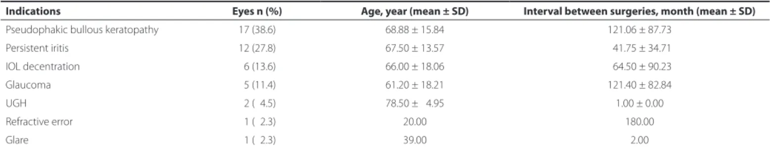
The most common reasons for explantation of the AC IOLs were pseudophakic bullous keratopathy (PBK) (17 eyes, 38.6%) and persis-tent iritis (12 eyes, 27.8%) (Table 2). After AC IOL explantation, a scleral fixated PC IOL was placed in 12 eyes (27.3%), and a PC IOL was implan-ted in six eyes (13.6%) above the remnant of the capsule at the sulcus without suturing. Finally, 26 (59.1%) eyes were left aphakic (Table 3). The mean logMAR BCVA had improved from 2.00 preoperatively to 1.80 postoperatively, but the difference did not reach statistical sig-nificance (p=0.186). The BCVA improved in 21 eyes (47.7%), remained stable in 17 eyes (38.6%), and decreased in six eyes (13.6%). The BCVA improved in patients with PBK and persistent iritis in two eyes (11.8%) and eight eyes (66.7%), respectively. The mean intraocular pressure of all the subjects was within the normal range, with 15 (34.1%) patients requiring topical anti-glaucomatous medication. Intraoperative and postoperative complications are shown in table 5.
PC IOL
GROUPThe mean preoperative intraocular pressure was 16.69 ± 7.42 mmHg (range: 7-40). Three patients used timolol + dorzolamide (Cosopt, MSD, Turkey), while two patients used timolol + dorzolamide (Cosopt, MSD, Turkey) and brimonidine tartrate (Alphagan P, Abdi Ibrahim, Turkey).
The most common indications for explantation of the PC IOLs were dislocation/decentration (30 eyes, 61.2%) and postoperative residual refractive error due to incorrect IOL power calculation (nine eyes, 18.4%). Other indications were IOL opacification (six eyes, 12.2%), persistent iritis (three eyes, 6.1%), and uveitis glaucoma hyphema (UGH) syn-drome (one eye, 2%) (Table 4). After the PC IOL explantation, a new PC IOL could be implanted into the capsular bag in 15 eyes (30.6%) and above the remnant of the capsule without suturing in 13 eyes (26.5%). If the capsular remnant did not offer adequate support for a PC IOL, a scleral fixated IOL was placed (17 eyes, 34.7%). Finally, four eyes (8.2%) were left aphakic (Table 3). The mean logMAR BCVA had improved significantly from 1.30 preoperatively to 0.62 postope-ratively (p<0.001). The BCVA improved in 37 eyes (75.5%), remai ned stable in four eyes (8.2%), and decreased in eight eyes (16.3%). Although 12 patients required topical anti-glaucomatous me dications, the mean intraocular pressure of all the subjects was within the normal range. Intraoperative and postoperative complications are shown in table 5.
DISCUSSION
Cataract extraction ranks among the most commonly performed surgical procedures in the United States(2). As a consequence of the large number of operations performed worldwide, increased use of IOLs leads to an increase in the number of complications requiring explantation of the IOLs, despite the marked improvement in surgical procedures and IOL technologies.
Table 2. Indications for AC IOL explantation and relation to age and intervals between surgeries
Indications Eyes n (%) Age, year (mean ± SD) Interval between surgeries, month (mean ± SD)
Pseudophakic bullous keratopathy 17 (38.6) 68.88 ± 15.84 121.06 ± 87.73
Persistent iritis 12 (27.8) 67.50 ± 13.57 041.75 ± 34.71
IOL decentration 06 (13.6) 66.00 ± 18.06 064.50 ± 90.23
Glaucoma 05 (11.4) 61.20 ± 18.21 121.40 ± 82.84
UGH 02 (04.5) 78.50 ± 04.95 1.00 ± 0.00
Refractive error 01 (02.3) 20.00 180.00
Glare 01 (02.3) 39.00 2.00
AC IOL= anterior chamber intraocular lens; UGH= uveitis glaucoma hyphema syndrome.
Table 1. Characteristics of patients with AC and PC IOL explantation
Characteristics AC IOL group PC IOL group
Sex, n (%)
Male 19 (43%) 37 (75.5%)
Female 25 (57%) 12 (24.5%)
Age (y)
Mean ± SD 65.9 ± 17.0 52.84 ± 24.60
Range 20-83 years 3-86 years
Interval between surgeries
Mean ± SD 83.14 ± 83.40 55.14 ± 39.25
Range 1-276 months 1-168 months
Intraocular lens explantation or exchange: indications, postoperative interventions, and outcomes
1 5 6 Arq Bras Oftalmol. 2015;78(3):154-7
In a series of 102 patients who had IOL explantation or exchange, AC IOLs comprised 66.7% of the removed lenses. PBK, followed by UGH syndrome and cystoid macular edema were the most frequent indications for explantation or exchange(3). Similarly, PBK and UGH were the most common indications for AC IOL explantation (53.9%), followed by iris-fixated lenses (33.7%)(4). Marques et al. reported that their rate of PBK was only 6.7%, while the main indication was inflam-mation (UGH and persistent iritis) with a rate of 53.3%(5). In this study, PBK (17 eyes, 38.6%) was the most common indication, in accordance with Mamalis et al.(3) and Doren et al.(4), for AC IOL explantation, which had a rate of 47.3%. Preventing the need for penetrating keratoplas-ty, AC IOL explantation has been indispensable in eyes with signs of progressive corneal endothelial damage(6). In our series, intervals between surgeries in patients with PBK and persistent iritis were 126.7 ± 89.7 months (range: 6-276 months) and 41.4 ± 38.6 months (range: 2-120 months), respectively. Early explantation of the AC IOLs may prevent progressive endothelial cell loss, as observed in the fact that BCVA improved in only two eyes (11.8%) in patients with PBK who had a longer time interval between surgeries and improved in eight eyes (66.7%) in patients with persistent iritis who had a shorter time interval between surgeries(7,8).
In the latest survey update in 2007 of members of the American Society of Cataract and Refractive Surgeons and the European
Socie-ty of Cataract and Refractive Surgeons, Mamalis et al. reported that dislocation/decentration, incorrect IOL power calculation, glare/opti cal aberrations, and IOL calcification were the most common rea sons for PC IOL explantation(9). Furthermore, Jones et al. investigated indica-tions of IOL exchange and found that IOL dislocation (46%) was the most common indication and that PC IOLs accounted for 88.5% of all decentered IOLs(10).
IOL dislocation is a rare complication in which the patient com-plains of blurred vision, glare, and possibly diplopia. The visual symptoms can be potentially disabling to the patient, and the condition requi-res intervention in either repositioning or even removing the lens. Patients with PEX are at risk for IOL dislocation after uncomplicated cataract surgery. Although IOLs can be well secured in the capsular bag, the possibility of progressive loss of zonular integrity may cause late endocapsular subluxation of PC IOLs. In our series, nine patients with PEX had IOL extraction because of delayed dislocation; the mean interval between implantation and exchange was 78 months. The current study at a single institution demonstrated that PC IOL dislocation (61.2%) was the most common indication for extracting PC IOLs, followed by incorrect IOL power (18.4%). This was similar to the findings reported by Mamalis et al.(9) and Jones et al.(10). According to the time interval between cataract surgery and IOL dislocation, IOL dislocation can be classified as early dislocation if it occurs within 3 months and late dislocation if it occurs after more than 3 months. Im-proper fixation within the capsular bag and instability of the capsular bag-IOL complex are the major causes of IOL dislocation(10). The major causes of early IOL dislocation are improper support of the capsular bag and ciliary sulcus due to zonular or capsular damage, rupture, or both(11). Late dislocations are often accompanied by trauma or progressive zonular dehiscence caused by contraction of the capsu-lar bag many years after routine cataract surgery(12). In the present study, early IOL dislocation was present in six eyes after complicated cataract surgery with vitreous loss, in one eye after ocular trauma, and in one eye with a broken IOL haptic. Of the 22 eyes with late IOL dislocation, the major predisposing factors were PEX in nine eyes (40.9%), trauma in seven eyes (31.8%), and capsule contracture syn-drome in three eyes (13.6%). No predisposing factor could be found in the remainder (three eyes, 13.6%).
Unpredicted postoperative refractive error due to preoperative incorrect IOL power calculation is a disturbing complication for cata-ract surgeons. Improved IOL calculation formulas and preoperative measurement of axial length and corneal curvature reduce the risk of this complication. In our study, nine (18.4%) eyes required IOL ex plantation due to incorrect IOL power. The IOLs were exchanged because of postoperative myopia in five eyes and hyperopia in four eyes. Our results were in accordance with a recent study in which IOL dislocation (46%) followed by incorrect IOL power (23%) were the most common causes of IOL exchange(10).
IOL opacification is a rare but possible event. The exact reason for opacification is unknown. Using microscopic analyses of explanted hydrophilic acrylic IOLs, Werner et al. revealed multiple fine, calcified granular deposits of variable sizes within the lens optics(13). Neuhann et al. concluded that it was important to determine whether the calcium deposits formed because of a problem in IOL manufacturing (properties of the polymer, its surface, or the IOL packaging) or were the result of environmental causes that can catalyze calcification(14). In the present study, five of the six patients with IOL opacifications had a history of diabetes mellitus, which may have contributed to IOL opacifications by catalyzing calcification.
By using a proper IOL stabilizing technique, intraocular tissues should be protected from damage that could be caused by IOLs, and appro-priate refractive outcomes should therefore be achieved. Secondary scleral fixated IOL implantation after IOL removal was the dominant procedure used to avoid further corneal complications in both the AC IOL and PC IOL groups in our study.
Table 3. IOL ixation technique used after IOL explantation
Fixation technique AC-IOL group PC-IOL group
PC IOL in bag 00 (0%) 15 (30.6%)
PC IOL in sulcus 06 (13.6%) 13 (26.5%)
PC IOL with scleral fixation 12 (27.3%) 17 (34.7%)
Aphakia 26 (59.1%) 04 (08.2%)
AC IOL= anterior chamber intraocular lens; PC IOL= posterior chamber intraocular lens.
Table 4. Indications for PC IOL explantation and relation to age and intervals between surgeries
Indications Eyes n (%)
Age, year (mean ± SD)
Interval between surgeries, month (mean ± SD)
IOL dislocation/ decentration
30 (61.2) 58.60 ± 22.89 65.06 ± 41.21
Incorrect IOL power 09 (18.4) 31.44 ± 28.34 48.00 ± 32.86 IOL opacification 06 (12.2) 59.00 ± 08.79 40.00 ± 22.34 Persistent iritis 03 (06.1) 45.67 ± 27.75 32.67 ± 35.80
UGH 01 (02.0) 57.00 2.00
PC IOL= posterior chamber intraocular lens; UGH= uveitis glaucoma hyphema syndrome.
Table 5. Intraoperative and postoperative complications of intraocular lens explantation
AC IOL group PC IOL group Intraoperative complications
Vitreous loss 8 (18.2%) 6 (12.2%)
Bleeding to the anterior chamber 4 (09.1%) 3 (06.1%)
Suprachoroidal hemorrhage 2 (04.5%),
-Postoperative complications
Bullous keratopathy 2 (04.5%)
-Cystoid macular edema 1 (02.3%)
-Corneal melting requiring evisceration 1 (02.3%)
-Endophthalmitis - 1 (2.0%)
OltuluR, et al.
157
Arq Bras Oftalmol. 2015;78(3):154-7 Postoperative corneal decompensation after IOL explantation was
heavily dependent on the initial measurement of endothelial cell density(15,16). It is important to bear in mind that IOL explantation has a risk of additional damage to corneal endothelial cells. Coli et al. showed progression of corneal decompensation in 23.5% of eyes after AC IOL explantation(17). In the current study, only two eyes (4.5%) developed postoperative PBK in the AC IOL explantation group, and postope-rative PBK was not observed in the PC IOL explantation group. The low incidence of progression to PBK in the AC IOL explantation group, compared with Coli et al.(17) may be attributed to the higher propor-tion of patients that were left aphakic in our study. With the applica-tion of proper techniques, BCVA improved in 21 eyes (47.7%) in the AC IOL group and in 37 eyes (75.5%) in the PC IOL group.
This study had several shortcomings, including its retrospective nature and a lack of information on the IOL types that were explanted, lack of measurements of preoperative and postoperative endothelial cell density, and the highly variable follow-up times. Although the mean follow-up time was 7.2 ± 9.6 months and some of the cases had 48 months of follow-up, some cases had 1 month of follow-up, which was insufficient to detect some of the postsurgical complications.
In conclusion, the main indications for IOL explantation/exchan-ge in the AC IOL and PC IOL groups were PBK and IOL dislocation/ decentration, respectively. PC IOL explantations/exchanges have more favorable outcomes with an increase in BCVA than AC IOL explanta-tions/exchanges, in which inflammation and corneal complications were much more common.
REFERENCES
1. Pascolini D, Mariotti SP. Global estimates of visual impairment: 2010. Br J Ophthalmol. 2012;96(5):614-8.
2. American Academy of Ophthalmology Preferred Practice Pattern: Cataract in the Adult Eye. 2011. Available at: http://one.aao.org/asset.axd?id=8d66318f-ff50-408e-9bb1-73d277cf14ce.
3. Mamalis N, Crandall AS, Pulsipher MW, Follett S, Monson MC. Intraocular lens explan-tation and exchange. A review of lens styles, clinical indications, clinical results, and visual outcome. J Cataract Refract Surg. 1991;17(6):811-8.
4. Doren GS, Stern GA, Driebe WT. Indications for and results of intraocular lens explan-tation. J Cataract Refract Surg. 1992;18(1):79-85.
5. Marques FF, Marques DM V, Osher RH, Freitas LL. Longitudinal study of intraocular lens exchange. J Cataract Refract Surg. 2007;33(2):254-7.
6. Liarakos VS, Ham L, Dapena I, Tong CM, Quilendrino R, Yeh RY, et al. Endothelial kera-toplasty for bullous keratopathy in eyes with an anterior chamber intraocular lens. J Cataract Refract Surg. 2013;39(12):1835-45.
7. Rao GN, Stevens RE, Harris JK, Aquavella JV. Long-term changes in corneal endothelium following intraocular lens implantation. Ophthalmology. 1981;88(5):386-97. 8. Morrison LK, Waltman SR. Management of pseudophakic bullous keratopathy.
Ophthal-mic Surg. 1989;20(3):205-10.
9. Mamalis N, Brubaker J, Davis D, Espandar L, Werner L. Complications of foldable intrao-cular lenses requiring explantation or secondary intervention-2007 survey update. J Ca taract Refract Surg. 2008;34(9):1584-91.
10. Jones JJ, Jones YJ, Jin GJC. Indications and outcomes of intraocular lens exchange during a recent 5-year period. Am J Ophthalmol. 2014;157(1):154-62.
11. Mönestam EI. Incidence of dislocation of intraocular lenses and pseudophakodonesis 10 years after cataract surgery. Ophthalmology. 2009;116(12):2315-20.
12. Jehan FS, Mamalis N, Crandall AS. Spontaneous late dislocation of intraocular lens within the capsular bag in pseudoexfoliation patients. Ophthalmology. 2001;108(10): 1727-31.
13. Werner L, Apple DJ, Kaskaloglu M, Pandey SK. Dense opacification of the optical com-ponent of a hydrophilic acrylic intraocular lens: a clinicopathological analysis of 9 ex planted lenses. J Cataract Refract Surg. 2001;27(9):1485-92.
14. Neuhann IM, Kleinmann G, Apple DJ. A new classification of calcification of intrao-cular lenses. Ophthalmology. 2008;115(1):73-9.
15. Panton RW, Viana MG, Panton PJ, Panton JH. Long-term follow-up of leiske closed-loop anterior chamber intraocular lenses. J Cataract Refract Surg. 2000;26(4):590-6. 16. Lee DA, Price FW. Management of concurrent corneal diseases and cataract. Curr Opin
Ophthalmol. 1993;4(1):97-101.
17. Coli AF, Price FW, Whitson WE. Intraocular lens exchange for anterior chamber intrao-cular lens-induced corneal endothelial damage. Ophthalmology. 1993;100(3):384-93.