Comparison of clinical outcomes between
limbal relaxing incisions and toric intraocular
lenses in eyes with astigmatic corneas
Comparação de resultados clínicos entre incisões
relaxantes limbares e lentes intraoculares tóricas
em olhos com córneas astigmáticas
Giuliano de Oliveira Freitas
1, Joel Edmur Boteon
2,
Mario Jose Carvalho
3,
Rogério de Melo Costa Pinto
41Universidade Federal de Minas Gerais, Belo Horizonte, MG, Brazil; Staff of Cataract Surgery Department, Instituto de Saúde Ocular, Uberlândia, MG, Brazil.
2Universidade Federal de Minas Gerais, Belo Horizonte, MG, Brazil.
3Head of Cataract Surgery Department, Instituto de Saúde Ocular, Uberlândia, MG, Brazil. 4Universidade Federal de Uberlândia, Uberlândia, MG, Brazil.
Financial Contributors
Private: Alcon Labs. of Brazil (São Paulo-SP) provided all intraocular lenses, at no cost, for scientific purposes.
Public: Municipal Health Authority of Uberlândia (Uberlândia-MG) funded surgical procedures as regular governmental assistance policy.
The authors declare no conflicts of interest
Recebido para publicação em 27/2/2013 - Aceito para publicação em 24/10/2013
A
BSTRACTObjective: To compare refractive and vectorial outcomes of limbal relaxing incisions (LRI) versus toric intraocular lenses (IOL) in the treatment of preexisting corneal astigmatism at the time of phacoemulsification. Methods: This longitudinal observational case series assessed 62 eyes of 31 consecutive cataract patients with preoperative corneal astigmatism between 0.75 and 2.50 diopters in both eyes. Patients were randomly assorted in two groups: one assigned to receive AcrySof ToricTM IOL in both eyes, and another one assigned to have AcrySof NaturalTM IOL associated with LRI, also in both eyes. All patients were re-evaluated, postoperatively, at 1, 3 and 6 months, when refractive astigmatism analysis was performed using vectorial methods proposed by Thibos. Variability of outcomes within each group and between groups were assessed and compared. Results: Manifest refractive cylinder, in diopters (D), as means ±standard deviation, in the LRI group for 1-month, 3-month and 6-month re-evalutions were respectively -0.66 ± 0.30; -0.70 ± 0.21 and -0.74 ± 0.26 when compared to -0.58 ± 0.24; -0.63 ± 0.20, and -0.62 ± 0.17 in the toric IOL group. (p value ≥ 0.06). Vectorial analysis evidenced greater astigmatism reduction in the toric IOL group in the 6th postoperative month, when postoperative mean astigmatic power vector was 0.31 D, when compared to 0.37 D in the LRI group (p value = 0.00). Conclusions: A trend of slightly better refractive outcomes favoring toric IOL group was seen, although such a trend was not statistically significant. Vectorial analysis, however, suggests that the use of toric IOL may constitute a more advantageous approach in the treatment of pre-existing corneal astigmatism, simultaneously with phacoemulsification.
Keywords: Cataract; Astigmatism /therapy; Lens, Intraocular; Phacoemusification
R
ESUMOObjetivo: Comparar os resultados refracionais e de análise vetorial, das incisões relaxantes limbares (IRL) versus lentes intraoculares (LIO) tóricas no tratamento do astigmatismo corneano pré-existente por ocasião da facoemulsificação. Métodos: Estudo observacional longitudinal (série de casos) no qual foram avaliados 62 olhos de 31 pacientes consecutivos de catarata com astigmatismo corneano pré-operatório entre 0,75 e 2,50 dioptrias para ambos os olhos. Os pacientes foram aleatoriamente distribuídos entre 2 grupos: um submetido a implante de LIO AcrySof ToricTM em ambos os olhos e outo grupo no qual seriam implantas LIO AcrySof NaturalTM complementada por IRL, também em ambos os olhos. Todos os pacientes foram reavaliados com 1, 3 e 6 meses de pós-operatório, sendo feitas análises do astigmatismo refracional pelo métodos vetorial proposto por Alpins, interessando a variação de resultados dentro de cada grupo e entre os grupos. Resultados: O cilindro refracional manifesto, em dioptrias, expresso como média ±desvio padrão, para o grupo IRL, nas avaliações de 1, 3 e 6 meses, foram respectivamente -0,66 ± 0,30; -0,70 ± 0,21 e -0,74 ± 0,26 em comparação aos -0,58 ± 0,24; -0,63 ± 0,20 and -0,62 ± 0,17 do grupo LIO tórica (valor de p ≥ 0,06). A análise vetorial evidenciou maior redução no astigmatismo no grupo LIO tórica no 6o mês pós-operatório, para o qual vetor de poder astigmático médio foi de 0,31 D, comparado ao de 0,37 D do grupo IRL (valor de p = 0,00). Conclusões: Tendência a melhores resultados refracionais favorecendo o grupo LIO tórica foi encontrada, entretanto, significância estatística não foi evidenciada ao longo do estudo. A análise vetorial, sugere que o uso de LIO tóricas possa se constituir em modalidade vantajosa no tratamento do astigmatismo corneano pré-operatório por ocasião da facoemulsificação.
I
NTRODUCTIONC
orneal astigmatism is an issue of major concern in mod ern cataract surgery. (1) At least 15% to 20% of cataract patients have 1.50 diopters (D) or more of corneal astig-matism at preoperative evaluation.(2) Suboptimum vision, due to cataract and astigmatism, is associated with impaired quality of life and increased number of falls in the elderly.(3) One popular approach to correct corneal astigmatism simultaneously to cata-ract surgery is to treat pre-existing cylinder by creating limbal relaxing incisions (LRI).(4-6) Toric intraocular lens (IOL) implan-tation is another valuable option in the treatment of corneal astig-matism in cataract patients.(7) To ascertain which approach con-stitutes a better surgical option remains under debate. (8) This study compared both techniques by means of pre and postopera-tive cylinder refraction and Thibos vectorial analysis. (9, 10)M
ETHODSThis longitudinal observational case series, designed as part of an ongoing Doctorate Thesis of one of the authors (G.F.) at Universidade Federal de Minas Gerais (UFMG), assessed 31 con-secutive cataract patients with preoperative corneal astigmatism between 0.75 and 2.50 diopters (D) in both eyes. Patients were randomly assorted, employing Microsoft ExcelTM “=RANDBETWEEN (1;2)” function, in two phacoemulsification groups: “1” for toric IOL group, assigned to receive toric IOL in both eyes (model AcrySof ToricTM, AlconTM, Inc.), and “2” for LRI group, assigned to have spherical IOL (AcrySof NaturalTM, AlconTM, Inc.) associated with LRI, also in both eyes. All patients provided a written informed consent, after they had received an explanation about the nature of the study and its potential complications, in ac-cordance with the tenets of the Declaration of Helsinki and the UFMG’s institutional ethics committee protocol (ETIC 341/09). All surgeries were performed, between May 2010 and June 2012, at ISO Olhos, Instituto de Saúde Ocular, Uberlândia-MG, Brazil.
Inclusion criteria were age older than 40 years and, for both eyes, visually significant cataract (best corrected visual acu-ity worse than LogMAR 0.3), regular corneal astigmatism be-tween 0.75 D and 2.50 D, and pharmacologic mydriasis of at least 6.0 millimeters to allow proper intraoperative visualization of axis marks on the toric IOL.
The following were exclusion criteria: previous surgery in the eye under study, pterygium, ocular disease that would lead to poor postoperative corrected visual acuity (corneal scarring,
uveitis, advanced glaucoma, neuro-ophthalmic disease, significant macular disease or other retinopathy), zonule or pupil abnor-malities and any irregular corneal astigmatism.
Preoperatively, every patient had a complete ophthalmic evaluation performed by the surgeon (M.C.), including logMAR best distance corrected visual acuity, manifest refraction, slit lamp examination, applanation tonometry, and fundoscopy under phar-macological mydriasys, in addition to corneal topography (OrbscanTM II, Bausch&LombTM, Inc.) and ultrasound immersion biometry (OcuScanTM, AlconTM, Inc.). Hoffer Q formula was used in eyes with an axial length shorter than 22 mm, and SRK/T formula was used for all other cases.
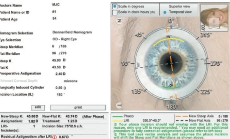
Toric IOL cylinder power and axis placement were deter-mined using the IOL manufacturer’s online calculator (www.acrysoftoriccalculator.com). Size and location of LRI were also determined via online application (www.lricalculator.com), ac-cording to Donnenfeld’s nomogram. For both Toric IOL and LRI groups, biometry, simulated keratometry (one reading per eye), main incision location, and surgeon’s expected surgically induced astigmatism (-0.50 D) were entered into the calculators, with em-metropia as the goal postoperative refraction, i.e., zero sphere and the smallest residual cylinder possible. (11, 12) Figures 1 and 2 show examples of toric IOL and LRI surgical plannnings, respectively.
Surgical Technique
The same surgeon (M.C.) performed all surgeries under mild sedation and topical anesthesia. Just before surgery, a ster-ile ink pen was used to make two marks on the corneal limbus at the 0-degree and 180-degree positions, with the patient sitting upright at the slit lamp, to avoid ocular torsion.
For both groups, phacoemulsification, followed by IOL im-plantation, was performed through a temporal 2.75 mm wide corneal incision.
In the toric IOL group, the IOL was rotated to align with the planned axis. LRI were created inside the limbus using a calibrated diamond knife with the blade depth set at 600 µm.
Postoperative follow up
In the postoperative period, patients were given an eye-drop combination of moxifloxacin and dexamethasone q.i.d. for a week and, then, prednisolone q.i.d. tapered throughout another 3 weeks. All patients were re-evaluated at 1, 3 and 6 months postoperatively by an examiner other than the surgeon (G.F.). Postoperative manifest refraction (sphere and cylinder) and visual acuity (uncorrected and corrected) were obtained. Calcu-lations of Thibos vectors (9,10), for refractive astigmatism, were
Figure 1: Example of toric IOL surgical planning (http://
performed using Microsoft ExcelTM for MacIntosh spreadsheets (version 12.2.7, Microsoft Corp.). Shapiro-Wilk normality tests of data set were performed using IBMTM SPSSTM for Microsoft WindowsTM software (version 20.0.0). A p value of 0.05 or less was considered statistically significant. (13) Wilcoxon test was used to analyze statistical non-parametric differences within the same group throughout the follow up period and Mann-Whitney U test was used to determine differences between Toric IOL and LRI groups at each reevaluation. (5)
R
ESULTSThe study enrolled 62 eyes of 31 consecutive eligible pa-tients. All surgeries were uneventful. None of the eyes required a second intervention. No potentially sight-threatening compli-cations, such as persistent corneal edema, pupillary block, reti-nal detachment or endophthalmitis were observed.
Patient demographics and preoperative data are presented in Table 1.
Group
LRI Toric IOL p-value*
Patients (n) 16 15
-Eyes (n) 32 30
-Sex (F/M) 8/8 11/4
-Age (y)
Mean ± SD 71.75 ± 8.87 65.67 ± 6.28 0.01
Topographic astigmatism (D)
Mean ± SD 1.32 ± 0.47 1.41 ± 0.54 0.60
Range 0.75 to 2.40 0.80 to 2.50
-Steepest topographic 180º-semimeridian angle (n)
0 to 30º or 151º to 180º 18 5
-61º to 120º 8 24
-31º to 60º or 121º to 150º 6 1
-All patients have accomplished the follow up period of 6 months
F = females; D = diopters; IOL = intraocular lens; M = males; LRI = limbal relaxing incisions; mm = millimeters; n = number; SD = standard deviation; y = years; (*) Mann-Whitney U test
Table 1
Patient demographics and preoperative data
Group
Cylinder diopters LRI Toric IOL p-value*
Preoperative
Mean ± SD -1.48 ± 0.60 -1.40 ± 0.73 0.73
Range -2.75 to -0.50 -2.75 to -0.25
-1-month postoperative
Mean ± SD -0.66 ± 0.30 -0.58 ± 0.24
Range -1.25 to 0.00 -1.00 to 0.00
0.25-p value1 0.00 0.00
-3-month postoperative
Mean ± SD -0.70 ± 0.21 -0.63 ± 0.20
0.17
Range -1.00 to 0.00 -1.00 to -0.25
-p value3 0.00 0.00
-6-month postoperative
Mean ± SD -0.74 ± 0.26 -0.62 ± 0.17 0.06
Range -1.25 to -0.25 -1.00 to -0.25
-p value6 0.00 0.00
-IOL = intraocular lens; LRI = limbal relaxing incisions; SD = standard deviation;Wilcoxon test – preoperative cylinder x 1-month(1), 3-month(3) and 6-month(6) postoperative cylinder; (*) Mann-Whitney U test
Table 2
All patients have accomplished the follow up period of 6 months.
Table 2 shows preoperative, 1-month, 3-month and 6-month postoperative manifest cylinder refraction of both groups.
Figure 3 compares the percentage of cumulative frequency of refractive astigmatism between LRI and toric IOL groups.
Figure 4 compares mean magnitudes of astigmatic power vectors (APV), preoperatively, 1-month, 3-month and 6-month between LRI and Toric IOL groups.
Figure 5 compares pre and 6-month postoperative APV in the LRI and toric IOL groups.
D
ISCUSSIONIn this study, both LRI and toric IOL groups presented comparable preoperative characteristics for most aspects of in-terest, as shown in Table 1, in accordance with randomization
Figure 3: compares the percentage of cumulative frequency of refractive astigmatism between LRI and toric IOL groups(IOL = intraocular lens; LRI = limbal relaxing incisions)
Figure 4: Mean magnitudes of preoperative, 1-month, 3-months and 6-months postoperative astigmatic power vectors (APV). Between groups, there was no statistical difference throughout the periods studied, except for the 6-m., when it was lower in the Toric IOL group*. Within each group, preoperative APV was greater than any postoperative APV, remaining stable thereafter† (APV = astigmatic power vector; IOL = intraocular lens; LRI = limbal relaxing incisions; m = n-month postoperative; Preop. = preoperative period; (*) Mann-Whitney U test, p value = 0.05; †Wilcoxon test – pre- and postoperative periods, p value = 0.00)
Figure 5: Scatterplot of astigmatic vectors J0 and J45 preoperatively and 6 months postoperatively in the LRI group (top), and the toric IOL group (bottom) (LRI = limbal relaxing incisions; IOL = intraocular lens)
B
A
A trend of lower mean values favoring the toric IOL group was observed, although such trend was, at most, close to statistical significance at 6th postoperative month. Toric IOL group, in the last postoperative visit, had 97% of eyes with refractive astig-matism between -0.75 D and zero; 100% of eyes between -1.00 D and zero. The LRI group had 69% of eyes between -0.75 D and zero of refractive astigmatism, and 94% of eyes between -1.00 and zero, as can be seen in figure 3. Again, a trend in out-comes predictability, favoring toric IOL group, can be noticed.
Thibos and coworkers (9,10) have proposed a scalar termed astigmatic power vector (APV) that may be used to determine sta-tistical differences between datasets, whenever astigmatism mag-nitude is the primary concern. (20) Such vectorial astigmatism analy-sis is gaining popularity in literature in recent years, as an increas-ing number of articles employ it as analytical instrument. (1, 4, 7, 8, 21, 22) Figure 4 compares mean magnitudes of pre and postoperative APV within each group and between groups. A statistically significant reduction in APV, considering preoperative and any postoperative APV, was found within each group (p value = 0.00). Between groups, toric IOL group exhibited lower APV magnitude mean at 6-month postoperatively; the difference to LRI group was statistically sig-nificant (p value =< 0.05). The trend suggested by non-vectorial analysis of refractive astigmatism, so far, is now highlighted by ob-jective data given by APV vectorial differences between groups.
Figure 5 shows components of APV, J0 and J45, plotted on a two-dimensional Cartesian plane. Spread of 6-month postop-erative APV, in both groups, deviate nearly ±0.50 D from origin (x=0; y=0). However, APV (the vector between origin and each data point) is more homogeneously concentrated around origin in the toric IOL plot than in the LRI plot, which is suggestive of lower postoperative astigmatism in the toric IOL group. (20)
C
ONCLUSIONIn conclusion, satisfactory refractive astigmatism reduc-tion was obtained in both groups. However, our results suggest that the use of toric IOL may be slightly advantageous, from vectorial standpoint, in the treatment of pre-existing corneal astig-matism during phacoemulsification. The main limitation of our study was the greater amount of eyes with oblique or against-the-rule astigmatism present in LRI group, which introduced a bias to the analysis of LRI group of unknown extent. It is also possible that longer follow up periods might undercover statisti-cal significance in the differences of manifest refractive cylinder means between groups.
R
EFERENCES1. Srivannaboon S, Soeharnilla C, Chirapapaisan C, Chonpimai P. Com-parison of corneal astigmatism and axis location in cataract patients measured by total corneal power, automated keratometry, and simu-lated keratometry. J Cataract Refract Surg. 2012;38(12):2088-93. 2. Alió JL, Agdeppa M, Pongo VC, Kady BE. Microincision cataract
sur-gery with toric intraocular lens implantation for correcting moderate and high astigmatism: Pilot study. J Cataract Refract Surg. 2010;36(1):44-52.
3. Wolffsohn JS, Boghal G, Shah S. Effect of uncorrected astigmatism on vision. J Cataract Refract Surg. 2011;37(3):454-60.
4. Muftuoglu O, Dao L, Cavanagh HD, McCulley JP, Bowman RW. Limbal relaxing incisions at the time of apodized diffractive multifo-cal intraocular lens implantation to reduce astigmatism with or with-out subsequent laser in situ keratomileusis. J Cataract Refract Surg. 2010;36(3):456-64.
5. Carvalho MJ, Higashitani-Suzuki S, Lemes-Freitas L. Limbal relax-ing incisions to correct corneal astigmatism durrelax-ing phacoemulsification. J Refract Surg. 2007;23(5):499-504.
6. Kamiya K, Shimuzu K, Ohmoto F, Amano R. Evaluation of corneal biomechanical parameters after simultaneous phacoemulsification with intraocular lens implantation and limbal relaxing incisions. J Cataract Refract Surg. 2011;37(2):265-70.
7. Mendicute J, Irigoyen C, Aramberri J, Ondarra A, Montés-Micó R. Foldable toric intraocular lens for astigmatism correction in cataract patients. J Cataract Refract Surg. 2008;34(4):601-7.
8. Mingo-Botín D, Muñoz-Negrete F, Kim HRW, Morcillo-Laiz R, Rebolleda G, Oblanca N. Comparison of toric intraocular lenses and peripheral corneal relaxing incisions to treat astigmatism during cata-ract surgery. J Catacata-ract Refcata-ract Surg. 2010;36(10):1700-8.
9. Thibos LN, Wheeler W. Power vectors: an application of Fourier analy-sis to the description and statistical analyanaly-sis of refractive error. Optom Vis Scie. 1997;74(6):367-75.
10. Thibos LN, Horner D. Power vector analysis of the optical outcome of refractive surgery. J Cataract Refract Surg. 2001;27(1):80-5. 11. Behndig A, Montan P, Stenevi U, Kugelberg M, Zetterström, Lundström.
Aiming for emmetropia after cataract surgery: Swedish National Cata-ract Register Study. J CataCata-ract RefCata-ract Surg. 2012;38(7):1181-6. 12. Goggin M, Moore S, Esterman A. Outcome of Toric Intraocular Lens
Implantation After Adjusting for Anterior Chamber Depth and In-traocular Lens Sphere Equivalent Power Effects. Arch Ophthalmol. 2011;129(8):998-1003.
13. Razali NM, Wah Y. Power comparisons of Shapiro-Wilk, Kolmogorov-Smirnov, Lilliefors and Anderson-Darling tests. J Statistic Model Analytics. 2011;2(1):21-33.
14. Goto T, Klyce S, Zheng X, Maeda N, Kuroda T, Ide C. Gender- and Age-ralated differences in corneal topography. Cornea. 2001;20(3):270-6. 15. Gudmundsdottir E, Jonasson F, Jonsson V, Stefánsson E, Sasaki H, Sasaki K. “With the rule” astigmatism is not the rule in the elderly - Reykjavik Eye Study: A population based study of refraction and visual acuity in citizens of Reykjavik 50 years and older. Acta Ophthalmol Scand. 2000;78(6):642-6.
16. Silva EF, Trindade F. Surgical correction of astigmatism during cata-ract surgery. Arq Bras Oftalmol. 2007;70(4):609-14.
17. Ganekal S, Dorairaj S, Jhanji V. Limbal relaxing incisions during phacoemulsification: 6-month results. J Cataract Refract Surg. 2011(37):2081-2.
18. Visser N, Nuijits R, deVries NE, Bauer NJC. Visual outcomes and patient satisfaction after cataract surgery with toric multifocal intraocu-lar lens implantation. J Cataract Refract Surg. 2011;37(11):2034-42. 19. Alió JL, Piñero D, Tomás J, Alesón A. Vector analysis of astigmatic changes after cataract surgery with toric intraocular lens implanta-tion. J Cataract Refract Surg. 2011;37(6):1038-49.
20. Statham M, Apel A, Stephensen D. Comparison of the AcrySof SA60 spherical intraocular lens and the AcrySof Toric SN60T3 intraocular lens outcomes in patients with low amounts of corneal astigmatism. Clin Exper Ophthalmology. 2009;37(8):775-9.
21. Mendicute J, Irigoyen C, Ruíz M, Illarramendi I, T Ferrer-Blasco, Montés-Micó R. Toric intraocular lens versus opposite clear corneal incisions to correct astigmatism in eyes having cataract surgery. J Cataract Refract Surg. 2009;35(3):451 - 8.
22. Sheppard AL, Wolffsohn J, Bhatt U, Hoffmann PC, Scheider A, Hütz WW, Shah S. Clinical outcomes after implantation of a new hydropho-bic acrylic toric IOL during routine cataract surgery. J Cataract Re-fract Surg. 2013;39(1):41-7.
Corresponding author:
Giuliano O. Freitas