Research brief
Vasoactive intestinal peptide reduces the in
fl
ammatory pro
fi
le in mice
infected with
Trypanosoma cruzi
Pulch
eria Maria Silva Higyno
a, Priscila Fagundes Mendes
a, Marina Barcelos de Miranda
a,
Dario Elias Pereira
a, Ana Paula Lucas Mota
c, Katiane de Oliveira Pinto Coelho Nogueira
a,
Ivo Santana Caldas
a,b, Sandra Aparecida de Lima Moura
a,
Cristiane Alves da Silva Menezes
c,*aInstituto de Ci^encias Exatas e Biologicas, Departamento de Ci^encias Biologicas, Universidade Federal de Ouro Preto, Campus Morro do Cruzeiro S/N,
Ouro Preto, Minas Gerais, CEP: 35400-000, Brazil
bDepartamento de Patologia e Parasitologia, Universidade Federal de Alfenas, Rua Gabriel Monteiro da Silva, 700, Alfenas, Minas Gerais, CEP: 37130-000,
Brazil
cFaculdade de Farmacia, Departamento de Analises Clínicas e Toxicologicas, Universidade Federal de Minas Gerais, Avenida Presidente Ant^onio Carlos,
6627, Belo Horizonte, Minas Gerais, CEP: 31270-901, Brazil
h i g h l i g h t s
g r a p h i c a l
a b s t r a c t
The effect of VIP treatment on mice infected withT. cruziwas studied.
VIP treatment had no impact on parasitemia.
VIP treatment reduced heart mass
and heart inflammatory infiltrates.
VIP treatment reduced levels of IFN-gamma and IL-2 and increased IL-4 levels.
VIP treatment effects may be due to the switch from a Th1 to a Th2 profile.
a r t i c l e
i n f o
Article history:
Received 29 October 2014 Received in revised form 23 July 2015
Accepted 3 September 2015 Available online 7 September 2015
Keywords: Trypanosoma cruzi
vasoactive intestinal peptide Neuroimmunomodulation
a b s t r a c t
Vasoactive intestinal peptide (VIP) has gained great prominence because of its therapeutic potential, which is ascribed to its ability to regulate innate immunity, inhibit antigen-specific Th1 cell responses, and generate T regulatory cells. Additionally, VIP may act as a natural antimicrobial peptide, killing bacteria, fungi, and infective forms ofTrypanosoma brucei. Despite the possible relevance of VIP during the course of Chagas disease, studies regarding this in human and experimentalTrypanosoma cruzi in-fections remain poorly characterized. In this work, we evaluated the effects of VIP on systemic and cardiac immune responses during experimental acute infection. C57BL/6 mice were infected with 5000 trypomastigotes of the VL-10 strain ofT. cruziand treated with intraperitoneal VIP injection every other day for one month. After 30 days, we observed no reduction in parasitemia levels. However, we observed a reduction in serum levels of IFN-gamma and IL-2 and an increase in that of IL-4. These data suggest that VIP treatment modified immune responses to favor the Th2 response, which had no impact on para-sitemia levels although the serum level of IFN-gamma was reduced. However, this change in immune balance reduced heart damage, as noted by the smaller cardiac volume and the moderate inflammatory
*Corresponding author.
E-mail address:menezescristiane1@gmail.com(C.A.S. Menezes).
Contents lists available atScienceDirect
Experimental Parasitology
j o u r n a l h o m e p a g e :w w w . e l s e v i e r . c o m / l o c a t e / y e x p r
infiltrate observed in VIP-treated mice. Our results indicate that VIP treatment reduced the inflammatory response at the cardiac site of mice that were experimentally infected withT. cruzi. These data suggest a protective role for VIP in the heart of infected mice.
©2015 Elsevier Inc. All rights reserved.
1. Introduction
Trypanosoma cruzi is the causative agent of Chagas disease, which afflicts approximately 8e10 million people in Latin America, making it the most lethal endemic infectious disease in the West-ern Hemisphere. Parasite transmission frequently occurs through contact with the feces of infected triatomine bugs. However, infection can additionally be transmitted through blood trans-fusion, organ transplantation, from mother to child, and the con-sumption of contaminated food and beverages (WHO, 2012).
Chagas disease progression involves two successive stages: an acute phase and a chronic phase. The acute phase is generally asymptomatic and is characterized by the presence of the parasite in blood. The chronic phase is characterized by decreased para-sitemia and features four distinct clinical forms: the indeterminate, cardiac, digestive, and cardiodigestive forms. The factors that cause the development of different clinical forms in individuals infected with T. cruzi are not completely understood. However, host ge-netics, the host immune system, and parasite variability are involved in disease evolution (Dutra et al., 2014).
Contact between parasites and host cells is critical in Chagas disease. This contact triggers the initial immune response through cellular activation. However, cellular reactivity is possibly main-tained by the antigenic components of the parasite and host. The anti-parasite reactivity, which is essential for chronic disease development, leads to pathology if not properly modulated (Dutra et al., 2009). The identification of endogenous factors that control exacerbated immune responses is a key goal for the development of preventive and therapeutic approaches to infectious and infl am-matory diseases.
Neuropeptides play many roles in the immune system. Vaso-active intestinal peptide (VIP) is a neuropeptide comprised of 28 amino acids and exerts immunoregulatory and neuromodulatory effects on various organs and tissues (Igarashi et al., 2011). The biological functions of VIP occur through the binding of the neu-ropeptide to its two G protein-coupled receptors, VPAC1 and VPAC2. VIP receptors are expressed differentially in immunocom-petent cells. The cAMP/PKA pathway is the major signaling pathway for VIP anti-inflammatory action in macrophages, mono-cytes, DCs, and microglia and in the regulation of T lymphocyte responses (Correa et al., 2013^ ).
VIP-receptor engagement inhibits phagocytic activity, reduces free radical production and adhesion/migration activity, and de-creases costimulatory molecule expression (De La Fuente et al., 1996.). Additionally, VIP decreases inflammatory cytokine produc-tion and increases anti-inflammatory cytokine production (Delgado et al., 1996, 1999), reduces nitric oxide synthases and cyclo-oxygenase production, and inhibits toll-like receptor (TLR) expression (Gomariz et al., 2005). With respect to regulatory T cell biology, VIP treatment generates these cells by converting CD4þ
CD25 cells into CD4þ CD25þ
cells (Anderson and Gonzalez-Rey, 2010).
Recent results in experimental models have demonstrated positive effects of VIP treatment in different pathological conditions where this neuropeptide has emerged as a potent anti-inflammatory agent because of its ability to regulate innate
immunity by inhibiting Th1 cell responses and generating regula-tory T cells (Pozo et al., 2007). In a murineSchistosoma mansoni infection, VIP treatment was very effective in diminishing worm fecundity, hepatic granuloma size and number, and liver collagen content. Serum levels of IL-10 increased, while levels of IL-12 and TNF-alpha decreased, because of VIP administration. In addition, hepatic function improved after VIP administration. These results indicate that the administration of exogenous VIP can be effective in ameliorating immunopathological damage associated with schistosomiasis (Allam, 2007). In addition to their immunomodu-latory role, neuropeptides also have antimicrobial activity against a range of microorganisms from skin, oral, respiratory, and gastro-intestinal tract sites. VIP possesses direct antimicrobial activities against a range of pathogens, including Escherichia coli, Pseudo-monas aeruginosa,Candida albicans, andStreptococcus mutans(El Karim et al., 2008). Additionally, VIP displays strong antimicrobial activity againstTrypanosoma brucei, causing parasite death via the destruction of membrane integrity (Gonzalez-Rey et al., 2006). In experimental T. brucei infection, VIP treatment improved symp-toms in chronic trypanosomiasis by reducing parasite burden in many target organs (Delgado et al., 2009).
Until now, no studies have elucidated the effect of VIP on par-asitemia or cardiac inflammation during an experimentalT. cruzi infection. Thus, considering that cardiac Chagas disease is charac-terized by a prominent initial inflammatory response, the aim of this investigation is to evaluate the effects of VIP treatment on parasite persistence and the development of systemic and cardiac inflammation in experimental acute infection induced by VL-10, a cardiotropic strain ofT. cruzi(Caldas et al., 2008).
2. Material and methods
2.1. Animal infection and VIP treatment
Five-to-six-week-old female C57BL/6 mice were obtained from and housed in the animal facility at Universidade Federal de Ouro Preto (UFOP, MG, Brazil) underad libitumwater and feeding con-ditions. Groups of ten animals were inoculated with theT. cruzi VL-10 strain (5000 trypomastigote forms) intraperitoneally. The groups of infected and uninfected mice were then treated with intraperitoneal VIP every other day (1.5 nmol/animal; American Peptide Company) for 30 days (Delgado et al., 2009). Parasitemia was determined daily for 30 days by using a 5
m
L blood sampleobtained from the tail, according to the Brener protocol (Brener, 1962). Curves were plotted by using the mean parasitemia of ten mice. All experimental protocols described here were approved by the Ethical Committee for Experiments with Laboratory Animals (CEUA⁄UFOP No #2012/47) and performed in compliance with the guidelines issued by Colegio Brasileiro de Experimentaç~ao Animal.
2.2. Cytokines and chemokine ELISA
trifluoroacetic acid⁄1.35 M NaCl were mixed and left for 10 min at 25C. The samples were then centrifuged for 5 min at 3000
g, and
supernatants were adjusted for salt content (0.14 M sodium chlo-ride and 0.01 M sodium phosphate) and normalized to pH 7.4 to determine the concentrations of these soluble murine cytokines. IFN-gamma, IL-2, IL-4, and MIP-2 (PeproTech; Brasil FUNPEC) were then quantified by ELISA according to the manufacturer's in-structions. All samples were measured simultaneously in duplicate.
2.3. Heart mass measurement
Hearts were carefully excised ex vivo and gently blotted on absorbent paper to remove blood before wet weight measurements were calculated. Relative heart weight was calculated using the ratio of heart weight to body weight in milligrams (mg). This value was used to evaluate the cardiac mass obtained after 30 days of infection and after treatment with VIP.
2.4. Histopathological analysis
Animals were euthanized on day 30 afterT. cruziinfection, and ventricular heart tissue wasfixed by using 4% glutaraldehyde in PBS (0.1 M, pH 7.4), dehydrated, and embedded in glycol methacrylate medium (Historesin®
, Leica). Blocks were cut into 2-
m
m sectionsand stained with hematoxylin and eosin (H&E) for routine his-topathological analysis and for the qualitative evaluation of myocardial inflammation and the presence of amastigote nests. Twentyfields from each H&E-stained section were randomly cho-sen at a 40X magnification, resulting in 1.5106
m
m2of analyzedmyocardial area. Images were obtained using a Leica DM 5000 B microscope (Leica Application Suite, version 2.4.0 R1) and pro-cessed with Leica QWin (version 3) image analyzer software. The inflammatory process was evaluated using the correlation index between the number of cells observed in myocardial muscle of uninfected and infected animals (Caldas et al., 2008)
2.5. Statistical analysis
For all experiments, statistical significance was established at p <0.05. Results are presented as the mean ±SD. All statistical analyses were performed using GraphPad Prism (version 5.0) software. Normal distribution of data was assessed according to
D'Agostino-Pearson analysis. Serological analyses between groups were performed using a t-Test. Parasitemia was analyzed using analysis of variance (ANOVA) followed by the Tukey's post hoc test.
3. Results
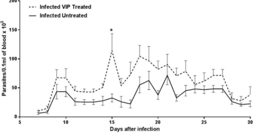
Wefirst evaluated the effects of VIP treatment on parasitemia in mice infected with the cardiotropic VL-10 strain ofT. cruzi. Parasite load in blood smears was monitored daily from day 7 until day 30 after infection. Assessment of parasitemia levels in all animals infected with T. cruzi showed that control (infected untreated) animals and VIP-treated animals had similar parasitemia levels (Fig. 1). However, on day 15, VIP-treated animals exhibited a drastic increase in circulating parasites. In subsequent days, parasitemia levels between groups remained similar (Fig. 1).
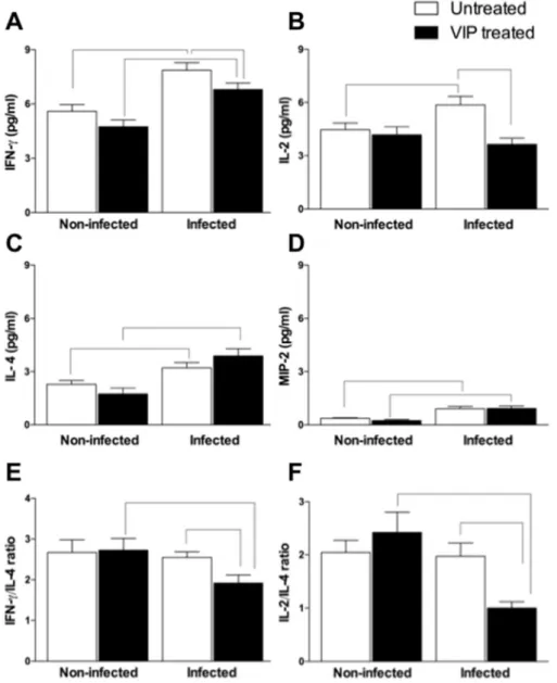
Considering that proinflammatory cytokine production is important to control initialT. cruzireplication and that VIP has been implicated in modulating the immune system, we evaluated possible alterations in the expression of cytokines (IFN-gamma, IL-2, and IL-4) and the chemokine MIP-2 after infection and treatment with VIP. In VIP-untreated mice, infection withT. cruzienhanced the production of IFN-gamma (Fig. 2A), IL-2 (Fig. 2B), IL-4 (Fig. 2C), and MIP-2 (Fig. 2D) by 41%, 31%, 39%, and 145%, respectively, when compared with uninfected VIP-untreated mice. In VIP-treated mice, infection with T. cruzi increased the production of IFN-gamma (Fig. 2A), IL-4 (Fig. 2C), and MIP-2 (Fig. 2D) by 44%, 124%, and 284%, respectively, when compared with uninfected VIP-treated mice. Compared withT. cruziinfected untreated group, adminis-tration of VIP inT. cruzi-infected mice led to a decrease in serum concentrations of IFN-gamma (Fig. 2A) and IL-2 (Fig. 2B) by 14% and 39%, respectively. Conversely, VIP treatment increased the expres-sion of IL-4 by 22% compared withT. cruziinfected untreated mice (Fig. 2C). MIP-2 expression was not affected by VIP administration (Fig. 2D). The administration of VIP or its vehicle in uninfected mice did not modify the expression profiles of IFN-gamma, IL-2, IL-4, or MIP-2 (Fig. 2). The balance between proinflammatory and anti-inflammatory cytokine production is essential to parasitemia and inflammation control in Chagas disease. We analyzed cytokine ra-tios and observed a decrease in the ratio between the expression of IFN-gamma and that of the Th2 cytokine IL-4 (1.9±0.2) inT. cruzi -infected VIP-treated mice compared with un-infected VIP-treated mice (2.7±0.3) (Fig. 2E). A similar result was observed in the
IL-Fig. 1.Parasitemia curve in animals infected withTrypanosoma cruzitreated with vasoactive intestinal peptide (VIP). C57BL/6 mice were inoculated with 5.000 trypomastigotes of
T. cruziVL-10 strain intraperitoneally and received VIP (13mg/mouse) or vehicle (saline solution) every other day for 30 days. Parasitemia was evaluated daily from days 7e30 after
2/IL-4 ratio in infected VIP-treated mice (1±0.1) compared with uninfected VIP-treated mice (2.4±0.4) (Fig. 2F). On comparison of
IFN-gamma/IL-4 and IL-2/IL-4 ratios in the infected VIP-untreated and infected VIP-treated groups, we observed that VIP treatment decreased IFN-gamma/IL-4 (1.9±0.2 vs. 2.6 ±0.1) and IL-2/IL-4 (1±0.1 vs. 2±0.3) (Fig. 2E and F). In control VIP-untreated mice,
infection did not alter the cytokine balance (Fig. 2E and F). Since we observed alterations in systemic immune responses through cytokine expression after VIP administration, we wondered if those alterations were reflected at cardiac sites. Heart mass of infected VIP-treated mice (4.9±0.1 mg) was significantly decreased compared to infected VIP-untreated mice (5.5±0.12 mg)
(Fig. 3A). Histological analysis of the myocardium was performed to determine if heart weight was related to cellular alterations in heart tissue. Cardiac tropism with typical inflammatory reactions to T. cruzi, with vascular and cellular alterations such as hyperemia and hydropic degeneration of cardiac cells, was observed. Addi-tionally, at the end of treatment (30 days after infection), VIP-treated infected animals (Fig. 3B) showed lesser inflammatory
infiltrate (131 ± 18.5 cells) than did the VIP-untreated infected control animals (198±22.3 cells). The VIP-untreated group
pre-sented with intense focal inflammatory infiltrates, while the VIP-treated group presented with more discrete and diffuse infl am-matory infiltrates (Fig. 3CeF). We did not observe any amastigote nests in analyzed tissue sections of VIP-treated and untreated mice. Uninfected mice treated with VIP did not present any histopatho-logical alterations in the heart (data not shown). Our results indi-cate that VIP affects the hearts of mice infected with T. cruziby decreasing immune cell proliferation and myocardial infiltration.
4. Discussion
The inflammatory response generated afterT.cruziinfection is an important factor in the control of parasitemia in the acute phase and the development of the chronic phase of human and experi-mental Chagas disease (Lannes-Vieira et al., 2009; Talvani and Teixeira, 2011). However, the persistence of inflammation may lead to the development of lesions in host organs since the immune
cells recognize and attack host tissues. Because of the possible deleterious effects of immune responses in Chagas disease, studies have been designed to modulate the inflammatory process and its harmful effects to the body by using drugs with anti-inflammatory profiles. The use of anti-inflammatory drugs reduced cardiac in-flammatory infiltrates,fibrosis, and circulating cytokines/chemo-kines and improved cardiac function inT. cruziinfection (Botoni et al., 2007; Coelho Dos Santos et al., 2010; Paula-Costa et al., 2010). The results obtained using anti-inflammatory compounds in Chagas disease motivated the search for new therapeutic targets that are efficient in controlling the inflammatory response observed in Chagas disease patients. Along these lines, vasoactive intestinal peptide (VIP), a neuropeptide endogenously produced in many tissues and organs that is nontoxic and has immunomodu-latory activities, is a good candidate as a natural immunomodula-tory factor. Additionally, studies have proven the therapeutic
potential of VIP in several diseases, especially in inflammatory, respiratory, and neurodegenerative disorders (Campos-Salinas et al., 2014). Furthermore, VIP exerts a direct antimicrobial activ-ity against bacteria (El Karim et al., 2008) and T. brucei(Delgado et al., 2009).
Based on the above-mentioned information, we evaluated the effects of VIP in acute experimental infection withT. cruzi. Our re-sults indicate that VIP did not alter parasitemia during the 30 days of infection. However, VIP shifted the immune response to a Th2 profile, which led to less aggressive myocarditis when compared to infected VIP-untreated mice.
VIP shares similarities with other neuropeptides, such as small size, cationic charge, and amphipathic design. Moreover, VIP is released during microbe-induced inflammation. These findings support a new role for VIP as a potential host defense peptide (Delgado et al., 2009, Delgado and Ganea, 2011). On the other hand, Fig. 3.Heart mass measurement and histological examination of hearts from mice infected withT. cruziand treated with vasoactive intestinal peptide (VIP). Hearts were carefully excisedex vivoand gently blotted on absorbent paper to remove blood before the wet weight measurements were calculated. The relative heart weight was calculated using the ratio of heart weight/body weight in milligrams (mg). This value was used to evaluate cardiac mass measurements after 30 days of infection in mice untreated (white bar) or treated with VIP (black bar) (A) (n¼10). Cellularity levels of the myocardial sections of mice infected withT. cruziare shown (B). Representative histopathology of infected control mice (C, E) and mice treated with vasoactive intestinal peptide (D, F). Hearts were obtained on day 30,fixed in 4% glutaraldehyde, and embedded in Historesin®
VIP is minimally stable after systemic administration, and its poor penetration at the desired site of action could influence its clinical application. In our model of study, VIP was unable to reduce blood parasitemia. In recent work that tested VIP and two analogues in experimental leishmaniasis infection, native VIP did not signifi -cantly reduce the viability of promastigote forms ofL. majorand thus failed as a leishmanicidal agent. Although VIP injection diminished paw swelling and lesion size, VIP was not able to significantly control the clinical manifestations and progression of this disease (Campos-Salinas et al., 2014). On the other hand, VIP displayed strong antimicrobial activity against T. brucei, causing parasite death by destroying membrane integrity (Gonzalez-Rey et al., 2006). In experimental infection with T. brucei, VIP treat-ment led to an improvetreat-ment of symptoms by reducing the parasite burden in many target organs (Delgado et al., 2009). Those results showed that the effects of VIP on trypanosomatid parasites and subsequent infections might vary. Considering these observations, VIP dose adjustment and/or the use of stable VIP analogues could clarify the effects of this neuropeptide onT. cruzi viability and confirm if there is a potential antimicrobial role for VIP inT. cruzi in vivoandin vitromodels.
In relation to cardiac effects, VIP treatment efficiently reduced the intensity of heart inflammation in animals inoculated with the VL10T. cruzistrain. This was confirmed by reduction of heart mass and cellular infiltrates in the myocardium of VIP-treated mice compared to infected VIP-untreated animals. VIP exerts its biolog-ical activities by binding to the closely related class II G protein-coupled receptors VIP receptor type 1 (VPAC1) and VIP receptor type 2 (VPAC2). Both receptors are coupled to adenylate cyclase activation and subsequent activation of the protein kinase A (PKA) in immune cells. Activation of this pathway leads to the inhibition of transcription factors that upregulate a large number of proin-flammatory genes (Smalley et al., 2009). The suppression of heart inflammation by VIP may be, in part, associated with a systemic reduction in cytokines such as IFN-gamma and IL-2 and a concomitant increase in IL-4 production. The protective response againstT. cruziinfection is influenced by innate and adaptive im-munity. These cells produce inflammatory cytokines, which play a central role in regulating parasite replication and immune re-sponses. In addition to the systemic effects of VIP on the immune system, infection increases the levels of IFN-gamma, IL-4, and MIP-2. VIP treatment reduced IFN-gamma and IL-2 production and increased 4 plasma levels. The modulation of IFN-gamma and IL-2 and upregulation of IL-4 observed in infected VIP-treated mice may be important to control cell mediated immunity and tissue damage elicited by T. cruzi infection because IL-4 regulates the development and effector functions of cell-mediated immunity (Talvani et al., 2000). Additionally, cytokines produced by T lym-phocytes afterT. cruziinfection, such as interleukin 2 (IL-2), trigger important effector mechanisms and act as important growth fac-tors for CD8þ
lymphocyte effector functions (Martin and Tarleton, 2004). Activated CD8þT cells that express granzyme A are abun-dant in cardiac tissue from Chagas disease patients, connecting these cells to tissue destruction (Reis et al., 1993). The reduction in IL-2 production may lead to a reduction of activated CD8þT cells and less heart damage. Studies show that production of proin-flammatory cytokines, such as IFN-gamma and TNF-alpha, plays a pathogenic role inT. cruzi-induced cardiomyopathy, and the dele-terious effects of these factors have already been reported in experimental models and in human disease (Cunha-Neto et al., 2009; Terasaki et al., 2008; Reifenberg et al., 2007). On the other hand, IFN-gamma production is essential for parasite elimination since this cytokine activates macrophages, which are thought to be a major cell population involved in parasite uptake through the production of several intracellular killing mediators such as nitric
oxide (Borges et al., 2013). In our model, the reduced of IFN-gamma production did not affect parasitemia, which was similar between VIP-treated and untreated mice. Therefore, the reduction in IFN-gamma production due to VIP treatment may limit cardiac dam-age without resulting in increased parasitemia. Given this, although treatment with vasoactive intestinal peptide did not prevent car-diac inflammation triggered by infection withTrypanosoma cruzi, the inflammation observed in infected VIP-treated mice was limited compared to that in VIP-untreated mice.
Histological analyses did not reveal amastigote nests in the cardiac tissue of infected mice either treated or untreated with VIP. Theories of parasite persistence and autoreactivity explain the establishment of Chagas disease (Dutra et al., 2009). Our under-standing is that parasite presence is required to trigger an immune response, which can control parasitemia in the transition period between acute and chronic phases of the disease. The immune response sustained by the host after attaining the chronic phase will determine the pathological manifestations of the disease. Thus, the inflammatory response is fundamental for parasite control in the early stages of infection, and the modulation of this immune response is essential to avoid an exacerbation of this response and the resulting tissue damage. Supporting this, VIP participates in the modulation of immune response.
Regarding human Chagas disease, data from our group show that patients with Chagas disease with cardiac disorders present lower plasma levels of VIP compared to those in individuals with the indeterminate form of the disease. This suggests that higher or lower VIP expression may be related to the maintenance of the indeterminate form or to the development of the cardiac disorders, respectively (Correa et al., 2013^ ). Altogether, results from the hu-man study and this work suggest a protective role of VIP in cardiac tissue afterT. cruziinfection. Future studies may indicate beneficial effects of VIP mediated immunomodulation, allowing for the cre-ation of new treatment strategies and therapeutics to improve the quality of life of patients with Chagas disease.
Acknowledgments
This work was supported by Pro-Reitoria de Pesquisa da UFMG
(PRPQ/UFMG) and by grants from the Fundaç~ao de Amparoa Pes-quisa do Estado de Minas Gerais and Conselho Nacional de Desenvolvimento Científico e Tecnologico (PRONEX-Chagas/ Demanda Universal e FAPEMIG and CNPq). We thank Dr. Luis Carlos Crocco Afonso (NUPEB⁄UFOP) for support with the C57BL/6 mice used in this work.
References
Allam, G., 2007. Vasoactive intestinal peptide inhibits liver pathology in acute murine schistosomiasis mansoni and modulates IL-10, IL-12 and TNF-alpha production. Immunobiology 212 (8), 603e612 (Epub).
Anderson, P., Gonzalez-Rey, E., 2010. Vasoactive intestinal peptide induces cell cycle arrest and regulatory functions in human T cells at multiple. Mol. Cell Biol. 30 (10), 2537e2551.
Borges, D.C., Araújo, N.M., Cardose, C.R., Lazo Chica, J.E., 2013 Feb. Different parasite inocula determine the modulation of the immune response and outcome of experimentalTrypanosoma cruziinfection. Immunology 138 (2), 145e156. Botoni, F.A., Poole-Wilson, P.A., Ribeiro, A.L., Okonko, D.O., Oliveira, B.M., Pinto, A.S.,
Teixeira, M.M., Teixeira, A.L.J.R., Reis, A.M., Dantas, J.B., Ferreira, C.S., Tavares, W.C.J.R., Rocha, M.O., 2007. A randomized trial of carvedilol after renin-angiotensin system inhibition in chronic Chagas cardiomyopathy. Am. Heart J. 153, e1e8.
Brener, Z., 1962. Therapeutic activity and criterion of cure on mice experimentally infected withTrypanosoma cruzi. Rev. Inst. Med. Trop. Sao Paulo 4, 389e396. Caldas, I.S., Talvani, A., Caldas, S., Carneiro, C.M., DE, L.M., Da Matta Guedes, P.M.,
Bahia, M.T., 2008. Benznidazole therapy during acute phase of Chagas disease reduces parasite load but does not prevent chronic cardiac lesions. Parasitol. Res. 103, 413e421.
vasoactive intestinal peptide against pathogens. J. Biol. Chem. 289 (21), 14583e14599.
Coelho Dos Santos, J.S., Menezes, C.A., Villani, F.N., Magalh~aes, L.M., Scharfstein, J., Gollob, K.J., Dutra, W.O., 2010. Captopril increases the intensity of monocyte infection byTrypanosoma cruziand induces human T helper type 17 cells. Clin. Exp. Immunol. 163, 528e536.
Corr^ea, M.V., Da Costa Rocha, M.O., De Sousa, G.R., Do Carmo Pereira Nunes, M., Gollob, K.J., Dutra, W.O., Da Silva Menezes, C.A., 2013. Low levels of vasoactive intestinal peptide are associated with Chagas disease cardiomyopathy. Hum. Immunol. 74 (10), 1375e1381.
Cunha-Neto, E., Nogueira, L.G., Teixeira, P.C., Ramasawmy, R., Drigo, S.A., Goldberg, A.C., Fonseca, S.G., Bilate, A.M., Kalil, J., 2009. Immunological and non-immunological effects of cytokines and chemokines in the pathogenesis of chronic Chagas disease cardiomyopathy. Mem. Inst. Oswaldo Cruz 104 (Suppl. I), 252e258.
De La Fuente, M., Delgado, M., Gomariz, R.P., 1996. VIP modulation of immune cell functions. Adv. Neuroimmunol. 6, 75e91.
Delgado, M., Anderson, P., Garcia-Salcedo, J.A., Caro, M., Gonzalez-Rey, E., 2009. Neuropeptides kill African trypanosomes by targeting intracellular compart-ments and inducing autophagic-like cell death. Cell Death Differ. 16 (3), 406e416. Delgado, M., Ganea, D., 2011. Vasoactive intestinal peptide: a neuropeptide with
pleiotropic immune functions. Amino Acids 3, 4e5.
Delgado, M., Munoz-Elias, E.J., Gomariz, R.J., Ganea, D., 1999. Vasoactive intestinal peptide and pituitary adenylate cyclase-activating polypeptide enhance IL-10 production by murine macrophages: in vitro and in vivo studies. J. Immunol. 162, 1707e1716.
Delgado, M., Munoz-Elias, E.J., Gomariz, R.J., Ganea, D., 1996. Vasoactive intestinal peptide and pituitary adenylate cyclase-activating polypeptide prevent induc-ible nitric oxide synthase transcription in macrophages by inhibiting NF-kB and IFN regulatory factor 1 activation. J. Immunol. 162 (8), 4685e4696.
Dutra, W.O., Menezes, C.A., Magah~aes, L.M., Gollob, K.J., 2014. Immunoregulatory networks in human Chagas disease. Parasite Immunol. 36 (8), 377e387. Dutra, W.O., Menezes, C.A., Villani, F.N., Da Costa, G.C., Da Silveira, A.B., Reis, D.D.,
Gollob, K.J., 2009. Cellular and genetic mechanisms involved in the generation of protective and pathogenic immune responses in human Chagas disease. Mem. Inst. Oswaldo Cruz 104 (Suppl. 1), 208e218.
El Karim, I.A., Linden, G.J., Orr, D.F., Lundy, F.T., 2008. Antimicrobial activity of neuropeptides against a range of micro-organisms from skin, oral, respiratory and gastrointestinal tract sites. J. Neuroimmunol. 200, 11e16.
Gomariz, R.P., Arranz, A., Abad, C., Torroba, M., Martinez, C., Rosignoli, F., Garcia-Gomez, M., Leceta, J., Juarranz, Y., 2005. Time-course expression of Toll-like receptors 2 and 4 in inflammatory bowel disease and homeostatic effect of VIP. J. Leukoc. Biol. 78, 491e502.
Gonzalez-Rey, E., Chorny, A., Delgado, M.V.I.P., 2006. An agent with license to kill infective parasites. Ann. N. Y. Acad. Sci. 1070, 303e308.
Igarashi, H., Fujimori, N., Ito, T., Nakamura, T., Oono, T., Nakamura, K., Suzuki, K., Jensen, R.T., Takayanagi, R., 2011. Vasoactive intestinal peptide (VIP) and VIP receptors: elucidation of structure and function for therapeutic applications. IJCM 2, 500e508.
Lannes-Vieira, J., Silverio, J.C., Pereira, I.R., Vinagre, N.F., Carvalho, C.M.E., Paiva, C.N., Silva, A.A., 2009. ChronicTrypanosoma cruzi-elicited cardiomyopathy: from the discovery to the proposal of rational therapeutic interventions targeting cell adhesion molecules and chemokines receptorsehow to make a dream come true. Mem. Inst. Oswaldo Cruz 104 (Suppl. I), 226e235.
Martin, D., Tarleton, R., 2004. Generation, specificity, and function of CD8þ T cells in
Trypanosoma cruziinfection. Immunol. Rev. 201, 304e331.
Paula-Costa, G., Silva, R.R., Pedrosa, M.C., Pinho, V., De Lima, W.G., Teixeira, M.M., Bahia, M.T., Talvani, A., 2010. Enalapril prevents cardiac immune-mediated damage and exerts anti-Trypanosoma cruzi activity during acute phase of experimental Chagas disease. Parasite Immunol. 32, 202e208.
Pozo, D., Gonzalez-Rey, E., Chorny, A., Anderson, P., Varela, N., Delgado, M., 2007. Tuning immune tolerance with vasoactive intestinal peptide: a new therapeutic approach for immune disorders. Peptides 28 (9), 1833e1846.
Reifenberg, K., Lehr, H.A., Torzewski, M., Steige, G., Wiese, E., Küpper, I., Becker, C., Ott, S., Nusser, P., Yamamura, K., Rechtsteiner, G., Warger, T., Pautz, A., Kleinert, H., Schmidt, A., Pieske, B., Wenzel, P., Münzel, T., L€ohler, J., 2007. Interferon-gamma induces chronic active myocarditis and cardiomyopathy in transgenic mice. Am. J. Pathol. 171 (2), 463e472.
Reis, D.D., Jones, E.M., Tostes, S.J.R., Lopes, E.R., Gazzinelli, G., Colley, D.G., McCurley, T.L., 1993. Characterization of inflammatory infiltrates in chronic chagasic myocardial lesions: presence of tumor necrosis factor-alphaþcells and dominance of granzyme Aþ, CD8þlymphocytes. Am. J. Trop. Med. Hyg. 48, 637e644.
Smalley, S.G., Barrow, P.A., Foster, N., 2009. Immunomodulation of innate immune responses by vasoactive intestinal peptide (VIP): its therapeutic potential in inflammatory disease. Clin. Exp. Immunol. 157 (2), 225e234.
Talvani, A., Teixeira, M.M., 2011. Inflammation and Chagas disease: some mecha-nisms and relevance. Adv. Parasitol. 76, 171e194.
Talvani, A.L., Ribeiro, C.S., Aliberti, J.C., Michailowsky, V., Santos, P.V., Murta, S.M., Romanha, A.J., Almeida, I.C., Farber, J., Lannes-Vieira, J., Silva, J.S., Gazzinelli, R.T., 2000. Kinetics of cytokine gene expression in experimental chagasic cardio-myopathy: tissue parasitism and endogenous IFN-gamma as important de-terminants of chemokine mRNA expression during infection withTrypanosoma cruzi. Microbes Infect. 2 (8), 851e866.
Terasaki, F., Ukimura, A., Tsukada, B., Fujita, S., Katashima, T., Otsuka, K., Otsuka, K., Kanzaki, Y., Shimomura, H., Fujita, M., Tanaka, T., Kitaura, Y., 2008. Enhanced expression of type 1 helper T-cell cytokines in the myocardium of active cardiac sarcoidosis. Circ. J. 72 (8), 1303e1307.