Trypanosoma cruzi
: Treatment with the iron chelator
desferrioxamine reduces parasitemia and mortality
in experimentally infected mice
Jerusa Marilda Arantes
a, Maria Lu´cia Pedrosa
a,b, Helen Rodrigues Martins
a,
Vanja Maria Veloso
a,b, Marta de Lana
a,c, Maria Terezinha Bahia
a,b,
Washington Luiz Tafuri
a, Cla´udia Martins Carneiro
a,c,*aNu´cleo de Pesquisas em Cieˆncias Biolo´gicas (NUPEB), Instituto de Cieˆncias Exatas e Biolo´gicas (ICEB), Universidade Federal de Ouro Preto (UFOP), Rua Costa Sena 171, 35 400-000 Ouro Preto, MG, Brazil
bDepartamento de Cieˆncias Biolo´gicas, ICEB, UFOP, MG, Brazil cDepartamento de Ana´lises Clı´nicas, Escola de Farma´cia, UFOP, MG, Brazil
Received 9 November 2006; received in revised form 18 February 2007; accepted 12 March 2007 Available online 24 March 2007
Abstract
The effects of prolonged treatment with iron chelator (desferrioxamine) on the development of infection in mice inoculated with Y
Trypanosoma cruziwere determined. Infected/treated mice presented lower levels of parasitemia and reduced mortality rate compared with infected/non-treated animals. The five out of twenty infected/treated mice that survived the acute phase of infection showed neg-ative hemoculture and positive ELISA in the acute and chronic phases and positive PCR in the acute phase: in the chronic phase, three of the animals presented negative PCR. The single surviving infected/non-treated animal exhibited positive hemoculture, PCR and ELISA in both phases of infection. Infected groups presented lower levels of iron in the liver compared with treated/non-infected or non-treated/ non-infected animals. The serum iron levels of the infected/non-treated group were higher on the 21st day post-infection in comparison with control and infected/treated groups. These results suggest that decrease of iron in the host leads toT. cruziinfection attenuation.
Ó2007 Elsevier Inc. All rights reserved.
Index Descriptors and Abbreviations: Trypanosoma cruzi; ANOVA, analysis of variance; DNA, deoxyribonucleic acid; DFO, desferrioxamine; d.p.i., day post-infection; ELISA, enzyme-linked immunosorbent assay; EDTA, ethylenediamine tetraacetic acid; i.p., intraperitoneal; LIT, live infusion tryptose medium; PCR, polymerase chain reaction; IT, treatment groups—infected withT. cruziand treated with DFO; INT, infected withT. cruzibut not treated with DFO; NIT, DFO-treated/non-infected control; NINT, non-treated/non-infected control; Mice; Iron chelator
1. Introduction
Iron is the most abundant of the heavy metal
micronu-trients in body fluids and tissues (Crichton and Ward,
1992), and is essential for the continued growth and
prolif-eration of almost all living cells. Moreover, iron
homeosta-sis is fundamental in the regulation of the human immune
system (Weinberg, 1984; Kent et al., 1990), affecting both
humoral and cellular immunity (Blakley and Hamilton,
1988; Galan et al., 1988). Modulation of the availability
of iron represents, therefore, a potential strategy for aug-menting host defence levels and restricting the development of disease.
In relation to the immune system, it has been demon-strated that the iron chelator desferrioxamine (DFO) blocks expression of the IL-12 receptor in human T cells, inhibits DNA synthesis through the inactivation
of ribonucleotide reductase (Carotenuto et al., 1986), and
0014-4894/$ - see front matter Ó2007 Elsevier Inc. All rights reserved.
doi:10.1016/j.exppara.2007.03.006
* Corresponding author. Address: Nu´cleo de Pesquisas em Cieˆncias Biolo´gicas (NUPEB), Instituto de Cieˆncias Exatas e Biolo´gicas (ICEB), Universidade Federal de Ouro Preto (UFOP), Rua Costa Sena 171, 35 400-000 Ouro Preto, Minas Gerais, Brazil Fax: +55 31 3559 1680/1694.
E-mail address:carneiro@ef.ufop.br(C.M. Carneiro).
up-regulates the expression of cyclooxygenase-2 and
pros-taglandin in human macrophages (Tanjii et al., 2001). In
vitro and in vivo studies have demonstrated that various malignant human and experimental animals tumours are
sensitive to therapies that include DFO (Wolfe et al.,
1988; Hann et al., 1992; DonFrancesco et al., 1996;
Rich-ardson, 1997; Wang et al., 1999). DFO also exhibits a
sig-nificant in vitro antiviral effect against cytomegalovirus
(Cinatl et al., 1994, 1995; Martelius et al., 1999) and is
effective in reducing trauma injuries mediated by free
radi-cals (Shadid et al., 1998).
Parasitic protozoa also depend on iron for their sur-vival, and iron chelators have been employed in various studies aimed to evaluating the relationship between the iron status of the host and the development of infection
(Dhur et al., 1989). Studies in human malaria have shown
that treatment with DFO, either alone or in combination with standard therapies, enhanced parasite clearance in
asymptomatic and in severe malaria (Traore et al., 1991;
Gordeuk et al., 1992; Mabeza et al., 1996). DFO has also
been shown to inhibit the growth ofPlasmodium falciparum
(Hershko and Peto, 1988) and of bloodstream forms of
Trypanosoma brucei(Breidbach et al., 2002) and this
chela-tor was considered to be a promising drug against
Toxo-plasma gondii(Mahmoud, 1999).
The first reports of a correlation between iron levels and
the development of infection by Trypanosoma cruzi were
published by Lalonde and Holbein (1984) and Loo and
Lalonde (1984). These authors observed that the depletion
of iron stores in mice that had been treated with DFO or maintained on an iron-deficient diet, reduced the
parasite-mia and mortality of the infection. Subsequently, Pedrosa
et al. (1990) evaluated the effects of iron deficiency on the
evolution of experimental T. cruzi infection in mice and
observed a strain-dependency. Compared with the control group, mice infected with the YuYu strain developed a less severe form of the disease when treated with DFO at a dose of 10 mg/mouse in the 5th d.p.i., but no differences were
observed in mice infected with Y and CL strains. Lima
and Villalta (1989)showed that amastigote forms ofT.
cru-zipossess receptors for human transferrin, the major iron
transport protein in mammalian plasma. It would thus appear that the iron essential for amastigote growth is delivered by receptor-mediated transferrin endocytosis.
Previous studies concerning the effect of DFO on the
development of T. cruzi infection have typically involved
short-term treatments, i.e. up to the 5th (Pedrosa et al.,
1990) or 5th and 6th (Lalonde and Holbein, 1984) d.p.i.
Considering that DFO is rapidly cleared from the
circula-tion (Brittenham, 1988), it is suggested that a longer period
of treatment with this chelator could give rise to a more
pronounced effect on the development ofT. cruziinfection.
In order to test this hypothesis, mice were experimentally
infected with T. cruzi Y strain that not affect the course
of infection when used a single dose of DFO (Pedrosa
et al., 1990), herein we used a daily treatment with a dose
(5 mg/mouse) of DFO from 14 days before infection up
until the 14th or 21st d.p.i. The effect of the iron chelator on parasite virulence and host survival was determined during the acute phase of the infection. Our results clarify that this DFO prolonged treatment resulting in powerful
protection of infection withT. cruziY leading attenuation
of parasitemia and mortality in infected mice.
2. Materials and methods
2.1. Animals and experimental design
All animal procedures were approved by the Committee on Ethics in Research of the Universidade Federal de Ouro Preto, MG, Brazil, and followed the guidelines for the use
and care of animals for research published by theCanadian
Council on Animal Care (1980, 1984).
Eighty Swiss male mice, each ca.30 days old, were fed
throughout the 45 days of the study on a commercial non-purified diet consisting of Purina Rodent Chow (Puri-na, Sa˜o Paulo, Brazil) provided in pellet form. Forty mice
received a daily dose (5 mg; 0.05 ml) of DFO (DesferalÒ
, Novartis, Basel, Switzerland) by i.p. injection during a per-iod commencing 14 days prior to infection and continuing up to 14 or 21 d.p.i. The second set of 40 animals received a daily i.p. injection of 0.05 ml of sterile water. On day 14 of the study period, 20 animals that had been receiving the
DFO treatment were infected withT. cruziY strain (Silva
and Nussenzweig, 1953) by i.p. injection of 500
blood-stream forms, thus forming the infected/treated (IT) group. Twenty animals that had not received DFO treatment were
similarly infected with T. cruzi Y strain forming the
infected/non-treated (INT) group. The remaining 40 mice
were not infected with T. cruzi and formed the
non-infected/treated (NIT) and the non-infected/non-treated (NINT) groups, respectively.
2.2. Parameters evaluated
Levels of hemoglobin and of iron in the liver and serum were evaluated in mice from all four groups on the 14th and 21st d.p.i. as appropriate. In all infected animals (i.e. those in groups IT and INT), parasitemia was measured on the 4th d.p.i. and daily thereafter according to the
method ofBrener (1962): the prepatent period, the patent
period, the maximum parasitemia, and the day of maxi-mum parasitemia were thus determined. Mortalities were recorded on a daily basis and expressed as a cumulative percentage up to the 32nd d.p.i. In the case of animals that survived the acute phase of the infection, blood samples were collected in the acute (60th d.p.i.) and chronic (240th d.p.i.) phases and submitted to parasitological (hemoculture and PCR) and serological (ELISA) tests.
2.3. Hemoculture
Hemocultures were carried out according to the method
orbi-tal sinus vein was inoculated into tubes containing 3 ml of
LIT medium (Camargo, 1964). Tubes were maintained at
28°C and examined for the presence of parasites after 30,
60, 90, and 180 days of incubation.
2.4. PCR amplifications
Blood samples were mixed in a proportion of 1:2 with 6 M guanidine in 0.2 M EDTA (pH 8.0) and stored at
room temperature (A´ vila et al., 1991). DNA was extracted
according to the method ofWincker et al. (1994)as
modi-fied byGomes et al. (1998). PCR amplifications were
per-formed according to Gomes et al. (1998) in 9ll of
reaction mixture containing 10 mM Tris–HCl (pH 9.0),
0.1% Triton X-100, 75 mM KCl, 3.5 mM MgCl2, 0.2 mM
of each deoxynucleotide (Sigma, St. Louis, MO, USA), 0.5 U of Platinum Taq DNA polymerase (Invitrogen, Sa˜o Paulo, Brazil), and 10 pmoles of each oligonucleotide
pri-mer (S35 and S36 described byA´ vila et al., 1991;
Invitro-gen). The reaction mixture was added to 2ll of the
sample and overlaid with 30ll of mineral oil in order to
avoid evaporation. Amplifications were performed in an MJ Research (Ramsey, MN, USA) model PTC-150 ther-mal cycler using the following protocol: initial
denatur-ation step of 5 min at 94°C, followed by 35 cycles
consisting of 1 min denaturation at 95°C, 1 min annealing
at 65°C and 1 min extension at 72°C, and a final extension
step of 10 min at 72°C. Amplicons were separated by
elec-trophoresis on 6.0% polyacrylamide gels and revealed by
silver staining (Santos et al., 1993). Positive, negative,
and reagent controls were processed in parallel with each assay.
2.5. ELISA assay
Serum samples were assayed at 1:80 dilution in
phos-phate-buffered saline using the technique described by
Vol-ler et al. (1976). ELISA assay plates were sensitised with
4.5lg/ml of T. cruziantigen prepared by alkaline
extrac-tion of Y strain parasites in exponential growth phase in LIT medium. Antibody binding was determined with per-oxidase-labelled anti-mouse immunoglobulin G (Sigma) at 1:5000 dilution, and absorbance was measured on a Bio-Rad (Hercules, CA, USA) model 3550 spectrophotom-eter equipped with a 490 nm filter. Positive and negative controls were processed in parallel with each assay.
Cut-off values were calculated for each plate asX 2r, where
X is the mean absorbance (n= 10) of negative control
serum andr is the standard deviation.
2.6. Iron levels in the liver
Liver samples were digested in HNO3at 100°C,
evapo-rated to dryness, and the iron quantified colorimetrically
using the orthophenanthroline method of theAssociation
of Official Analytical Chemists (1980). An external iron
standard solution of concentration 89.5lmol/l was
employed.
2.7. Iron levels in sera
Serum iron concentrations were determined spectropho-tometrically in non-hemolysed serum samples using the
FerrozineÒ
dye-binding method (Labtest Kit catalogue no. 38; Labtest Diagnostica and Bioclin Quı´mica Ba´sica, Belo Horizonte, MG, Brazil). An external iron standard
solution of concentration 89.5lmol/l was employed.
2.8. Levels of hemoglobin
Hemoglobin concentrations were determined in blood
samples collected from the tails of mice (Henry et al.,
1974) immediately prior to infection and on the 14th and
21st d.p.i using Labtest Kit catalogue no. 43: a solution containing 10 g/l of cyanmethemoglobin (Labtest Standard catalogue no. 47) was used as standard.
2.9. Statistical analysis
Statistical analyses of the data were carried out using MINITAB 13 Statistical Software (Minitab, State College, PA, USA). Data were initially assessed by two-way ANOVA: when interactions were significant, the Tukey test was used to determine the specific differences between mean values. The Kolmogorov–Smirnov test was employed to compare parasitemia between treated and not-treated/ infected groups. One-way variance analysis or Mann– Whitney U tests were used to compare values of the prepatent period, the patent period and the maximum par-asitemia, and the day of maximum parpar-asitemia, between different groups. Values are expressed as mean ± standard deviation: a difference in mean values was considered
sig-nificant at thep< 0.05 level.
3. Results and discussion
3.1. Effect of treatment with DFO on parasitemia and mortality
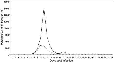
The parasitemia curves ofT. cruziinfected mice that had
either been treated with DFO (the IT group) or had received
no such treatment (the INT group) are shown in Fig. 1.
Whilst significant differences were detected in the patent period between the IT and the NIT groups (21 and 11 days, respectively), no differences were observed in the prepatent period (7 days for both groups) or in the day of the peak of parasitemia (10th and 11th d.p.i., respectively). The average parasitemia value in IT mice (274,666 trypomastigotes/ 0.1 ml of blood) was five times lower than in INT animals
(1,397,000 trypomastigotes/0.1 ml of blood) (p< 0.05).
The cumulative mortality rates for DFO-treated and
non-treated mice that had been infected with T. cruziare
in the IT group died, with mortality commencing on the 14th d.p.i., whilst the death rate in the NIT group was 93.75%, with mortality commencing on the 13th d.p.i.
If the hypothesis that iron is important for the
prolifera-tion of T. cruzi is valid, then removal of the heavy metal
through the use of a chelator could moderate the prolifera-tion of the parasite and hence reduce its levels in the blood. In the present study, a reduction in the parasitemia level, accompanied by an increase in the patent period, of animals in the IT group was observed, indicating that the severity of infection by the Y strain was attenuated by treatment with
the iron chelator. This finding is in accord with that of
Lal-onde and Holbein (1984)who demonstrated that depletion
of iron supplies, either by treatment with DFO or by admin-istration of a diet deficient in iron, promoted the reduction
of mortality in mice infected withT. cruzi.The
demonstra-tion that parasite reproducdemonstra-tion is negatively correlated with the level of iron in the plasma would appear to support the original hypothesis. In contrast to the results outlined
above,Pedrosa et al. (1990)reported thatT. cruziY strain
was not sensitive to DFO treatment, and that an effect on parasite replication could only be observed when mice were fed on an iron-free diet. The authors explained this result in terms of the more rapid multiplication rate of the Y strain compared with the YuYu strain, which permitted earlier attainment of the parasitemia peak. However, the inoculum used in this study (i.e. 1400 trypomastigotes forms/animal) and principal the DFO treatment administered (one dose of DFO on the 5th d.p.i.) were very different from those pres-ently employed and could explain the divergence in the results obtained.
The beneficial effects of DFO treatment have not only
been observed with respect to parasite infections.Weinberg
(1994)demonstrated thein vivouse of the chelator against
Pneumocystis cariniiby establishing that daily injections of the drug reduced the intensity of infection in rats and mice and produced alterations in the morphology. These authors also demonstrated that daily administration of DFO led to a more pronounced reduction in the replication of trophozoites compared with weekly i.p. injections.
0 200 400 600 800 1000 1200 1400 1600
1 2 3 4 5 6 7 8 9 10 11 12 13 14 15 16 17 18 19 20 21 22 23 24 25 26 27 28 29 30 31 32
P
a
ras
ites/0.1 ml
of
blood
(x
10
3)
Days post-infection
0 200 400 600 800 1000 1200 1400 1600
1 2 3 4 5 6 7 8 9 10 11 12 13 14 15 16 17 18 19 20 21 22 23 24 25 26 27 28 29 30 31 32
P
a
ras
ites/0.1 ml
of
blood
(x
10
)
Days post-infection
Fig. 1. Parasitaemia curves of Swiss mice that had been inoculated with 500 blood trypomastigotes ofTrypanosoma cruziY strain and had received no treatment (INT — ) or had been treated with desferrioxamine (ITÆ Æ Æ).
Percentage
morta
lit
y
0 20 40 60 80 100
1 3 5 7 9 11 13 15 17 19 21 23 25 27 29 31
Days post-infection
lit
y
0 20 40 60 80 100
1 3 5 7 9 11 13 15 17 19 21 23 25 27 29 31
3.2. Effect of treatment with DFO on hemoculture, PCR, and ELISA assays
The results of the parasitological and serological tests
are displayed inTable 1. All of theT. cruziinfected mice
that had been treated with DFO (IT group) presented neg-ative hemoculture, while the infected/non-treated (INT) group exhibited positive hemoculture in the acute and chronic phases of infection. This result was surprising since
Filardi and Brener (1987)had previously found that 50% of
animals infected with T. cruzi Y strain and treated with
benznidazole presented positive hemoculture, while the untreated group exhibited 100% positive hemoculture.
In order to determine if infection was still present in an animal, both PCR and ELISA assays were also performed in parallel. Only one mouse from the INT group survived the infection and this animal presented, PCR- and ELISA-positive in both the acute and chronic phases. All of the mice in the IT group that survived the acute infection
(n= 5) were PCR- and ELISA-positive in the acute phase,
whilst in the chronic phase, three animals were PCR-nega-tive although all 5 remained ELISA-posiPCR-nega-tive. Considering the higher sensitivity of the PCR assay and that ELISA serology can remain positive for a considerable period
fol-lowing effective and specific etiological treatment (Andrade
et al., 1991a,b), these results suggest that treatment with
DFO could have eliminated the infection in these animals
(Miyamoto et al., 2006), however other evaluated have
been necessary of this affirmation. The lower frequencies of hemoculture- and PCR-positive results, tests commonly positive in infected mice, taken together with the signifi-cantly reduced parasitemia curve and decreased mortality rate recorded in the IT animals, indicates that DFO treat-ment certainly plays an important protective role during the course of the infection.
3.3. Effect of treatment with DFO on iron levels in the liver
Significantly (p< 0.05) lower concentrations of iron
were detected on the 14th and 21st d.p.i in the liver of infected mice (IT and INT groups) in comparison with
non-infected animals (NIT and NINT groups) (Fig. 3).
Pedrosa et al. (1990) reported that treatment of mice with
DFO led to a reduction in the iron concentration in the
liver of animals infected with T. cruzi Y and CL strains,
together with a reduction in iron concentration in the spleen of those infected with the Y strain. These authors further established that the levels of iron in the liver and spleen of germfree mice remained unaltered when DFO
was administered 15 days postT. cruziinfection (Pedrosa
et al., 1993). Moreover, in a study of the effect of iron on
T. cruzi infection in mice, Lalonde and Holbein (1984) demonstrated a reduction in the iron supplies (48% in the liver and 15% in the spleen) of non-infected animals follow-ing treatment with DFO or the administration of an iron deficient diet.
According to Dallman (1986), iron deficiency in an
organism occurs in three overlapping stages. The first stage is characterised by a reduction in the concentration of serum ferritin, reflecting a loss in the supply of iron in the spleen, liver and bone marrow. The second stage is associated with an increasing capacity of iron linkage and a reduction in the concentration of serum iron, whilst in the final stage the concentrations of hemoglobin and hematocrit decrease and the iron reserves in the spleen and liver became exhausted. In the present study, the reduced concentration of iron found in the liver of infected mice on the 14th and 21st d.p.i. was probably associated with the regulation of iron within the organism as a
response to infection byT. cruzi.
3.4. Effect of treatment with DFO on iron levels in sera
Significantly (p< 0.05) higher concentrations of iron
were detected on the 21st d.p.i in serum samples of infected/non-treated mice (INT group; mean value
273.47lg/dl) in comparison with the NINT (mean value
100.9lg/dl) and IT (mean value 119.28lg/dl) groups
(Fig. 4). Whilst iron levels in the liver of infected animals
(IT and INT groups) had decreased on 14th d.p.i., the serum iron levels remained normal and increased serum concentrations could only be observed on the 21st d.p.i.
Table 1
Results of hemoculture, PCR and ELISA assays conducted during the acute (60th d.p.i.) and chronic (240th d.p.i.) phases of infection in mice that had survived inoculation withTrypanosoma cruziY strain
Treatment Animal number Assaysa
Hemoculture PCR ELISA
Acute phase Chronic phase Acute phase Chronic phase Acute phase Chronic phase
1 + + +
2 + + +
ITb 3 + + + +
4 + + +
5 + + + +
INTc 6 + + + + + +
It appears that some parasite forms provoke a large release of iron from the liver, leading subsequently to an increase in the level of the heavy metal in the serum of infected ani-mals (IT and INT). The lower serum iron levels in the IT group compared with the INT group would then be corre-lated with the chelation of plasma iron by DFO.
Letendre (1985)observed that during infection there was
a decrease in the levels of circulating iron, occasioned by its diminished release from the mononuclear phagocyte sys-tem, which restricted the amount of iron available to extra-cellular parasites. However, the beneficial aspects of this process are questionable in the case of intracellular
para-sites such asT. cruzithat lodge in the mononuclear
phago-cyte cells of the endothelial reticulum system. In the study
conducted by Pedrosa et al. (1990), a reduction of serum
iron in mice treated with DFO and infected with T. cruzi
strains Y, CL and YuYu could not be confirmed.
Further-more, Lalonde and Holbein (1984) demonstrated that no
significant changes occurred in the serum iron levels of non-infected mice that had been treated with DFO, while
treated/T. cruzi infected animals presented iron supplies
that were sufficient to maintain a normal immune response. It is important to note, however, that the protocols regard-ing inoculum and DFO administration used by these
authors were different from those employed in the present work.
A reduction in serum iron concentration was not observed in the present study presumably because amasti-gote forms of the intracellular parasite, established in the cells of the mononuclear phagocyte system but unable to obtain the necessary iron for survival, stimulated a release of heavy metal into the serum. Iron present in the serum would then bind to transferrin and become available to the intracellular parasite by receptor-mediated endocytosis.
3.5. Effect of treatment with DFO on hemoglobin levels
Significant (p< 0.05) differences were observed on the
21st d.p.i. in the hemoglobin levels between the IT
(7.14lg/dl) and NIT (10.9lg/dl) groups, but no significant
differences were observed between the other groups
(Fig. 5).
According to Bothwell et al. (1979), the primary iron
pool in the blood of a vertebrate host is associated with
hemoglobin, and hemoglobin levels are commonly
employed in order to evaluate iron deficiency in hosts that have been submitted to iron deficient diets, or to infection, *
*
0 1 2 3 4 5 6
7 *
*
Concentr
ation of
iron (mg/0.1 g of tissue)
Treatment groups
NINT NIT INT IT NINT NIT INT IT
Fig. 3. Levels of iron, measured (a) on 14th d.p.i and (b) on 21 d.p.i, in the liver of Swiss mice that had been inoculated with 500 blood trypomastigotes of
Trypanosoma cruziY strain and had received no treatment (INT) or had been treated with desferrioxamine (IT), together with the corresponding non-infected control groups (NINT and NIT, respectively). Values shown are means ± SD (n= 20). Results of two-way ANOVA-infection *p< 0.05; treatment: NS; ironxtreatment: NS, both 14 and 21 days.
0 100 200 300 400 500 600 700
*
NINT NIT INT IT *
Treatment groups
Level
of
i
ron
i
n
s
e
ru
m
(µ
/dL)
0 100 200 300 400 500 600 700
*
NINT NIT INT IT *
Treatment groups
Level
of
i
ron
i
n
s
e
ru
m
(µ
/dL)
Fig. 4. Levels of iron, measured on 21 d.p.i, in serum of Swiss mice that had been inoculated with 500 blood trypomastigotes ofTrypanosoma cruzi
Y strain and had received no treatment (INT) or had been treated with desferrioxamine (IT), together with the corresponding non-infected control groups (NINT and NIT, respectively). Values shown are means ± SD (n= 12). Results of two-way ANOVA-infection *p< 0.05; treatment: NS; ironxtreatment: NS, both 14 and 21 days.
Hem
ogl
obin
(
µ
g/dL)
0 5 10 15 20 25
*
Treatment groups NINT NIT INT IT 0
5 10 15 20 25
*
Treatment groups NINT NIT INT IT
or to treatments that trigger alterations in iron levels. Changes in hemoglobin levels have been reported in
vari-ous trypanosome infections (Esievo et al., 1982; Igbokwe
and Anosa, 1989), including infection byT. cruzi, and are
associated with a reduction in the number of platelets (
Car-doso and Brener, 1980; Ruiz et al., 1989), erythrocytes
(Cardoso and Brener, 1980) and hematocrit values (
Lal-onde and Holbein, 1984). Moreover, Marcondes et al.
(2000)observed that the acute phase of experimental
infec-tion byT. cruziis characterised by anaemia,
thrombocyto-penia, leucopoenia and bone marrow hypoplasia.
However, the mechanisms responsible for these hematolog-ical alterations are not fully understood.
Pedrosa et al. (1990) evaluated iron deficiency in mice
and correlated the effect of this deficiency with the evolu-tion of Chagas’ disease. The authors showed that the hyp-ohemoglobinemia presented by the host was permanent in animals fed on an iron-free diet, probably because the low supplies of the heavy metal were not enough to compensate for the erythropoiesis that follows anaemia. In animals infected with CL and Y strain and treated with DFO, the haemoglobin levels recovered suggesting that this treat-ment does not interfere with the supplies required for erythropoiesis. Hence, the reduction in hemoglobin levels observed in the IT group compared with the NIT group
may be explained by the presence of infection by T. cruzi
in association with the action of DFO in the dose employed in the present work.
The data reported in this study suggest that the
pro-longed treatment with DFO of T. cruzi infected mice
reduces the availability of iron to the infecting parasites leading to a reduction in parasitemia and mortality. Fur-ther study of this system could be of significant value in
understanding the effect of iron reduction duringT. cruzi
infection.
Acknowledgments
This work was supported by the Conselho Nacional de Desenvolvimento Cientı´fico e Tecnolo´gico (CNPq), the Fundac¸a˜o de Amparo a` Pesquisa do Estado de Minas Ger-ais (FAPEMIG) and by the Universidade Federal de Ouro Preto (UFOP).
References
Andrade, S.G., Freitas, L.A., Peyrol, S., Pimentel, A.R., Sadigursky, M., 1991a. Experimental chemotherapy of Trypanosoma cruzi infection: persistence of parasite antigens and positive serology in parasitolog-ically cured mice. Bulletin of the World Health Organization 69, 191– 197.
Andrade, S.G., Stocker-Guerret, S., Pimentel, A.S., Grimaud, J.A., 1991b. Reversibility of cardiac fibrosis in mice chronically infected with
Trypanosoma cruziunder specific chemotherapy. Memo´rias do Insti-tuto Oswaldo Cruz 86, 187–200.
Association of Official Analytical Chemists, 1980. Official methods of analysis of the AOAC, third ed. AOAC, Washington.
A´ vila, H.A., Sigman, D.S., Cohen, L.M., Millikan, R.C., Simpson, L., 1991. Polymerase chain reaction amplification ofTrypanosoma cruzi
kinetoplast minicircle DNA isolated from whole blood lysates: diagnosis of chronic Chagas disease. Molecular and Biochemical Parasitology 48, 211–222.
Blakley, B.R., Hamilton, D.L., 1988. The effect of iron deficiency on the immune response in mice. Drug Nutrient Interactions 5, 249–255. Bothwell, H., Charlton, R.W., Cook, J.D., Finch, C.H., 1979. Iron
Metabolism in Man. Blackwell, Oxford, pp. 284–306.
Breidbach, T., Scory, S., Krauth-Siegel, R.L., Steverding, D., 2002. Growth inhibition of bloodstream forms ofTrypanosoma bruceiby iron chelator deferoxamine. International Journal of Parasitology 32, 473–479.
Brener, Z., 1962. Therapeutic activity and criterion of cure on mice experimentally infected withTrypanosoma cruzi. Revista do Instituto de Medicina Tropical de Sa˜o Paulo 4, 389–396.
Brittenham, G.M., 1988. Iron chelating agents. Current Therapy in Hematology and Oncology 3, 149–153.
Camargo, E.P., 1964. Growth and differentiation inTrypanosoma cruzi. I. Origin of metacyclic trypanosomes in liquid media. Revista do Instituto de Medicina Tropical de Sa˜o Paulo 12, 93–100.
Canadian Council on Animal Care, 1980. Guide to the care and use of experimental animals. CCAC, Ottawa.
Canadian Council on Animal Care, 1984. Guide to the care and use of experimental animals. CCAC, Ottawa.
Cardoso, J.E., Brener, Z., 1980. Hematological changes in mice experi-mentally infected with Trypanosoma cruzi. Memo´rias do Instituto Oswaldo Cruz 75, 97–104.
Carotenuto, P., Pontesilli, O., Cambier, J.C., Hayward, A.R., 1986. Desferoxamine blocks IL-2 receptor expression on human T lympho-cytes. Journal of Immunology 136, 2342–2347.
Cinatl Jr., J., Cinatl, J., Rabenau, H., Gumbel, H.O., Kornhuber, B., Doerr, H.W., 1994. In vitro inhibition of human cytomegalovirus replication by desferrioxamine. Antiviral Research 25, 73–77. Cinatl Jr., J., Cinatl, J., Weber, B., Rabenau, H., Gumbel, H.O., Chenot,
J.F., Scholz, M., Encke, A., Doerr, H.W., 1995.In vitroinhibition of human cytomegalovirus replication in human foreskin fibroblasts and endothelial cells by ascorbic acid 2-phosphate. Antiviral Research 27, 405–418.
Crichton, R.R., Ward, R.J., 1992. Structure and molecular biology of iron binding-proteins and the regulation of free iron pools. In: Lauffer, R.B. (Ed.), Iron and Human Diseases. CRC Press, Boca Raton, pp. 23–75. Dallman, P.R., 1986. Biochemical basis for the manifestations of iron
deficiency. Annual Review in Nutrition 6, 13–40.
Dhur, A., Galan, P., Hercberg, S., 1989. Iron status, immune capacity and resistance to infections. Comparative Biochemistry and Physiology 94, 11–19.
DonFrancesco, A., Deb, G., De Sio, L., Cozza, R., Castellano, A., 1996. Role of desferrioxamine in tumor therapy. Acta Haematologica 95, 66–69.
Esievo, K.A., Saror, D.I., Ilemobade, A.A., Hallaway, M.H., 1982. Variation in erythrocyte and free sialic acid serum concentrations during experimentalTrypanosoma vivaxinfection in cattle. Research in Veterinary Science 32, 1–15.
Filardi, L.S., Brener, Z., 1987. Susceptibility and natural resistance of
Trypanosoma cruzistrains to drugs used clinically in Chagas disease. Transactions of the Royal Society of Tropical Medicine and Hygiene 81, 755–759.
Galan, P., Davila, M., Mekki, N., Hereberg, S., 1988. Iron deficiency, inflammatory processes and humoral immunity in children. Interna-tional Journal of Vitamin and Nutrition Research 58, 225–230. Gomes, M.L., Macedo, A.M., Vago, A.R., Pena, S.D., Galva˜o, L.M.,
Chiari, E., 1998.Trypanosoma cruzi: optimization of polymerase chain reaction for detection in human blood. Experimental Parasitology 88, 28–33.
Hann, H.W.L., Stahlhut, M.W., Rubin, R., Maddrey, W.C., 1992. Antitumor effect of deferoxamine on human hepatocellular carcinoma growing in athymic nude mice. Cancer 70, 2051–2056.
Henry, R.J., Winkelman, J.W., Cannon, D.C. (Eds.), 1974. Clinical chemistry—principles and techniques, second ed. Harper & Row, New York.
Hershko, C., Peto, T.E.A., 1988. Deferroxamine inhibition of malaria is independent of host iron status. Journal of Experimental Medicine 168, 375–387.
Igbokwe, I.O., Anosa, V.O., 1989. Response to anaemia in experimental
Trypanosoma vivax infection of sheep. Journal of Comparative Pathology 100, 111–118.
Kent, S., Weinberg, E.D., Stuart-MacAdam, P., 1990. Dietary and prophylactic iron supplements: helpful and harmful. Human Nature 1, 55–81.
Lalonde, R.G., Holbein, B.E., 1984. Role of iron inTrypanosoma cruzi
infection of mice. Journal of Clinical Investigation 23, 470–476. Letendre, E.D., 1985. The importance of iron in the pathogenesis of
infection and neoplasia. Trends in Biochemical Sciences 12, 166–168. Lima, M.F., Villalta, F., 1989.Trypanosoma cruzitrypomastigote clones
differentially express a parasite adhesion molecule. Molecular and Biochemical Parasitology 33, 159–170.
Loo, V.G., Lalonde, R.G., 1984. Role of iron in intracellular growth of
Trypanosoma cruzi. Infection and Immunity 45, 726–730.
Mabeza, G.F., Biemba, G., Gordeuk, V.R., 1996. Clinical studies of iron chelators in malaria. Acta Haematologica 95, 78–86.
Mahmoud, M.S., 1999. Effect of deferoxamine alone and combined with pyrimethamine on acute toxoplasmosis in mice. Journal of the Egyptian Society of Parasitology 29, 791–803.
Marcondes, M.C.G., Borelli, P., Yoshida, N., Russo, M., 2000. Acute
Trypanosoma cruziinfection is associated with anemia, thrombocyto-penia, leukothrombocyto-penia, and bone marrow hypoplasia: reversal by nifurti-mox treatment. Microbes and Infection 2, 347–352.
Martelius, T., Scholz, M., Krogerus, L., Hockerstedt, K., Loginov, R., Bruggeman, C., Cinatl Jr., J., Doerr, H.W., Lautenschlager, I., 1999. Antiviral and immunomodulatory effects of desferrioxamine in cyto-megalovirus-infected rat liver allografts with rejection. Transplanta-tion 68, 1753–1761.
Miyamoto, C.T., Gomes, M.L., Marangon, A.V., Araujo, S.M., Bahia, M.T., Lana, M., Toledo, M.J., 2006.Trypanosoma cruzi: sensitivity of the polymerase chain reaction for detecting the parasite in the blood of mice infected with different clonal genotypes. Experimental Parasitol-ogy 112, 198–201.
Pedrosa, M.L., Silva, M.E., Silva, M.E., Silva, M.E.C., Nicoli, J.R., Vieira, E.C., 1990. The effect of iron deficiency and iron overload on the evolution of Chagas’ disease produced by three strains of
Trypanosoma cruzi in CFW mice. Comparative Biochemistry and Physiology 97, 235–243.
Pedrosa, M.L., Nicoli, J.R., Silva, M.E., Silva, M.E., Silva, M.E.C., Vieira, L.Q., Bambirra, E.A., Vieira, E.C., 1993. The effect of iron
nutritional status on Trypanosoma cruzi infection in germfree and conventional mice. Comparative Biochemistry and Physiology 106, 813–821.
Richardson, D.R., 1997. Iron chelators as effective anti-proliferative agents. Canadian Journal of Physiology and Pharmacology 75, 1164– 1180.
Ruiz, R.C., Fernandes, M., Irulegui, I., 1989. Observation of marked thrombcytopenia during the course of acute experimental Chagas’ disease. Memo´rias do Instituto Oswaldo Cruz 84, 26.
Santos, F.R., Pena, S.D.J., Epplen, J.T., 1993. Genetic and population study of a Y-linked tetranucleotide repeat DNA polymorphism with a simple non-isotopic technique. Human Genetics 90, 655–656. Shadid, M., Buonocore, G., Groenendaal, F., Moison, R., Ferrali, M.,
Berger, H.M., Van Bel, F., 1998. Effect of deferoxamine and allopurinol on non-protein-bound iron concentrations in plasma and cortical brain tissue of newborn lambs following hypoxia-ischemia. Neuroscience Letters 248, 5–8.
Silva, L.H.P., Nussenzweig, V., 1953. Sobre uma cepa deTrypanosoma cruzialtamente virulenta para o camundongo branco. Folia Clinica et Biologica 20, 191–203.
Tanjii, K., Imaizumi, T., Matsumiya, T., Itaya, H., Fujimoto, K., Cui, X.F., Toki, T., Ito, E., Yoshida, H., Wakabayashi, K., Satoh, K., 2001. Desferrioxamine, an iron chelator, upregulates cyclooxygenase-2 expression and prostaglandin production in a human macrophage cell line. Biochemistry Biophysics Acta 1530, 227–235.
Traore, O., Carnevale, P., Kaptue-Noche, L., Bede, J., Desfontaine, M., Elion, J., Labie, D., Nagel, R.L., 1991. Preliminary reports in the use of desferrioxamine in the treatment ofPlasmodium falciparummalaria. American Journal of Hematology 37, 206–208.
Voller, A., Bidwell, D.E., Bartlett, A., 1976. Enzyme immunoassays in diagnostic medicine. Theory and practice. Bulletin of the World Heath Organization 53, 55–65.
Wang, F., Elliot, R.L., Head, J.F., 1999. Inhibitory effect of deferoxamine mesylate and low iron diet on the 13762 NF rat mammary adenocar-cinoma. Anticancer Reserach 19, 445–450.
Weinberg, E.D., 1984. Iron withholding: a defence against infection and neoplasia. Physiological Reviews 64, 65–102.
Weinberg, G.A., 1994. Iron chelators as therapeutic agents against
Pneumocystis carinii. Antimicrobial Agents and Chemotherapy 38, 997–1003.
Wincker, P., Britto, C., Pereira, J.B., Cardoso, M.A., Oelemann, W., Morel, C.M., 1994. Use of a simplified polymerase chain reaction procedure to detectTrypanosoma cruziin blood samples from chronic chagasic patients in a rural endemic area. American Journal of Tropical Medicine and Hygiene 51, 771–777.