i
UNIVERSIDADE FEDERAL DE SÃO CARLOS
DEPARTAMENTO DE CIÊNCIAS BIOLÓGICAS
PROGRAMA DE PÓS-GRADUAÇÃO EM ECOLOGIA E RECURSOS NATURAIS
PHYSIOLOGICAL RESPONSE OF CHLORELLA VULGARIS TO
CADMIUM, PHOSPHORUS AND NITROGEN
(RESPOSTA FISIOLOGICA DE CHLORELLA VULGARIS SOB
CONCENTRAÇÕES VARIÁVEIS DE CÁDMIO, FÓSFORO E NITROGÊNIO)
MATHIAS AHII CHIA
Thesis presented as a part of the fulfillment of the requirement for Doctor of Science degree, Area: Ecology and Natural Resources.
Supervisor: Profa. Dra. Ana Teresa Lombardi
Ficha catalográfica elaborada pelo DePT da Biblioteca Comunitária/UFSCar
C532rf
Chia, Mathias Ahii.
Resposta fisiologica de Chlorella vulgaris sob
concentrações variáveis de cádmio, fósforo e nitrogênio / Mathias Ahii Chia. -- São Carlos : UFSCar, 2012.
103 f.
Tese (Doutorado) -- Universidade Federal de São Carlos, 2012.
1. Ficologia. 2. Microalga. 3. Metais. 4. Composição bioquímica. 5. Nutrientes. 6. Energia da biomassa. I. Título.
iv Look deep, deep into nature, and then
you will understand everything better –
v This work is dedicated to my lovely
wife Estyluv for her unending love and
vi
DECLARATION
This doctorate degree program commenced at the beginning of March 2009 in the Postgraduate Program in Ecology and Natural Resources (PPGERN) of the Federal University of São Carlos (UFSCar), Brazil.
This study was made possible via the doctoral scholarship jointly provided by the Third World Academy of Science (TWAS) and the Conselho Nacional de Desenvolvimento Científico e Tecnológico (CNPq). The scholarship permits doctoral studies for up to 48 months in a Brazilian institution. Taking advantage of this program, all the research work was done in Brazil under the supervision of Profa. Dra. Ana Teresa Lombardi.
vii
ACKNOWLEDGEMENT
I would like to specially thank Profa Ana Teresa Lombardi for the supervision of this project. You are the best. Also not forgetting is Profa. Maria da Graça Gama Melão who has been patient with me and have guided me throughout this study, to you I say am very grateful.
Prof Christopher C. Parrish is graciously acknowledged for his profound contribution to this work ranging from data treatment (chemometrics) to fatty acid analysis that made a major contribution to the success of this work.
I will like to express my sincere thanks to João Augusto da Silva Affonso, Roseli Aparecida Gonçalves, Profa. Dra. Rocha Odete and Profa. Dra. Dalva Maria da Silva Matos of the Postgraduate Program for all their help during my stay in Brazil.
I am grateful to Marcelo M. Nogueira for his help during the analysis of my samples for nitrate content.
To my head of department (Biological Sciences, Ahmadu Bello University, Zaria, Nigeria), Prof. Jehu Auta, and my colleagues at ABU: Prof S.P. Bako, Prof S.J. Oniye, Prof E.A. Ezealor, Prof I.H. Nock, Dr Ladan, Mr John Sow Gudzan, Aliyu Habibu, and Mr Tanimu Yahuza, your help rendered to me during my stay in Brazil is acknowledged.
I am indebted to my lovely wife Esther O Chia for her support throughout the period of this study. For your endurance and steadfastness, despite the whole period I spent in Brazil away from you. For this, I say I love you very much and deeply appreciate you.
To my colleagues in the Laboratory especially Jaqueline, Giseli, Bruna, Rodrigo, Daniela, Patrícia F.M. Nogueira and Alex, I say thank you.
I also like to acknowledge my friends Gustavo and Andrés. You guys are wonderful.
viii
The Third World Academy of Science (TWAS) and National Counsel of Technological and Scientific Development (CNPq) are acknowledged for the doctoral scholarship provided for this study.
ix
TABLE CONTENTS
1 INTRODUCTION………..……….…….1
1.1 Microalgae lipids…….……….……….……3
1.2 Effect of nutrient stress on microalgal biochemical composition ……….………..…...7
1.3 Effect of trace metals on the physiology of microalgae….………….…..10
1.4 Justification………...11
1.5 Research hypotheses……….14
1.6 Objectives of study………...14 2 MATERIALS AND METHODS………..….…………15
2.1 Algal species……….….……..……15 2.2 The effect of media composition……….……..…...15
2.3 Cadmium, phosphorus and nitrogen experiment………...…... .17
2.4 Experimental cultures………...19
2.5 Growth and biomass determinations……….………..20
2.5.1 Absorbance………. 20
2.5.2 Dry Weight………. 21
2.5.3 Chlorophyll a……….21
2.5.4 Density (cells ml-1)……….…… 22
2.5.5 Productivity………..…...22 2.6 Biochemical composition analyses……….…… .23
2.6.1 Total intracellular carbohydrates……….….23
2.6.2 Total proteins……….24
2.6.3 Calorific value of Chlorella vulgaris……….25
2.6.4 Lipids………..…………...25
2.7 Chemical Analyses……….………..33
2.7.1 Nitrogen……….……33
x
2.8 Data analyses………36
3 RESULTS AND DISCUSSION……….37
3.1 Growth, biomass production and biochemical composition of Chlorella
vulgaris grown in different culture media…..………...……37
3.2 Calorific values of Chlorella vulgaris cultured under varying phosphate
concentrations using semi-continuous culture……….….47
3.3 Cadmium, nitrogen and phosphorus induced changes in growth and
biochemical composition of Chlorella vulgaris………...…53
3.4 Changes in Chlorella vulgaris lipid composition as a function of different
cadmium and phosphorus concentrations………65
3.5 Effect of cadmium and nitrogen on the lipid composition of Chlorella
vulgaris.………77
4 GENERAL CONCLUSION………...89
xi
LIST OF TABLES
TABLE 2.1: Growth media composition used in this current study…………..16
TABLE 2.2: Cd speciation in the growth media as calculated using MINEQL
software. Where CD1 = 2.0x10⁻⁸ mol L-1 Cd and CD2 = 10-7 mol L-1 Cd, P =
phosphorus and N = nitrogen. pCd and pLabile are negative natural log values
for free Cd ion and labile Cd concentrations………18
TABLE 2.3: Experimental combinations of Cd and P to which Chlorella vulgaris
was exposed……….……….19
TABLE 2.4: Experimental combinations of Cd and N to which Chlorella
vulgariswas exposed………...19
TABLE 2.5: Lipid standards that were resolved in a three-step separation for
lipid class determination in this study………..28
TABLE 3.1: MINEQL+ metal speciation results for selected elements in the growth media. Values are in pCu, pFe and pZn, where p = negative base log10 of
the molar concentrations………..……….37
TABLE 3.2: A summary of the biomass, biochemical and calorific production of
Chlorella vulgaris as a function of phosphorus concentration in semicontinuous
cultures. Values represent plus or minus standard error for n = 3. Rows with the same superscripts letters are not significantly different at 95% significance level
(P < 0.05)………..48
TABLE 3.3: Lipid class composition (pg cell-1) of Chlorella vulgaris at different phosphate regimes (mol L-1) using semicontinuous cultures. Values are mean plus or minus standard error for n = 3. Rows with the same superscript letters are
not significantly different at 95% significance level (P <
xii
TABLE 3.4: Analysis of variance results for the effect of phosphorus, nitrogen and cadmium on the growth and biomass of Chlorella vulgaris. Values represent
F values and those in parenthesis represent P value. Note: values with P < 0.05
are significant………..….55
TABLE 3.5: Percent organic contribution of total carbohydrates, proteins and lipids (Carb:PR:L) and specific growth rate (d-1) in parenthesis of Chlorella
vulgaris acclimated to different Cd, P and N concentrations in mol L-1…….…59
Table 3.6: Chlorophyll a concentration(pg cell-1), percentage chlorophyll a (total lipids) and total lipids (pg cell-1) of Chlorella vulgaris as a function of different Cd and P concentrations (mol L-1). Values are plus or minus standard deviation
for n = 3………...….66
TABLE 3.7: Analysis of variance results for the effect of phosphorus and cadmium on the lipid composition of Chlorella vulgaris. Values represent F value and those in parenthesis represent P value. Note: values with P < 0.05 are
significant.………...…72
Table 3.8: Chlorophyll a concentration(pg cell-1), percentage chlorophyll a (total lipids) and total lipids (pg cell-1) of Chlorella vulgaris as a function of different Cd and N concentrations (mol L-1). Values are plus or minus standard deviation
for n = 3………..……….78
TABLE 3.9: Analysis of variance results for the effect of nitrogen and cadmium on the lipid composition of Chlorella vulgaris. Values represent F value and those in parenthesis represent P value. Note: values with P < 0.05 are
xiii
LIST OF FIGURES
FIGURE 1.1: Fatty acid de novo synthesis pathway in chloroplasts. Acetyl CoA
enters the pathway as a substrate for acetyl CoA carboxylase (Reaction 1) as well as a substrate for the initial condensation reaction (Reaction 3). Reaction 2, which is catalyzed by malonyl CoA:ACP transferase, transfers malonyl from CoA to form malonyl ACP. Malonyl ACP is the carbon donor for subsequent
elongation reactions (adapted from Hu et al. 2008)………4
FIGURE 1.2: Simplified schematic showing the triacylglycerol biosynthesis pathway in algae. (1) Cytosolic glycerol-3-phosphate acyl transferase, (2) lyso-phosphatidic acid acyl transferase, (3) lyso-phosphatidic acid phosphatase, and (4)
diacylglycerol acyl transferase (adapted from Hu et al. 2008)……….………..7
FIGURE 2.1: Carbohydrate calibration curve used in the present study……….23
FIGURE 2.2: The protein calibration curve used in this study………24
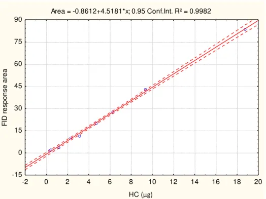
FIGURE 2.3: Aliphatic hydrocarbon (HC) calibration curve for lipid class
determination………29
FIGURE 2.4: Internal standard (Hexadecan-3-one, KE) calibration curve for lipid
class determination………..29
FIGURE 2.5: Wax ester (WE) calibration curve for lipid class determination
………...30
FIGURE 2.6: Triacylglycerol (TAG) calibration curve for lipid class determination………...30
FIGURE 2.7: Free fatty acid (FFA) calibration curve for lipid class
determination. ………31
FIGURE 2.8: Free sterol (ST) calibration curve for lipid class determination.
xiv
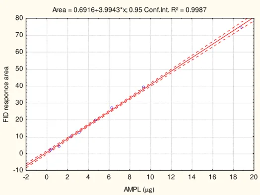
FIGURE 2.9: Acetone mobile polar lipid (AMPL) calibration curve for lipid
class determination………...32
FIGURE 2.10: Phospholipid (PL) calibration curve for lipid class determination
……….32
FIGURE 2.11: Nitrate nitrogen calibration curve for nitrate determination
………34
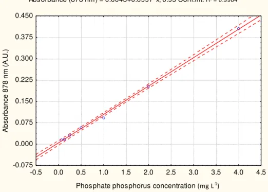
FIGURE 2.12: Phosphate phosphorus calibration curve for phosphate
determination………..35
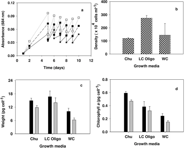
FIGURE 3.1 (a) Dry weight (pg cell-1), and (b) chlorophyll a production (pg cell -1
) (black bars) and yield (gray bars) (pg cell-1) for Chlorella vulgaris grown in Chu (Chu medium), WC (WC medium), LC Oligo (LC Oligo medium). Error
bars represent standard deviation for n= 3………..38
FIGURE 3.2: Total carbohydrate (a), total protein (b) and total lipid (c) production (black bars) and yield (gray bars) (pg cell-1) of Chlorella vulgaris as a function of media type using semi-continuous culture system. Error bars
represent standard deviation for n = 3. Chu = Chu medium, WC = WC medium,
LC Oligo = LC Oligo medium………...40
FIGURE 3.3: Lipid class composition (%) of Chlorella vulgaris grown in different growth media (black bars: Chu; white bars: LC Oligo; gray bars: WC). Neutral lipids are represented by HC, SE/WE, TAG, FFA, ALC and ST, while polar lipids are AMPL and PL. Error bars represent standard deviation for n = 3. Chu = Chu medium, WC = WC medium, LC Oligo = LC Oligo medium. Right
axis apply to AMPL and PL classes……..………..……41
FIGURE 3.4: PCA biplot for the various parameters measured for Chlorella
vulgaris cultured with different growth media using semi continuous culture.
xv
FIGURE 3.5: Principal components analyses to show the relationship between and within phosphate levels and physiological parameters analyzed for Chlorella
vulgaris grown as a function of different phosphate concentrations (mol L-1)…50
FIGURE 3.6: Growth curve for Chlorella vulgaris at different Cd, N and P concentrations reported as absorbance (684 nm) vs time (days). Stars represent 2.3x10⁻⁴ mol L-1 P or 1.1x10⁻³ mol L-1 N, empty squares 2.3x10⁻⁶ mol L-1 P or 1.1x10⁻⁵ mol L-1 N, and filled squares 6.0x10⁻⁷ mol L-1 P or 2.9x10⁻⁶ mol L-1 N. a. P without Cd; b. P with 10-8 mol L-1 Cd; c. P with 10-7 mol L-1 Cd; d. N without
Cd; e. N with 10-8 mol L-1 Cd; f. N with 10-7 mol L-1Cd………..56
FIGURE 3.7: Effect of Cd on cell density, chlorophyll a and dry weight at different P and N concentrations (mol L-1). Black bars represent treatments without Cd, white bars 10-8 mol L-1 Cd and checked bars 10-7 mol L-1 Cd. Error bars represent standard deviation for n = 3. Bars with same letters are not significantly different at p < 0.05...57
FIGURE 3.8: Total carbohydrate, proteins and lipids production as a function of different Cd, P and N concentrations (mol L-1). Black bars represent treatments without Cd, white bars 10-8 mol L-1 Cd and checked bars 10-7 mol L-1 Cd. Error bars represent standard deviation for n = 3. Bars with same letters are not
significantly different at p < 0.05………58
FIGURE 3.9: PCA biplot of different parameters analyzed for Chlorella vulgaris
at different Cd and P concentrations (mol L-1)………60
FIGURE 3.10: PCA analysis biplot of different parameters analyzed for
Chlorella vulgaris as a function of Cd and N concentrations to which the alga
was exposed. ………...61
FIGURE 3.11: Lipid class composition of Chlorella vulgaris at different P and Cd concentrations. Black bars represent the controls, white bars 10-8 mol L-1 Cd and checked bars 10-7 mol L-1 Cd. Error bars represent standard deviation for n =
xvi
FIGURE 3.12: The production of total SAFA, MUFA, PUFA, ω3 and
PUFA/SAFA ratios of Chlorella vulgaris as a function of different P and Cd concentrations. Black bars represent the controls, white bars 10-8 mol L-1 Cd and checked bars 10-7 mol L-1 Cd. Error bars represent standard deviation for n = 3
………...69
FIGURE 3.13: Saturated fatty acids (SAFA) and monounsaturated fatty acids (MUFA) composition of Chlorella vulgaris exposed to different P and Cd concentrations. Black bars represent the controls, white bars 10-8 mol L-1 Cd and checked bars 10-7 mol L-1 Cd. Error bars represent standard deviation for n = 3
………...70
FIGURE 3.14: Polyunsaturated fatty acids (PUFA) composition of Chlorella
vulgaris as a function of different P and Cd concentrations. Black bars represent
the controls, white bars 10-8 mol L-1 Cd and checked bars 10-7 mol L-1 Cd. Error
bars represent standard deviation for n= 3………...71
FIGURE 3.15: PCA biplot for lipid composition of Chlorella vulgaris at
different P and Cd concentrations……….73
FIGURE 3.16: Lipid class composition of Chlorella vulgaris at different N and Cd concentrations. Black bars represent the controls, white bars 10-8 mol L-1 Cd and checked bars 10-7 mol L-1 Cd. Error bars represent standard deviation for n =
3………...79
FIGURE 3.17: The production of total SAFA, MUFA, PUFA, ω3 and
PUFA/SAFA ratios of Chlorella vulgaris as a function of different N and Cd concentrations. Black bars represent the controls, white bars 10-8 mol L-1 Cd and checked bars 10-7 mol L-1 Cd. Error bars represent standard deviation for n = 3
……….81
xvii
concentrations. Black bars represent the controls, white bars 10-8 mol L-1 Cd and checked bars 10-7 mol L-1 Cd. Error bars represent standard deviation for n = 3
……….82
FIGURE 3.19: Polyunsaturated fatty acids (PUFA) composition of Chlorella
vulgaris as a function of different Nand Cd concentrations. Black bars represent
the controls, white bars 10-8 mol L-1 Cd and checked bars 10-7 mol L-1 Cd. Error
bars represent standard deviation for n= 3……….…………83
FIGURE 3.20: PCA biplot for lipid composition of Chlorella vulgaris at
xviii
ABBREVIATIONS AND SYMBOLS
ω3 = Omega 3
µ = Specific growth rate
ANOVA = Analysis of variance
ABS = Absorbance
ALC = Free aliphatic alcohol(s)
AMPL = Acetone mobile polar lipid(s)
BBM = Bold basal medium
C = Carbon
CaCl2 = Calcium chloride
Carb = Total carbohydrates
Cd = Cadmium
CDCA1 = Carbonic anhydrase
CH2Cl2 = Dichloromethane
Chl = Chlorophyll
CHU = Chu 10 medium
CoA = Coenzyme-A
Cu = Copper
DAG = Diacylglycerol(s)
DMSO = Dimethyl sulfoxide
DNA = Deoxyribonucleic acid
DTPA = Diethylenetriaminepentaacetic acid
DW = Dry weight
EDTA = Ethylenediamine tetracetic acid
xix
FAME = Fatty acid methyl ester
Fe = Iron
FFA = Free fatty acid(s)
FID = Flame Ionization detector
HC = Aliphatic hydrocarbons
HCl = Hydrochloric acid
HUFA = Highly-unsaturated fatty acid(s)
KE = Ketone(s)
L = Lipids
LC Oligo = LC Oligo medium
MeOH = Methanol
MUFA = Monounsaturated fatty acid(s)
N = Nitrogen
NaOH = Sodium hydroxide
NTA = Nitrilotriacetic acid
OD = Optical density
P = Phosphorus
PA = Phosphoric acid
Pb = Lead
PCA = Principal component analysis
PL = Phospholipid(s)
PPS = Partial pyrolysis scan
PR = Total proteins
PUFA = Polyunsaturated fatty acid(s)
xx
RNA = Ribonucleic acid
SAFA = Saturated fatty acid(s)
SGR = Specific growth rate
ST = Free sterol(s)
TAG = Triacylglycerol(s)
UV-VIS = Ultraviolet-Visible light
WC = WC medium
WE/SE/ME = Wax ester(s)/Steryl ester(s)/Methyl ester
W/W = Wet weight
X = Biomolecule concentration
xxi
ABSTRACT
Microalgae are capable of adapting themselves to changes in environmental conditions through the production of different biomolecules. This study investigated the effects of the metal, cadmium on the ecophysiological response of microalgae under varying nutrient conditions. In the first phase of the study, we examined the growth, biomass production and biochemical composition of Chlorella vulgaris using semi-continuous cultures employing three growth media (LC Oligo, Chu 10 and WC media) to enable the selection of the most appropriate growth medium. The highest cell density, chlorophyll a, carbohydrate, protein and lipid concentration were found in Chu and LC Oligo media. Due to the cost effectiveness of using LC Oligo medium, it was the chosen as the preferred medium among the three tested media.
The calorific values of C. vulgaris at different phosphorus (P) levels were investigated in the second phase of this study. Calorific value reported under replete concentration (13.78 kJ g-1) was less than those found under P limitation (30.47-33.07 kJ g-1). The highest calorific values without growth retardation were obtained at the 10-6 mol L-1 P.
The third phase of this research involved determinations of growth, biomass production and biochemical composition of C. vulgaris under varying
cadmium (Cd), phosphorus (P) and nitrogen (N) concentrations. Three P (6.0x10
-7, 2.3x10⁻⁶ and 2.3x10⁻⁴ mol L-1) and N (2.9x10⁻⁶, 1.1x10⁻⁵, 1.1x10⁻³ mol L-1)
xxii
Cd were significantly reduced. Combined P limitation and Cd exposure stimulated higher total lipid production than only P or N limitation.
Triacylglycerols (TAG) was the most accumulated lipid class under P and N limitation and Cd stress among all the neutral and polar lipid classes. Increasing P and N limitation and Cd exposure resulted in higher SAFA and MUFA concentrations. 16:1n - 11 was an exception among the MUFA as its
levels decreased at low N. Total PUFA and ω3 PUFA levels, and PUFA:SAFA
xxiii
RESUMO
A geração de resíduos industriais e a possibilidade de produção biomassa de microalgas em águas residuais estimulam a pesquisa relacionada com os efeitos do cádmio na resposta fisiológica dos microorganismos. Este estudo investigou os efeitos de metais sob condições variaveis de nutrientes sobre a resposta ecofisiológica de Chlorella vulgaris. Na primeira fase do estudo, examinamos o crescimento, produção de biomassa e composição bioquimica de
Chlorella vulgaris nos meios nutritivos LC Oligo, Chu 10 and WC para
selecionar o meio mais adequado. A densidade celular, clorofila a, proteínas, carboidratos e lipídios determinados e os resultados mostraram maiores concentrações das biomoléculas nos meios Chu e LC Oligo. Devido à relação custo-eficácia do uso, o meio LC Oligo foi escolhido entre os três meios testados. Esta segunda fase da pesquisa investigou o poder calorífico da C. vulgaris em concentrações diferentes do fósforo (P). O valor calorifico no controle (13.78 kJ g-1) foi menor do que sob limitação do P (30.47-33.07 kJ g-1). Os melhores valores caloríficos com menor retardo de crescimento foram obtidos no tratamento com 2.3x10⁻⁶ mol L-1 P.
-xxiv
1 Cd. Sob condições repleta de nutrientes, os efeitos inibitórios do Cd sobre o
crescimento e produção de biomassa algal foram significativamente reduzidos. Limitação de P e estresse Cd combinados estimulou maior produção lipidica totais do que apenas N.
Triacilgliceróis (TAG) foi a classe de lipídios mais acumulada em limitação do P e N e com estresse de Cd, entre todas as classes de lipídios neutros e polares. Limitação do P e N, e a exposição ao Cd resultaram em maiores
concentrações de SAFA e MUFA. 16:1n – 11 foi uma exceção entre os MUFA,
uma vez que seus níveis diminuiram em baixo N. PUFA total e ω3 PUFA, e
proporção de PUFA:SAFA aumentaram sob alta P sem Cd. Uma exceção entre
os PUFA foi 18:2n – 6, cuja concentração aumentou com aumento da limitação
1
1. INTRODUCTION
The biosynthesis of biomolecules by microalgae is influenced by changes in physical and chemical conditions of their immediate environment. Abiotic variables like temperature, light intensity, pH, nutrients and pollutants affect the physiology, biomass production, and biochemical composition of algae (GRESSLER et al., 2011; PINTO et al., 2011). Thus, investigations into the physiology of microalgae involved in their biochemical composition generate important knowledge that can be used to manipulate and optimize the production of lipids, proteins or carbohydrates.
Contamination of aquatic ecosystems with trace metals has increased worldwide since the industrial revolution (RUANGSOMBOON and WONGRAT, 2006). Cadmium (Cd) is a potent environmental contaminant from batteries, anti-corrosive metal coating and pigment industry residues (BAJGUZ 2011) that can accumulate in food webs (CHAN et al., 2003). Cadmium toxicity to microalgae is well reported in literature. DUONG et al. (2010) showed a
decrease in diatom cell density in Cd contaminated cultures.
RUANGSOMBOON and WONGRAT (2006), BAUMANN et al. (2009) and MONTEIRO et al. (2011) showed that Cd inhibits growth, biomass production and affects microalgae biochemical composition. Although its nutritional role has not been confirmed for the majority of microalgae, Cd can substitute for Zn in
Thalassiosra weissflogii, T. pseudonana, Nitszchia cf. pusilla and
Asterionellopsis glaciallis (PARK et al., 2007). Cadmium has been proposed as a
nutrient for marine diatoms with the evidence of a novel carbonic anhydrase (CDCA1) having Cd as its metal center (PARK et al., 2007).
2
The water soluble ascorbate (AsA) and glutathione (GSH), and the water
insoluble α-tocopherol and carotenoids are the non-enzymatic agents that scavenge ROS (MUNNÉ-BOSCH and ALEGRE, 2002).
The antioxidative responses to Cd stress are different between algal species (WU et al., 2009). Cd increased H2O2 concentrations, lipid peroxidation, and the activities of specific enzymes (APX, POX and CAT) in the marine microalga Nannochloropsis oculata, but decreased the activities of SOD and GR (LEE and SHIN, 2003). In the marine red macroalga Gracilaria tenuistipitata, Cd increased CAT activity but did not affect SOD or APX activities (COLLÉN et al., 2003). In the marine dinoflagellate Gonyaulax polyedra, acute exposure to Cd generated oxidative stress in chloroplasts while under chronic exposure, the antioxidant system was able to provide protection (OKAMOTO et al., 2001a,b; WU et al., 2009).
Nutrients are one of the most important factors regulating phytoplankton growth in natural and artificial systems. Changes in nutrient regimes can have drastic effects on aquatic ecosystems (CHIA et al., 2009, 2011a, 2011b); phosphorus is critical to the support of life and, excess phosphorus can cause eutrophication, which results in severe environmental impact in both fresh and marine ecosystems. It is well documented that algal blooms are caused by over abundance of nutrients and phosphorus, mostly in the form of phosphates, is often the reported as the limiting nutrient in the growth of algae (BUTZLER, 2002; CHIA et al., 2009). In biogeochemical cycles, phosphorus is considered as one of the most critical single factor that acts upon its maintenance.
3
concentrations and structure in aquatic environments, and can be patchily distributed (BEARDALL et al., 2001).
As a response to nutrient availability and other environmental conditions, microalgal cells may undergo a series of metabolic acclimations that can be referred altogether as physiological plasticity. These often result in variations of the cellular composition, considering macromolecules such as carbohydrates, lipids, amino acids, nucleic acids, pigments and proteins (BERDALET et al., 1994; LATASA and BERDALET, 1994; ZHAO et al., 2009). Changes in these
molecules and their specific ratios, including chlorophyll/protein,
chlorophyll/RNA, RNA/DNA, protein/carbohydrate, carbohydrate/protein/lipid under different nutrient conditions can help to indicate the algal physiological state and nutritional status (LAI et al., 2011). Phytoplankton pigment abundance or composition has also been suggested as indicators of the physiological status of phytoplankton (STOLTE et al., 2000; LAI et al., 2011).
Thus, microalgae have attributes that can be used with success in biotechnological research, focusing on the production of specific molecules. This research focused on the physiological response of Chlorella vulgaris, a freshwater Chlorophyceae to several environmental stresses as to delineate its response through modifications of its biochemical composition. Cadmium was chosen as a toxic agent so we could infer its nutrient role as well.
1.1 Microalgae lipids
4
unsaturated, and unsaturated fatty acids may vary in the number and position of double bonds on the carbon chain backbone.
FIGURE 1.1 Fatty acid de novo synthesis pathway in chloroplasts. Acetyl CoA
enters the pathway as a substrate for acetyl CoA carboxylase (Reaction 1) as well as a substrate for the initial condensation reaction (Reaction 3). Reaction 2, which is catalyzed by malonyl CoA:ACP transferase and transfers malonyl from CoA to form malonyl ACP. Malonyl ACP is the carbon donor for subsequent elongation reactions (adapted from HU et al., 2008).
Polyunsaturated fatty acids (PUFAs) contain two or more double bonds. Based on the number of double bonds, individual fatty acids are named dienoic, trienoic, tetraenoic, pentaenoic and hexaenoic fatty acids (HU et al., 2008). In addition, relying on the position of the first double bond from the terminal
methyl end (ω) of the carbon chain, unsaturated fatty acids are designated with
5
third carbon from the end of the fatty acid) or an ω6 PUFA (i.e. the sixth carbon
from the end of the fatty acid: HU et al., 2008). Studies on major algal groups
show that the predominant PUFAs are 20:5n – 3 and 22:6n – 3 in
Bacillarilophyceae; 18:2 and 18:3n – 3 in Chlorophyceae; 18:2 and 18:3n– 3 in Euglenophyceae; 20:5, 22:5 and 22:6 in Chrysophyceae; 18:3n – 3, 18:4 and 20:5 in Cryptophyceae; 20:3 and 20:4n– 3 in Eustigmatophyceae; 18:3n– 3 and 20:5 in Prasinophyceae; 18:5n – 3 and 22:6n – 3 in Dinophyceae; 18:2, 18:3n –
3 and 22:6n– 3 in Prymnesiophyceae; 18:2 and 20:5 in Rhodophyceae; 16:3 and
20:5 in Xanthophyceae; and 16:0, 18:2 and 18:3ω3 in Cyanobacteria
(COBELAS and LECHADO, 1989; BASOVA, 2005; PETKOV and GARCIA, 2007; HU et al., 2008). Unlike what is observed in higher plants where there is constancy in fatty acid composition, significant variations in fatty acid composition are found in algal taxa (HU et al., 2008; HARWOOD and GUSHINA, 2009).
Different lipid classes are produced by microalgae ranging from biogenic hydrocarbons to phospholipids. Hydrocarbons are often thought of as being indicators of pollution. However, hydrocarbons can account for a significant proportion of lipids in aquatic organisms. An important example is seen in
Botryococcus braunii, a fresh- and brackish water organism, which is
predisposed to hydrocarbon synthesis. It can produce hydrocarbons in amounts between 20 and 80% of its dry mass (HU et al., 2008). Generally, the amount of this lipid class is found in small quantities (<2%) in most algae (LOMBARDI and WANGERSKY, 1991, 1995; HU et al., 2008).
Free fatty acids are those fatty acids that exist in chemically uncombined
6
Triacylglycerols, diacylglycerols and monoacylglycerols are important classes of algal lipids. Triacylglycerols act as energy reservoirs, buoyancy control and/or as thermal insulators (BASOVA, 2005). The exact proportion of triacylglycerols in the cell is regulated by nutrient availability (MERZLYAK et al., 2007). Mono- and diacylglycerols are usually minor constituents in cells, but they are important intermediates in anabolic and catabolic fatty acid ester pathways (PARRISH, 1988).
7 FIGURE 1.2: Simplified schematic figure showing the triacylglycerol biosynthesis pathway in algae. (1) Cytosolic glycerol-3-phosphate acyl transferase, (2) lyso-phosphatidic acid acyl transferase, (3) phosphatidic acid phosphatase, and (4) diacylglycerol acyl transferase (adapted from HU et al., 2008).
1.2 Effect of nutrients on microalgal biochemical composition
Under nutrient limitation microalgae exhibit variations in biochemical composition, which depend on the type of limiting nutrient and degree of limitation (KILHAM et al., 1997). Phosphorus and nitrogen limitations result in higher carbohydrate and lipid production as storage products than in non-nutrient limited cells (KILHAM et al., 1997; ALCOVERRO et al., 2000; GRANUM et al., 2002; URBANI et al., 2005; BHOLA et al., 2011). Production of carbohydrates and lipids by microalgae are known to be highly variable depending on the species, growth stage and environmental conditions (KILHAM et al., 1997; URBANI et al., 2005; GUSHINA and HARWOOD, 2006a; MATA et al., 2010). While some species increase their lipid content under nutrient limitation, others may have opposite behavior. MATA et al. (2010) showed that
Dunaliella bardawil and Dunaliella salina presented a 10% decrease of its lipid
fraction (W/WDW), and a metabolic shift was observed towards carbohydrate synthesis and storage.
PRATT and JOHNSON (1963) reported that the accumulation of lipids in excess of about 25% of total dry weight is indicative of waning vigor in cultures
of Chlorella, and a lipid content that equals or exceeds the protein content is
8
conditions (nitrogen and phosphorus) the polar lipids in the form of acetone mobile polar lipids (AMPL) and phospholipids (PL) make up the major lipid
classes in most green algae(LOMBARDI and WANGERSKY, 1995;
KHOZIN-GOLDBERG and COHEN, 2006). When one or more important nutrients
become limiting, microalgae resort to accumulation of neutral lipids (mainly TAGs) resulting in an increased total lipid production (MUTLU et al., 2011).
The overall lipid yield by microalgae depends not only on the concentration of biomass, but also on the oil content of the individual cells (AMARO et al., 2011). Lipid productivity and content exhibit a negative relationship with each other (KHOZIN-GOLDBERG and COHEN, 2006; RODOLFI et al., 2009). According to ILLMAN et al. (2000), lipid content of
Chlorella vulgaris grown under nitrogen-sufficient conditions ranges from 14 to
30%, while SCOTT et al. (2010) showed values up to 70%. In addition,
Chlorella emersonii and Chlorella minutissima increased their lipid content by
63% and 56%, respectively, under nitrogen limitation (AMARO et al., 2011). The general principle is that when there is insufficient nitrogen for the protein synthesis required for growth, excess carbon from photosynthesis is channeled into such storage molecules such as triacylglycerols or starch (SCOTT et al., 2010). RODOLFI et al. (2009) proposed a two step process that led to 0.2 kg oil m3 d-1 in the case of photosynthetic microalgae; in their setup, cells were first
9
Phosphorus (P) is an essential nutrient for algal nucleic acid and ATP production among other essential molecules, and it is often the growth limiting nutrient for phytoplankton in aquatic systems (SPIJKERMAN, 2008; SPIJKERMAN and WACKER, 2011). One of the responses to a P-limitation in plants and algae is membrane lipid remodelling: upon P-deficiency, a significant
10
1.3 Effect of trace metals on the physiology of microalgae
A number of trace metals are used by living organisms to stabilize protein structures, facilitate electron transfer reactions and catalyze enzymatic reactions (ASH and STONE, 2003). For instance, copper (Cu), zinc (Zn) and iron (Fe) are essential as constituents of the catalytic sites of several enzymes (ALLAN, 1997; TORRES, et al. 2008). Other metals, however, such as lead (Pb), mercury (Hg) and cadmium (Cd) may displace or substitute for essential trace metals and interfere with proper functioning of enzymes and associated cofactors (TORRES et al., 2008; QIAN et al., 2009; BAJGUZ, 2011). These metals however occur at low or very low (~ 10-9 - 10-12 mol L-1) concentrations in aquatic ecosystems. The exposure of microalgae to trace metals occurs in mainly chronic exposure, e.g, exposure to low concentrations of trace metal for long periods of time. This can lead to metabolism modification/adaptation and persistence of the cells, enabling them to survive or eliminate the algae that are unable to adapt themselves to the new conditions. Finally, this will affect the biodiversity of the environment.
It is recognized that stress-induced changes at the ecosystem level are of concern. However, such changes are generally too complex and are often omitted from the list of indicators used for early detection and prediction of environmental stress (DEPLEDGE et al., 1993). A probable solution to this problem lies in the effective characterization of distress signals at the molecular and cellular levels (biomarkers) that can provide early warning prognostics of reduced performance (MOORE et al., 2004; TORRES et al., 2008). Doing this, possible linkages to higher ecological levels are possible and precise prediction of its consequences can be made. Typically, biomarkers are defined as quantitative measures of changes in the biological system that can be related to exposure to the toxic effect of environmental chemicals (TORRES et al., 2008). According TORRES et al. (2008) the use of the term biomarker or biomarker
response is often restricted to cellular, biochemical, molecular or physiological
11
biochemical composition variation like total proteins, carbohydrates and lipids will implicate the effect of metals on microalgae. Metal toxicity is usually accompanied by an inhibition of protein production by algae (KUMAR et al., 2010). An increased production of carbohydrates is also common when microalgae are exposed to toxic metal concentrations (PISTOCCHI et al. 2000). However, the production of proteins and carbohydrates by microalgae when exposed to different metal concentrations vary depending on their degree of tolerance and species.
Changes in lipid class and fatty acid composition occur with varying concentrations of metals in the environment. A study by EINICKER-LAMAS et al. (1996) showed that 2 mg L-1 (1.8x10⁻⁵ mol.L-1) Cd was capable of increasing total lipid content of Euglena gracilis in autotrophic, heterotrophic and mixotrophic cultures. They showed that among the membrane lipids, sterol content was lower in Cd-treated cells cultivated under illumination. However, no apparent changes were noticed in total phospholipid content, although the cardiolipin levels were altered in all three culture types.
It has generally been shown that exposure to metals leads to increases in 18:1 content, alteration of the relative proportions of 18:2 and 18:4 (with the
changes being metal-specific) in microalgae (GUSHINA and HARWOOD,
2006a; PINTO et al., 2011).
1.4 Justification
12
maintenance, algal biomass yield and physiological/growth experiments are a good way to search for ideal production conditions.
It is known that growth rates give a general index of algal health status and physiological condition in the cultures since it reflects algal metabolism, as a response to all of its cellular cycles (LOMBARDI and MALDONADO, 2011). From this rationale, it is clear that batch cultures can offer adequate growth conditions for microalgae only during a short period of time, after which cell metabolism begins to collapse and photosynthesis is reduced (LOMBARDI and MALDONADO, 2011). Thus, physiological investigations with microalgae are better performed using continuous or semi-continuous cultures, which offer more constant and stable growth conditions than batch cultures.
Most studies have considered the influence of either metals or nutrients on the growth, biomass production and biochemical composition of microalgae. Very few investigations consider both together, metals at sub-lethal concentrations and limitation of major nutrients. It is known that the availability and uptake of trace metals, including Cd, are influenced by the concentrations of nutrients like phosphorus and nitrogen (SIVAKUMAR et al., 2010). According to SIVAKUMAR et al. (2010) phosphorus (as phosphate) and nitrogen (as nitrate) are the most common reactive forms in aquatic ecosystems. It has been shown that there is an association of high levels of Cd with high nutrient levels, e.g., artificial eutrophication. The presence of nutrient replete conditions aids microalgae to cope with the toxicity of trace metals in the environment (SATHYA and BALAKRISHNAN, 1988; SERRA et al., 2010).
13
trace metal binding ligands, the PCs and their producing pathway evolved very early in order to maintain the endurance of plants and algae growing under potentially toxic environments (PAL and RAI, 2010).
In many aquatic systems, growth of phytoplankton is not only phosphorus and/or nitrogen limited (or in excess), but also controlled by the presence of trace metals (SPIJKERMAN and WACKER, 2011). This in turn poses a risk to the survival of herbivores that depend on phytoplankton, which is due to P limitation, fatty acid limitation and Cd toxicity (LUKAS et al., 2011). Therefore there is a need for studies that contribute to the limited knowledge about the combined effect of trace metals and major nutrients, such as nitrogen and phosphorus on the physiology of freshwater microalgae.
14
1.5 Research hypotheses
The research hypotheses for this study were that:
1. Cd influences the growth and biochemical composition of Chlorella
vulgaris.
2. P and N with structural function for the cells control the growth and biochemical composition of C. vulgaris.
3. Cd interacts with N and P to determine the growth, biomass production and biochemical composition of C. vulgaris.
1.6 Objectives
The objectives of this study were to:
1. Select for the least expensive growth media that would still result in high growth rates. Hence, we have tested the effect of media composition on growth rate, biomass production, lipid classes, carbohydrates and proteins
in Chlorella vulgaris grown in semi-continuous cultures;
2. Evaluate the influence of Cd and P on the growth rate, biomass production, biochemical (lipid classes, carbohydrates and proteins) and fatty acid compositions in C. vulgaris;
3. Determine the influence of Cd and N on the growth rate, biomass production, biochemical (lipid classes, carbohydrates and proteins) and fatty acid compositions in C. vulgaris;
15
2. MATERIALS AND METHODS
2.1 Unialgal species
The alga used for this study was Chlorella vulgaris. Stock cultures were maintained in LC Oligo medium (table 2.1) (AFNOR 1980) in batch system. Culture media sterilization was performed through autoclaving at 121oC for 20 min, which was done 24 h before use. Cultures were kept under controlled conditions of light intensity (150 µmol m-2 s-1), light/dark cycle (16:8 hours), and temperature (20 + 2 °C). The reagents used for culture media preparations were of analytical grade.
2.2 The effect of media composition
This experimental phase was carried out by culturing C. vulgaris in three different growth media: half-strength Chu 10 (Chu) (NALEWAJKO and
O‘MAHONY, 1989), WC medium (WC) (GUILLARD and LORENZEN, 1972), and LC Oligo medium (LC Oligo) (AFNOR 1980). The different media compositions used are shown in table 2.1. The experiments were performed in 500 mL polycarbonate Erlenmeyer flasks containing 200 mL of culture kept semi-continuously (see section 2.4).
The choice of the different growth media was due to their use in culture of
a wide array of microalgal species including Chlorella. In addition, the LC Oligo
medium is recommended in Brazil (ABNT) for ecotoxicity evaluation with
phytoplankton. The culture media investigated had varied composition such as
the presence of metal complexing ligands (EDTA and citrate) and vitamins,
which are required for microalgal growth. However, the presence of EDTA and
vitamins increases the cost of cultivation. Therefore, the selection of cheap and
16 TABLE 2.1: Growth media composition for LC Oligo, WC and half-strength Chu 10 culture media used in this study. Final concentrations
are reported in mol L-1.
LC Oligo WC Chu 10
Reagent Concentration Reagent Concentration Reagent Concentration Ca (NO3)2. 4H2O 1.7 x10⁻⁴ NaNO3 1.0 x 10⁻³ Ca(NO3)2 1.2 x 10⁻⁴
KNO3 1.0 x 10⁻³ CaCl2.2H2O 2.5 x 1-4 K2HPO4 1.4 x 10⁻⁵
MgSO4. 7H2O 1.2 x 10⁻⁴ MgSO4.7H20 1.5 x 10⁻⁴ MgSO4.7H2O 5.1 x 10⁻⁵
K2HPO4 2.3 x 10⁻⁴ NaHCO3 1.5 x 10⁻⁴ Na2CO3 9.4 x 10⁻⁵
CuSO4.5H2O 6.0 x 10⁻⁸ Na2SiO3.9H2O 1.0 x 10⁻⁴ Na2SiO3 1.0 x 10⁻⁴
(NH4)6Mo7O24.4H2O 2.4 x 10⁻⁸ K2HPO4 5.0 x 10⁻⁵ FeCl3 2.5 x 10⁻⁶
ZnSO4.7H2O 1.0 x 10⁻⁷ Na2EDTA.2H20 1.2 x 10⁻⁵ H3BO3 4.0 x 10⁻⁸
Mn(NO3)2.4H2O 1.2 x 10⁻⁷ FeCl3.6H2O 1.2 x 10⁻⁵ MnSO4.H2O 8.7 x 10⁻⁹
H3C6H5O7.H2O 1.4 x 10⁻⁷ CuSO4.5H2O 4.0 x 10⁻⁸ ZnSO4.7H2O 8.0 x 10⁻¹⁰
H3BO3 4.9 x 10⁻⁷ ZnSO4.7H2O 7.7 x 10⁻⁸ CuSO4.5H2O 4.0 x 10⁻¹⁰
C6H5FeO7.5H2O 1.5 x 10⁻⁶ CoCl2.6H2O 4.2 x 10⁻⁸ (NH4)6Mo7O24.4H2O 5.7 x 10⁻¹¹
FeSO4.7H2O 1.1 x 10⁻⁶ MnCl2.4H2O 9.1 x 10⁻⁷ Co(NO3)2.6H2O 4.8 x 10⁻¹⁰
FeCl2.4H2O 1.6 x 10⁻⁶ Na2MoO4.2H2O 2.5 x 10⁻⁸ Vitamin B1 1.5 x 10⁻⁷
NaHCO3 1.79 x 10⁻⁴ H3BO3 1.6 x 10⁻⁵ Biotin 1.0 x 10⁻⁸
Vitamin B1 3.0 x 10⁻⁷ Cyanocobalamin 1.8 x 10⁻⁹ Biotin 2.1 x 10⁻⁹
17
2.3 Cadmium, phosphorus and nitrogen experiments
These experiments were performed using the selected culture medium, LC Oligo. Phosphorus (P) was provided as K2HPO4 at 6.0x10⁻⁷, 2.3x10⁻⁶ and 2.3x10⁻⁴ mol L-1 (control) (table 2.3), and nitrogen (N) at 2.9x10⁻⁶, 1.1x10⁻⁵, 1.1x10⁻³ mol L-1 (control) (table 2.4) concentrations. Because the LC Oligo medium N is provided in four different reagents, for the experiments nitrogen concentrations were varied using Ca(NO3)2.4H2O (~ 10-3 mol L-1) and KNO3 (~ 10-4 mol L-1). Due to their higher concentrations, the concentrations of the other two sources, Mn(NO3)2.4H2O and (NH4)6Mo7O24.4H2O that are ~ 10-7 and 10-8 mol L-1 respectively, are considered insignificant compared to the other two. Nitrogen and P were altered one at a time, not simultaneously, and when pertinent, Cd was added as Cd(NO3)2 at 2.0x10⁻⁸ and 1.0x10⁻⁷ mol L-1 (table 2.3 and 2.4).
The chosen N and P concentrations reflect conditions of natural environments that range from eutrophic to nutrient limited environments. The Cd concentrations used here are environmentally relevant as the lowest concentration (2.0x10⁻⁸ mol L-1) approximates to the maximum allowed level of Cd in water. However, prior to the selection of these concentrations, laboratory trials were made with the microalga to determine concentrations within which their growth was inhibited.
Free Cd2+ ions concentrations were obtained through calculations using the chemical equilibrium software MINEQL+ 4.62.3 (Environmental Research Software, Hallowell, ME, USA). These results showed that approximately 98% of the added Cd remained as free Cd2+ ions in the cultures (table 2.2).
18 TABLE 2.2:Cd speciation in the growth media as calculated using MINEQL software.
Where Cd1 = 2.0x10⁻⁸ mol L-1 Cd and Cd2 = 10-7 mol L-1 Cd, P = phosphorus, N =
nitrogen, pCd and pLabile are negative natural log values for free Cd ion and labile Cd
concentrations.
Treatment Cd speciation
pCd2+ pLabile Cd
Control No Cd 18.01
Cd1 7.71 9.37
Cd2 7.01 9.93
P 2.3x10⁻⁶ No Cd 28.01
Cd1 7.71 10.63
Cd2 6.71 8.43
P 6.0x10⁻⁷ No Cd 18.01
Cd1 7.71 9.27
Cd2 7.01 9.43
N 1.1x10⁻⁵ No Cd 18.01
Cd1 7.71 10.00
Cd2 7.01 9.13
N 2.9x10⁻⁶ No Cd 18.01
Cd1 7.70 9.95
19 TABLE 2.3: Experimental combinations of Cd and P to which Chlorella vulgaris was
exposed.
Cadmium (mol L-1)
P (mol L-1) No Cd 2.0x10⁻⁸ (Cd1) 1.0x10⁻⁷ (Cd2)
2.3x10⁻⁴ (P1) P1 P1Cd1 P1Cd2
2.3x10⁻⁶ (P2) P2 P2Cd1 P2Cd2
6.0x10⁻⁷ (P3) P3 P3Cd1 P3Cd2
TABLE 2.4:Experimental combinations of Cd and N to which Chlorella vulgaris was
exposed.
Cd (mol L-1)
N (mol L-1) No Cd 2.0 x 10⁻⁸ (Cd1) 1.0 x 10⁻⁷ (Cd2)
1.1 x 10⁻³ (N1) N1 N1Cd1 N1Cd2
1.1 x 10⁻⁵ (N2) N2 N2Cd1 N2Cd2
2.9x 10⁻⁶ (N3) N3 N3Cd1 N3Cd2
2.4 Experimental cultures
20
10% HCl and finally rinsed with deionized water and ultrapure water (18.2 mΩ -cm, EasyPure II, Thermo Scientific, USA) before use.
Algal cells were acclimated to the specific N or P concentrations that would be used for the experimental treatments prior to the beginning of the experiments. This was performed through culture transfers at exponential growth phase (~ 106 cell mL-1). This acclimation process is important to assure that algae metabolism reflects the culture media condition. During each acclimation, growth rates were measured for each new culture, and after 3 statistically similar and consecutive growth rates, Chlorella vulgaris was considered to be acclimated and reflecting the specific N or P condition to be tested.
For the experiments, performed in semi-continuous cultures, an inoculum was obtained from the acclimated batch cultures and daily dilutions through culture removal and replacement with fresh and sterile culture media were made according to the growth rates, so cell density was kept within a known range throughout the experiments. These daily dilutions continued for about 16-19 days, when samples were obtained for the biochemical composition determination.
2.5 Growth and biomass determinations
2.5.1 Absorbance
On each sampling day, aliquots were taken for biomass and growth kinetics determinations before daily dilutions were made. Monitoring of growth was done using optical density at 684 nm using a HACH DR 5000 (HACH Company, USA) spectrophotometer. Specific growth rates (µ) were determined according to equations 1 and 2, as described in LOMBARDI and MALDONADO (2011).
21 (1)
Where
(2)
Note: ABS = absorbance at 684 nm, t = time, Tvol = total volume and Rvol =
removed/replaced volume.
Three experimental replicates were performed for each treatment.
2.5.2 Dry Weight
Dry biomass was determined using either Millipore or Sartorius cellulose acetate filter (0.45 µM pore size) that were dried to constant weight at 60°C in an oven
(FANEM – Orion 515, Brazil) for 24 h and weighed on a Sartorius MC21S micro
analytical balance with 1 µg readability (Precision Weighing Balances; Bradford, MA). Twenty milliliters of algal culture was filtered to retain algal biomass using a Millipore Swinnex® filter holder. Afterwards the filters were dried at 60°C for 24 h, removed and placed in a dessicator for at least 10 min and weighed to the nearest microgram (µg).
2.5.3 Chlorophyll a
22
returned to the dark for another 15 min. The absorbance was read at the appropriate wavelengths using a HACH DR5000 spectrophotometer (Loveland, Co., USA) and the chlorophyll a concentration calculated through equation 3, as described in JEFFREY and HUMPHREY (1975).
Chl a (mgL-1) = (11.47 * OD664) – (0.4 * OD630) x/y (3)
Where x is the total volume of extraction solvent used and y represents the volume of culture filtered.
Note: OD is optical density at 664 nm and 630 nm wavelengths.
2.5.4 Density (cells ml-1)
Direct microscopic counts using a Leica microscope were used to determine cell density with the aid of an improved Neubauer haemocytometer.
2.5.5 Productivity
Biomolecule productivity was obtained from the product of biomass productivity at the moment of sampling and the biomolecule content according to the equation 4 below (GRIFFITHS and HARRISON, 2009; LV et al., 2010):
(4)
Where µX is the biomass productivity per day and b is biomolecule content. Biomolecule productivities (Qv) are reported in grams per liter per day (g L-1 d -1
23
2.6 Biochemical composition analyses
2.6.1 Total intracellular carbohydrates
Analysis for intracellular carbohydrates was performed according to the modified phenol-sulfuric acid technique (LIU et al., 1973) using glucose as standard. Five milliliters aliquots were taken from the culture and centrifuged at 1,500 rpm for 10 min using an Eppendorf Centrifuge 5702 R (Eppendorf; AG, Hamburg) to pellet the cells. The pellets were stored at -20oC until the time of analysis. The pellet obtained was re-suspended in 1 mL of distilled water and 1 mL phenol solution was added to it. After thoroughly mixing, 5ml of concentrated H2SO4 was quickly added, directing the flow at the liquid surface to obtain a good mixing. The mixture was left to stand at room temperature for 10 min and centrifuged at 3000 g for 10 min. The supernatant was read at 485 nm against a reagent blank. Carbohydrate concentrations were obtained from a calibration curve of glucose with concentrations from 10 µg mL-1 to 150 µg mL-1 (FIGURE 2.1).
Absorbance (485 nm) = -0.0012+0.0123*x; 0.95 Conf.Int. R² = 0.995
0 20 40 60 80 100 120 140 160
Carbohydrate concentration (µg mL-1)
0.0 0.4 0.8 1.2 1.6 2.0
Ab
so
rb
a
n
ce
4
8
5
n
m
(A.
U
.)
24 2.6.2 Total proteins
To determine the total intracellular protein content of the microagal cells, the procedure of BRADFORD (1976) with bovine serum albumin (BSA) as protein standard was used.
The extraction procedure used was performed according to RAUSCH (1981). Five milliliters of algal culture was centrifuged at 1500 rpm and the pellet formed resuspended in 1.5 mL of 0.5 N NaOH. This sample was then extracted for 120 min at 100 oC in an oven. The extracted proteins were obtained by centrifuging at 4,400 rpm for 10 min and collecting the supernatant.
To every mililiter of the supernatant, 4 mL of Bradford reagent (0.01% Coomassie blue, 4.7% ethanol, and 8.5% phosphoric acid) prepared just prior to the assay, were added and allowed to stand for 5 min at room temperature. The absorbance of the solution was then read at 595 nm. The BSA used as standard was prepared in 0.5 N NaOH solution with concentrations ranging from 10 to 150 µg mL-1. These values were used to plot the total protein standard curve (FIGURE 2.2).
Absorbance (595 nm) = 0.6043+0.0012*x; 0.95 Conf.Int. R² = 0.9969
-20 0 20 40 60 80 100 120 140 160
Protein concentration (µg mL-1)
0.60 0.64 0.68 0.72 0.76 0.80
Ab
so
rb
a
n
ce
5
9
5
n
m
(A.
U
.)
25
2.6.3 Calorific value
Calorific values of C. vulgaris under each phosphorus treatment was determined using an IKA C200 bomb calorimeter (IKA, Heitersheim, Germany). Algal
samples (150 mL) were filtered through cellulose acetate filters (Sartorius) and
dried at 60°C for 24 h prior to the analysis. These samples were then weighed before combustion in the calorimeter. The calorific values of blank filters were obtained by combusting several filters without the microalga. This value was
subtracted from filters with algae (ILLMAN et al., 2000). The instrument was
calibrated according to the manufacturer‘s instructions using pre-weighed tablets provided.
2.6.4 Lipids
26
Analytical equipment and conditions of operation: Lipid class analysis was performed using an Iatroscan MK-6 (Mitsubishi Kagaku Iatron, Inc. Tokyo, Japan) according to the procedure described in PARRISH (1999). The Iatroscan was connected to a computer using a Model 333 Single Channel USB Chromatography A/D alongside an Auto Start Controller TU-600. On the computer, a PeakSimple version 6.78 for windows was used to record the chromatograms resulting from the scans.
Standards and Calibrations: A composite lipid stock solution containing nine different lipid standards was made in dichloromethane (table 2.5) and stored in a freezer (-20oC) under N2. Serial dilutions were made from this composite standard solution and stored in 5 mL glass reaction vials fitted with Teflon caps. The concentrations of standards used on a daily basis ranged from 0.2 to 20 µg µL-1 and were regularly renewed from new dilutions of the stock solution. Each lipid class standard was individually chromatographed through the entire development procedure and scanned in the Iatroscan to determine its purity before the final composite standard solution was made. When a large number of samples were to be analyzed, calibration curves (FIGURES 2.3 to 2.10) were compiled throughout the whole period of sample analysis. One rod was chosen at random and used for analysis of the standards on each day of analysis. This was used to confirm the retention values, which depending on environmental conditions may vary within the development procedure in thin layer chromatography.
27
Chromarod Conditioning: After spotting and before each development, the rods were conditioned for 5 min in a constant humidity chamber (~30%), which was a desiccator containing a saturated water solution of CaCl2.
Developments and Partial Scans: Four different solvent systems were used to obtain three chromatograms per rod. The first solvent system, hexane:diethyl ether:formic acid (99:1:05), was used for a double development of 25 min, followed by a 20 min development. In-between the two developments, the rods were kept in the constant humidity chamber for 5 min. Each rod was then scanned to the lowest point behind the KET peak to obtain the first chromatogram (PPS 32). The second chromatogram was obtained after a 40 min development in the solvent system constituted by hexane:diethyl ether:formic acid (80:20:0.1); this time the scan continued to the lowest point behind the ST peak (PPS 15). For the last chromatogram, a complete scan was obtained after two double developments: the first was done in 100% acetone (15 min for each
development) and the second in CH2Cl2:MeOH:water (5:4:1). The last
28 TABLE 2.5: Lipid standards that were resolved in a three-step separation for lipid class determination in this study.
Class Abbreviation Principal representative Compound
Source
Aliphatic Hydrocarbon
HC n-Nonadecane Sigma
Wax Ester WE Octadecyl hexadecanoate
Sigma
Ketone KET Hexadecan-3one K+KLABS
Triacylglycerol TG Glyceryl
trihexadecanoate
Sigma
Free Fatty Acids FFA Hexadecanoic acid Supelco
Free Aliphatic Alcohol
ALC Hexadecan-1-ol Sigma
Free Sterol ST Cholesterol Sigma
Acetone Mobile
Polar Lipids AMPL Glyceryl-1-monohexadecanoate Sigma
Phospholipids Pl Phosphatidyl choline Sigma
29
Area = -0.8612+4.5181*x; 0.95 Conf.Int. R² = 0.9982
-2 0 2 4 6 8 10 12 14 16 18 20
HC (µg)
-15 0 15 30 45 60 75 90
F
ID
re
sp
o
n
se
a
re
a
FIGURE 2.3: Aliphatic hydrocarbon (HC) calibration curve for lipid class determination.
Area = -1.7832+4.5392*x; 0.95 Conf.Int. R² = 0.9976
-2 0 2 4 6 8 10 12 14 16 18 20
KE (µg)
-20 0 20 40 60 80 100
F
ID
re
sp
o
n
ce
a
re
a
30
Area = -2.5772+5.1935*x; 0.95 Conf.Int. R² = 0.9981
-2 0 2 4 6 8 10 12 14 16 18 20
WE (µg) -20 0 20 40 60 80 100 F ID r e sp o n se a re a
FIGURE 2.5:Wax ester (WE) calibration curve for lipid class determination.
Area = 3.9786+2.4604*x; 0.95 Conf.Int. R² = 0.9197
-2 0 2 4 6 8 10 12 14 16 18 20
TAG (µg) -5 0 5 10 15 20 25 30 35 40 45 50 F ID r e sp o n se a re a
FIGURE 2.6: Triacylglycerol (TAG) calibration curve for lipid class
31
FFA = 1.0651+2.397*x; 0.95 Conf.Int. R² = 0.9507
-2 0 2 4 6 8 10
FFA (µg)
-4 0 4 8 12 16 20 24 F ID re sp o n ce a re a
FIGURE 2.7: Free fatty acid (FFA) calibration curve for lipid class
determination.
Area = -3.9591+7.6356*x; 0.95 Conf.Int. R² = 0.998
-2 0 2 4 6 8 10 12 14 16 18 20
ST (µg) -15 0 15 30 45 60 75 90 105 120 135 150 F ID re sp o n ce a re a
32
Area = 0.6916+3.9943*x; 0.95 Conf.Int. R² = 0.9987
-2 0 2 4 6 8 10 12 14 16 18 20
AMPL (µg)
-10 0 10 20 30 40 50 60 70 80 F ID re sp o n ce a re a
FIGURE 2.9: Acetone mobile polar lipid (AMPL) calibration curve for lipid class determination.
Area = -5.2416+7.2298*x; 0.95 Conf.Int. R² = 0.9925
-2 0 2 4 6 8 10 12 14 16 18 20
PL (µg)
-15 0 15 30 45 60 75 90 105 120 135 150 F ID re sp o n ce a re a
33
Fatty acid analysis: Samples for fatty acid analysis were dried under ultrapure N2, transesterified to fatty acid methyl esters with 14% boron trifluoride at 85oC for 1.5 h. Analyses of the fatty acid methyl esters were carried out with a HP Agilent 6890 gas chromatograph (GC) equipped with 7683 autosampler and flexible fused-silica column (30 m x 0.32 mm internal diameter x 0.25 µm film thickness) coated with polyethylene glycol (ZBwax+, Phenomenex). Column temperature began at 65oC for 0.5 min, which was then raised to 195 oC at a rate of 40oC/min and held for 15 min. Temperature was then increased to 220oC at a rate of 2oC/min and maintained for 0.75 min. Injector temperature started at 150oC and was ramped at a rate of 120oC min-1 to a final temperature of 250oC, while the FID detector remained constant at 260oC. Fatty acid retention times were determined with Supelco‘s 37 component FAME mix (product number 47885-U; Sigma Aldrich), bacterial acid methyl ester mix (product number 47080-U), PUFA 1 (product number 47033) and PUFA 3 (product number 47085-U).
2.7 Chemical Analyses
2.7.1 Nitrogen
34
Nitrate calibration curves were made using previously dried KNO3 for 1 h at 105oC. The KNO3 (0.36 g) was dissolved in deionized water and diluted to 500 mL. Nitrate concentrations used to obtain the calibration curve ranged from 10 to 400 µg L-1 (FIGURE 2.11).
Absorbance (543 nm) = 0.0034+0.0019*x; 0.95 Conf.Int. R² = 0.9952
-50 0 50 100 150 200 250 300 350 400 450
Nitrate nitrogen concentration (µg L-1)
-0.2 0.0 0.2 0.4 0.6 0.8
Ab
so
rb
a
n
ce
5
4
3
n
m
(A.
U
.)
FIGURE 2.11:Nitrate calibration curve for nitrate determination.
2.7.2 Phosphorus
35
than 30 min, absorbance of each sample was determined at 878 nm, using reagent blank as the reference solution.
Calibration curves were made using K2HPO4 ranging from 0.06 to 4 mg L -1
. To make sure that all treatments were within the range of the calibration curve, where P concentrations were higher, dilutions were made. Ultrapure water was used as blank with the combined reagent to make photometric readings for the calibration curve (FIGURE 2.12).
Absorbance (878 nm) = 0.0043+0.0997*x; 0.95 Conf.Int. R² = 0.9984
-0.5 0.0 0.5 1.0 1.5 2.0 2.5 3.0 3.5 4.0 4.5
Phosphate phosphorus concentration (mg L-1)
-0.075 0.000 0.075 0.150 0.225 0.300 0.375 0.450
Ab
so
rb
a
n
ce
8
7
8
n
m
(A.
U
.)
36
2.8 Data analyses
The data were subjected to the Levene‘s test for homogeneity of variances. Factorial analysis of variance (ANOVA) and Tukey‘s HSD multiple range
37
3. RESULTS AND DISCUSSION
3.1 Growth, biomass production and biochemical composition of
Chlorella vulgaris grown in different culture media
Results: The MINEQL+ chemical equilibrium calculations for Cu, Fe and Zn showed that the lowest free and labile Cu concentrations were present in the WC medium, which can be expected due to the presence of the synthetic organic ligand, EDTA. In general, our results showed that for the speciation of Cu, Zn and Fe, LC Oligo had the highest values and Chu 10 medium presented intermediate concentrations of both labile and free metal ions (TABLE 3.1).
TABLE 3.1: MINEQL+ metal speciation results for selected elements in the growth media. Values are in pCu, pFe and pZn, where p = negative base log10 of the molar
concentrations.
Biomass results are shown in FIGURE 3.1. The highest growth (FIGURE 3.1a) and cell density (FIGURE 3.1b) were obtained in the LC Oligo medium. C.
vulgaris growth rates obtained for the culture media tested were: LC Oligo: 0.84
d-1, Chu: 0.79 d-1 and WC: 0.62 d-1 media.
Element Speciation
Media
Chu 10 LC Oligo WC
Copper Free 9.56 7.76 12.84
Labile 9.91 8.03 8.72
Iron Free 5.62 5.52 5.89
Labile 7.09 6.05 6.96
Zinc Free 9.11 7.03 10.03
38
Time (days)
0 2 4 6 8 10 12
A bso rbance (684 nm) 0.00 0.03 0.06 0.09 0.12 Growth media Chu LC Oligo WC
W eight (p g ce ll -1) 0 6 12 18 24 Growth media Chu LC Oligo WC
C hloro ph y ll a (p g ce ll -1) 0.0 0.2 0.4 0.6 0.8 Growth media Chu LC Oligo WC
D
en
sit
y
( x 10
4 cells
ml -1) 0 100 200 300 400 a b c d
FIGURE 3.1: (a) Growth curve (star: WC; open square: LC Oligo; filled square: Chu) and specific growth rates (d-1) for Chu (0.79), LC Oligo (0.84) and WC (0.62 d-1); (b) density (cells mL-1); (c) dry weight (pg cell-1), and (d) chlorophyll a production (black bars) and yield (gray bars) (pg cell-1) for Chlorella vulgaris grown in Chu (Chu