e84
Mailing Address: Raquel Benedetto •
Rua Coronel Praes, 305 - Cidade Nova - 31035-590 - Belo Horizonte, MG, Brasil
E-mail: [email protected]
Manuscript received April 14, 2009; revised manuscript received May 15, 2009; accepted May 15, 2009
Key Words
Atherosclerosis; Fluorodeoxyglucose F18; Positron-Emission Tomography.
Background
The glucose analogue, 18F-FDG, can be used to image
inflammatory cell activity non-invasively by PET. In the presentstudy, we investigate the possibility of using 18F-FDG
to characterize atheroscleroticplaques.
A 77-year-old man with symptomatic carotidatherosclerosis was imaged using 18F-FDG-PET and co-registeredMRI.
A plaque with intense fibrotic and necrotic content was obtained. Due to the fact that the tissue showed up as inactive, according to the metabolic activity, it was not possible to observe 18F-FDG uptake.
Our aim was to confirm that it could be clinically used to predict the inflammatory activity of the plaque.
Preface
Stroke is a serious public health problem, representing the leading cause of death (4.4 million deaths per year) and disability worldwide. Of the different causes of stroke, attention has been given to carotid atherosclerosis1. Atherothrombosis of
the carotid bifurcation is responsible for approximately 30% of cerebral ischemic episodes2.
The atherosclerotic plaque composition, rather than the degree of arterial stenosis, appears to be a critical determinant of atherosclerotic plaque vulnerability and thrombogenicity3. The
vulnerability of an atherosclerotic plaque to rupture is believed to be related to its intrinsic composition, such as the size of the lipid core and the presence of intraplaque hemorrhage.
The inflammatory process is significant in both the pathogenesis and outcome of atherosclerosis. Unstable plaques, containing numerous inflammatory cells, have a high risk of rupture4,5.
The current “gold-standard” imaging technique for atherosclerosis is the x-ray contrast angiography, which provides high-resolution definition of the site and severity of luminal stenoses, but no information about plaque
constitution. The information about the size of the plaque is not enough to differentiate between unstable and stable plaques and, therefore, it is unable to predict the risk of plaque rupture6,7.
In order to examine the presence, extent and composition of atherosclerotic lesions in patients, there is a clinical need for a non-invasive diagnostic imaging technique which can be used to check the vulnerability of atherosclerotic plaques6,8.
[18F]-fluorodeoxyglucose (18F-FDG) is a glucose analogue
that is taken up by cells in proportion to their metabolic activity. 18F-FDG is a substrate for hexokinase, but it is not
further metabolized and accumulates in the cells, allowing a measure of metabolic activity9. Inflammatory cells, mainly
macrophages, have a high uptake of 18F-FDG, therefore their
activity can determine plaque`s vulnerability6.
This is a preliminary study that aims at investigating the possibility of using 18F-FDG to characterize atherosclerotic
plaque within carotid artery through PET (Positron Emission Tomography) technology with MRI (Magnetic Resonance Imaging) fusion.
Case Report
A 77-year-old non-diabetic, hypertensive, sedentary man, with coronary heart disease and family history of Carotid Vascular Disease (CVD), was submitted to a PET/MRI and Doppler examinations, previous to carotid surgery.
The Doppler was performed on an Acuson Antares equipment (Siemens, Mountain View, California, USA), with a linear transducer of 5-10Mz and color Doppler DR:55dB. The PET assessment was carried out using an ECAT EXACT 921/47 (Siemens, Knoxville, TN-USA) PET scanner, with a BGO crystal. We administered 8,92 mCi 18F-FDG
intravenously. The PET image was acquired in 3D mode, 2 bed position (6 min/bed), at 60 minutes after 18F-FDG
administration.
The examination was accomplished to allow a visualization of the carotid, and thus, the image was obtained from the top of the skull down to the emergence of the great vessels.
The PET image was reconstructed using an interactive algorithm with attenuation corrections using 68Ga source.
This patient had an internal carotid artery stenosis of 76%, measured by MRI (using NASCET). The patient underwent MRI imaging for the purpose of locating the anatomical structures in the PET images. The MRI studies were performed on a 3.0T scanner (Magnetom Trio, Siemens–Erlangen, Germany). The
18
F-FDG in distinction of atherosclerotic plaque: Innovation in PET/
MRI Technology
Raquel Benedetto
1, Michel Pontes Carneiro
2, Flávia Albuquerque Junqueira
2, Antônio Coutinho Jr.
2,
Arno von Ristow
3, Lea Mirian Barbosa da Fonseca
1,2Universidade Federal do Rio de Janeiro – UFRJ1; CDPI – Centro de Diagnóstico por Imagem2; Centervasc3, Rio de Janeiro, RJ, Brazil
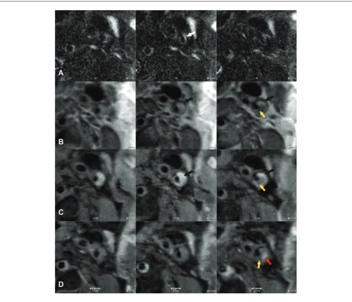
protocol included cardiac trigger, T1.TSE-DARK blood fat sat 2D sequence (TR: 750ms, TE: 15ms, FOV: 80mm, matrix: 192x192), followed of T1.TSE-DARK blood 2D sequence (TR: 750ms, TE: 15ms, FOV: 80mm, matrix: 192x192). In addition, we acquired T2. TSE-DARK blood fat sat 2D sequence TR: 800 ms, TE 123ms, FOV 80mm, 192x192 of matrix and a 19 turbo factor.
Ten minutes after the intravenous administration of 0.2mmol/kg of gadodiamide (Dotaren™, Gerbet, France) images were obtained of the proximal portion of the left internal carotid artery in T1FS sequence (Figure 1).
The PET was co-registered with MRI imaging through a software system. Thus, anatomical structures identified by MRI imaging were correlated with 18F-FDG PET images by a
process of image co-registration.
This work was approved by Ethical Committee of our Institution. The patient authorized the study and signed the Informed Consent Form.
An extensive uniform plaque with abnormal surfaces, which extended from the bifurcation to the proximal portion of the internal segment, was identified at the Doppler examination. Increased turbulence and high peak systolic velocity, which exceeded the maximum limit of Nyquist, suggested stenosis > 70%. The lumen diameter at the stenosis was 1.2 mm and 5.0 mm distal to the stenosis (post-plaque).
The MRI examination was carried out to provide anatomical information.
About 20 days after the examinations, the patient was submitted to carotid endarterectomy, with removal the atherosclerotic plaque. A plaque with intense fibrotic and necrotic content was obtained.
Due to the fact that the tissue appeared inactive, according to the metabolic activity, it was not possible to observe 18F-FDG
uptake at the PET examination.
Figure 1 - In vivo transverse imaging of a left internal carotid artery. A - T2-weighted image shows a high-signal intensity region (arrow white); B and C - T1 and T1-FS -weighted images show a high-signal intensity region (arrow black and yellow); D - Post contrast T1-FS -weighted image shows enhancement of a small region of the plaque (yellow and red arrows).
Arq Bras Cardiol 2009; 93(6) : e84-e87
Benedetto et al 18F-FDG in distinction of atherosclerotic plaque
e85
Discussion
Plaque composition has been associated with the onset of cerebral vascular disease. Pathological studies suggest that the development of stroke in carotid artery disease events depend, principally, on the composition and vulnerability of the plaques and at a lesser degree, on the severity of the stenosis. Rupture occurs preferentially in plaques containing a soft, lipid-rich core that is covered by a thin cap of fibrous tissue5–7.
The use of 18F-FDG has been validated in Oncology9.
The development of techniques to apply this radioisotope to others areas, such as Cardiology, has been the study purpose of many research centers.
Several studies with animal models have suggested that 18
F-FDG can accumulate in atherosclerotic plaque macrophages. Thus, 18F-FDG might be used as a marker to quantify
macrophages in atherosclerotic lesions and discriminate between unstable and stable plaques6,10.
The fusion of the PET/MRI images has proven to be feasible, from the clinical viewpoint, due to the fact that carotids are fixed structures, in contrast to mobile organs, such as the liver and the lungs, which require manual adjustments (Figure 2).
In this case report, the necrotic process of the atherosclerotic plaques was already installed and an intense fibrotic content was observed, reducing the inflammatory process and the metabolic activity of the tissue.
It was not possible to observe the uptake of 18F-FDG in
the macrophages due to the ignoble number of inflammatory cells and, consequently, there was no development of image through the PET methodology.
This was a preliminary study. Our research will be extended to other patients in order to evaluate different kinds of plaques, including those with the presence of macrophages and intense inflammatory processes.
18F-FDG PET may be capable of imaging the plaque and
potentially quantifying its inflammation degree. Moreover,
18F-FDG PET could be used to predict the risk of future plaque
rupture and to monitor the effects of atheroma-modifying therapies.
Authors` contributions
MPC carried out the PET studies and participated in the discussion. FAJ and ACC Jr carried out the MRI assessments and participated in the discussion. AvR performed the validation studies, through the carotid endarterectomy procedure. RB developed the initial concept and drafted the manuscript. LMBF and RB participated in the design, discussion and edited the manuscript.
Acknowledgements
This study was supported by Centro de Diagnóstico por Imagem (CDPI) and Instituto de Engenharia Nuclear (IEN), Rio de Janeiro. The authors thank Dr. Romeu Cortês Domingues and Ana Maria Braghirolli. Without their contribution, this work would not have been possible.
Potential Conflict of Interest
No potential conflict of interest relevant to this article was reported.
Sources of Funding
There were no external funding sources for this study.
Study Association
This article is part of the thesis of master submitted by Raquel Benedetto, from Universidade Federal do Rio de Janeiro - UFRJ.
Figure 2 - No metabolic activity was found in 18F-FDG PET and PET/MRI fusion images in the neck, due to the ignoble number of macrophages. A – MRI; B - FDG PET images; C - PET/MRI Fusion.
Arq Bras Cardiol 2009; 93(6) : e84-e87
Benedetto et al
18F-FDG in distinction of atherosclerotic plaque
e86
References
1. Biasi GM, Froio A, Diethrich EB, Deleo G, Galimberti S, Mingazzini P, et al. Carotid plaque echolucency increases the risk of stroke in carotid stenting: the Imaging in Carotid Angioplasty and Risk of Stroke (ICAROS) Study. Circulation. 2004; 110: 756-62.
2. Albuquerque LC, Narvaes LB, Maciel AA, Staub H, Friedrich M, Hoefel JRF, et al. Intraplaque hemorrhage assessed by high-resolution magnetic resonance imaging and C-reactive protein in carotid atherosclerosis. J Vasc Surg. 2007; 46: 1130-7.
3. Ballotta E, Giau G, Renon L. Carotid plaque gross morphology and clinical presentation: a prospective study of 457 carotid artery specimens. J Surg Res. 2000; 89: 78-84.
4. Cappendijk VC, Cleutjens KBJM, Kessels AGH, Heeneman S, Schurink GWH, Welte RJTJ, et al. Assessment of human atherosclerotic carotid plaque components with multisequence MR imaging: initial experience. Radiology. 2005; 234: 487-92.
5. Davies MJ, Richardson PD, Woolf N, Katz DR, Mann J. Risk of thrombosis in human atherosclerotic plaques: role of extracellular lipid,
macrophage, and smooth muscle cell content. Br Heart J. 1993; 69: 377-81.
6. Ogawa M, Ishino S, Mukai T, Asano D, Teramoto N, Watabe H, et al. 18F-FDG
accumulation in atherosclerotic plaques: immunohistochemical and PET imaging study. J Nucl Med. 2004; 45: 1245-50.
7. Rudd JHF, Warburton EA, Fryer TD, Jones HA, Clark JC, Antoun N, et al. Imaging atherosclerotic plaque inflammation with [18
F]-fluorodeoxyglucose positron emission tomography. Circulation. 2002; 105: 2708-11.
8. Wu KC, Lima JAC. Noninvasive imaging of myocardial viability current techniques and future developments. Circ Res. 2003; 93: 1146-58.
9. Yu S. Review of 18F-FDG synthesis and quality control. Biomed Imaging Interv
J. 2006; 57: 1-11.
10. Zhang Z, Machac J, Helft G, Worthley SG, Tang C, Zaman AG, et al. Non-invasive imaging of atherosclerotic plaque macrophage in a rabbit model with F-18 FDG PET: a histopathological correlation. BMC Nucl Med. 2006; 6: 3.
Arq Bras Cardiol 2009; 93(6) : e84-e87
Benedetto et al 18F-FDG in distinction of atherosclerotic plaque