Evaluation of patients with Alzheimer
’
s
disease before and after dental treatment
Avaliação de pacientes com doença de Alzheimer antes e depois do tratamento dentário
Thaís de Souza Rolim1, Gisele Maria Campos Fabri2, Ricardo Nitrini3, Renato Anghinah3,Manoel Jacobsen Teixeira3, José Tadeu T. de Siqueira2, José Augusto Ferrari Cesari1, Silvia Regina Dowgan Tesseroli de Siqueira4
ABSTRACT
Oral infections may play a role in Alzheimer’s disease (AD).Objective:To describe the orofacial pain, dental characteristics and associated factors in patients with Alzheimer’s Disease that underwent dental treatment. Method: 29 patients with mild AD diagnosed by a neurologist were included. They fulfilled the Mini Mental State Exam and Pfeffer’s questionnaire. A dentist performed a complete evaluation: clinical questionnaire; research diagnostic criteria for temporomandibular disorders; McGill pain questionnaire; oral health impact profile; decayed, missing and filled teeth index; and complete periodontal investigation. The protocol was applied before and after the dental treatment. Periodontal treatments (scaling), extractions and topic nystatin were the most frequent.Results: There was a reduction in pain frequency (p=0.014), mandibular functional limitations (p=0.011) and periodontal indexes (p,0.05), and an improvement in quality of life (p=0.009) and functional impairment due to cognitive compromise (p,0.001) after the dental treatment. Orofacial complaints and intensity of pain also diminished.Conclusion:The dental treatment contributed to reduce co-morbidities associated with AD and should be routinely included in the assessment of these patients.
Keywords:Alzheimer’s disease, oral infections, orofacial pain, periodontal disease, dental treatment.
RESUMO
Infecções orais podem ter um papel na doença de Alzheimer (DA).Objetivo:Descrever as características orofaciais, dor, odontológicas e fatores associados em doentes com DA submetidos a tratamento dentário.Método:29 doentes diagnosticados com DA por neurologista foram avaliados através do Mini Exame do Estado Mental e questionário Pfeffer. O exame odontológico foi realizado antes e depois do tratamento dentário e incluiu: questionário clínico; critérios diagnósticos de pesquisa para disfunção temporomandibular; questionário de dor McGill; protocolo de impacto de saúde oral; dentes cariados, perdidos e obturados; e avaliação periodontal. Os procedimentos mais frequentes foram raspagem periodontal, exodontias e prescrição de nistatina tópica.Resultados:Houve uma redução na frequência de dor (p=0,014), limitações mandibulares (p=0,011), índices periodontais (p,0.05), e melhora na qualidade de vida (p=0,009) e no
comprometimento funcional e cognitivo (p,0,001) após o tratamento dentário. Queixas orofaciais e intensidade de dor também
diminuíram.Conclusão:O tratamento dentário contribuiu para reduzir comorbidades associadas à DA e deveria ser incluído na rotina de avaliação desses pacientes.
Palavras-chave:doença de Alzheimer, infecções orais, dor orofacial, doença periodontal, tratamento dentário.
In the last century the world population got older, and since then there has been a growing interest in maintaining health and an active and functional life in the third age. The frequency of elderly people with chronic diseases is high (20%-35%), and many of them are fragile (2%-10%)1.
Neurodegenerative diseases cause severe morbidity, and among them Alzheimer’s Disease (AD) are progressive and still do not have an effective treatment; therefore, there is an interest in preventing it, improving the quality of life of these patients and reducing the speed of progression2.
1Hospital das Clínicas, Faculdade de Medicina, Universidade de São Paulo, Sao Paulo SP, Brazil; 2Divisão de Odontologia, Faculdade de Medicina, Universidade de São Paulo, Sao Paulo SP, Brazil; 3Departamento de Neurologia, Faculdade de Medicina, Universidade de São Paulo, Sao Paulo SP, Brazil; 4Escola de Artes, Ciências e Humanidades, Universidade de São Paulo, Sao Paulo SP, Brazil.
Correspondence:Thaís de Souza Rolim; Alameda dos Anapurus, 883 / ap. 112; 04087-002 São Paulo SP, Brasil; E-mail: [email protected]
Conflict of interest:There is no conflict of interest to declare.
Support:This manuscript was supported by FAPESP (Foundation of Research of the State of Sao Paulo, Brazil)–2007/04930-1 and 2008/05078-0. Received 05 November 2013; Received in final form 01 August 2014; Accepted 21 August 2014.
DOI:10.1590/0004-282X20140140
AD is the most common degenerative cerebral disease and the main cause of dementia in Western countries (50%-66%)3. It seriously interferes in personal, social and work activities of the patients4. Its pathophysiology includes chronic neuronal and inflammatory abnormalities5, and dental infections are common in these patients6. These infections are not often assessed during the treatment of AD but they need to be considered due to the risk of dissem-ination and the severe complications that they might cause7. Besides, dental infections are a cause of orofacial pain, which is a frequent complaint among the elderly. Despite recent evidence that oral infections such as periodontitis may be asso-ciated with AD, to our knowledge no study prior to this inves-tigated the effects of dental treatment to patients with AD8.
Thus, the objective of this study was to evaluate patients with AD before and after dental treatment about their oro-facial characteristics, as well as emotional, functional and cognitive aspects.
METHOD
In this descriptive not controlled open study, 29 (twenty-nine) patients with mild AD according to the diagnostic criteria of the National Institute for Communi-cative Disorders and Stroke – Alzheimer’s Disease and Related Disorders Association (NINCDS-ADRDA)9were eval-uated. They had been observed by the Grupo de Neurologia Cognitiva e do Comportamento (Hospital das Clínicas, Faculdade de Medicina, Universidade de São Paulo). All patients / relatives / guardians were informed about the pur-poses of the study and all signed the informed consent. The protocol was approved by the local Ethics Committee. This study was supported by FAPESP (Foundation for Research of the State of Sao Paulo–2007/04930-1 and 2007/06852-8).
Exclusion criteria: moderate or severe dementia
accord-ing to the NINCDS-ADRDA criteria. Diagnosis of other neu-rodegenerative or neuroendocrine diseases, neuroinfections.
Inclusion criteria: diagnosis of AD according to the
NINCDS-ADRDA9, score between 18 and 26 by the Mini Mental State Exam (MMSE) characterizing mild AD10. The diagnosis was performed by a trained neurologist.
A dentist performed a complete orofacial assessment which included:
(i) Clinical questionnaire of orofacial pain, for the diagnosis of orofacial pain and oral infections, including a complete dental exam and the evaluation of the oral mucosa, tongue, and pain characteristics (location, intensity by the visual analogue scale – VAS, associated factors including tinnitus, bruxism, alleviating and worsening factors)11;
(ii) Research diagnostic criteria for temporomandibular disorders (RDC/TMD): diagnosis of TMD and
evaluation of emotional and functional aspects related to the mandibular function12;
(iii) McGill pain questionnaire for the assessment of the quality of pain13;
(iv) Dental and periodontal evaluation: DMFT (decayed, missing and filled teeth), plaque and gingival bleeding indexes (PI, BI), probing pocket depth (PPD), cementoenamel junction (CEJ) and clinical attach-ment level (CAL). The PI evaluated the oral hygiene, which was calculated according to the number of dental surfaces stained by a dental plaque disclosing agent multiplied by 100 and divided by the total number of surfaces, with reference value of#30%14. BI evaluated gingival inflammation, and was expressed by the number of bleeding surfaces after probing with a periodontal probe, multiplied by 100 and divided by the total number of surfaces, the reference value of which was #20%. The PPD was determined by the distance from the bottom of the pocket to the gingival margin, with reference value of
.3 mm. By the periodontal evaluation, gingival hyperplasia or recession was identified. The CAL is calculated by the sum of PPD and CEJ, and its reference value is#3 mm (The American Academy of Periodontology, 1999)14,15;
(v) Oral health impact profile (OHIP): validated ques-tionnaire to investigate the impact of oral health on quality of life16.
The cognitive evaluation by the neurologist included the Mini Mental State Exam (cognitive deficit)10 and the Questionnaire of Pfeffer for Functional Activity (performed by the caregiver to determine functionality)17. These ques-tionnaires are part of the protocol of periodic evaluation of these patients to investigate respectively the progression of cognitive impairment and its impact on daily functional activities.
All patients were evaluated at three distinct stages: 1) First evaluation: before the dental treatment; 2) Second evaluation: after one month of the dental treatment;
3) Third evaluation: after 6 months of the second evaluation.
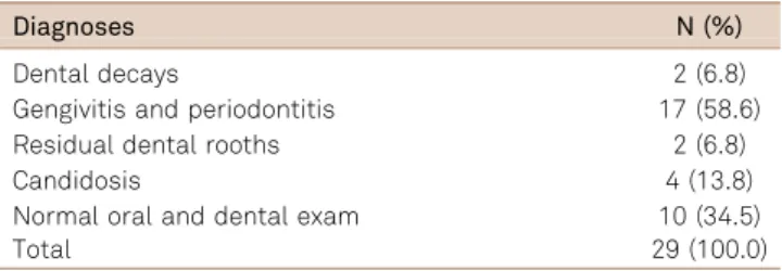
Table 1.Dental diagnoses in the initial evaluation of the patients (N=29).
Diagnoses N (%)
Dental decays 2 (6.8)
Gengivitis and periodontitis 17 (58.6)
Residual dental rooths 2 (6.8)
Candidosis 4 (13.8)
Normal oral and dental exam 10 (34.5)
For ethical reasons, in the post-treatment evaluations the patients received further dental treatment if it was necessary.
Dental treatments
The dental diagnoses obtained after the dental evaluation previous to the treatment of the 29 patients are listed in Table 1. Twenty four patients completed the treatment and the first evaluation after the dental treatment (could not come due to sickness not related to AD or severe aggravation of cognitive status due to other reasons not related to the oral condition, and one died). At 6 months, 14 patients were re-evaluated (8 could not come due to sick-ness not related to AD and 2 died). The treatments per-formed are shown in Table 2. They were perper-formed by the same dentist, and the most frequent were periodontal treat-ments (scaling, root planing and periodontal surgery), dental extractions and topic prescription of nystatin. Some of the treatments were made in the same patient or repeated in the post-treatment evaluations.
Statistical analysis
Data were tabled and initially analyzed according to the distribution of quantitative variable by the Shapiro-Wilk test and Q-Q plots. Variables with normal distribution were analyzed by the analysis of repetitive measurements. The non parametric test for the rest of the variables was the MacNemar test. Correlation among the variables with nor-mal distribution was tested with Pearson’s for the variables with normal distribution. The level of significance was 5%.
RESULTS
After the initial dental treatment, frequency of pain and its intensity reduced [p=0.014 and p=0.040 respectively, (Table 3)]. There were no differences among the evaluations in bruxism, uncomfortable bite, tinnitus, generalized pain, headache or side of mastication. There was also a reduction in the chronic pain severity index (p=0.014) and of mandibular functional limitations (p=0.011) (Table 4). The oral health Table 2. Dental treatments performed at each evaluation period.
Dental treatment after the first evaluation (N=29)
After 1 month of the first treatment (N=24)
After 6 months of the first treatment (N=14)
Topic nystatin 4 (13.8%) 4 (16.7%) 0 (0.0%)+
Periodontal treatment* 15 (51.7%) 14 (58.3%) 3 (21.4%)+
Dental extraction 6 (20.7%) 6 (25.0%) 0 (0.0%)+
Restoration 1 (3.4%) 1 (4.2%) 0 (0.0%)
Inferior prosthesis 2 (6.9%) 2 (8.3%) 0 (0.0%)
Periodontal surgery** 1 (3.4%) 1 (4.2%) 0 (0.0%)
Prosthesis rebasing 2 (6.9%) 2 (8.3%) 0 (0.0%)
Oral hygiene instructions 29 (100.0%) 24 (100.0%) 14 (100%)
*Periodontal scaling and root planning carried out in a minimum two sessions and a maximum of four; **Periodontal surgery by Widman modified procedure;
+McNemar test, p=0.002 (comparison between the last and the first evaluation).
Table 3.Orofacial pain characteristics and associated factors according to the clinical questionnaire of orofacial pain, Research diagnostic criteria for temporomandibular disorder (RDC/TMD) and McGill pain questionnaire.
Initial evaluation (N=29) After 1 month (N=24) After 6 months (N=14) p
Frequency of pain 6 (20.7%) 0 (0.0%)+ 0 (0.0%)+ 0.014
Pain intensity (VAS*) 1.37±3.2 (0-10) 0 (0.0%)+ 0 (0.0%)+ 0.040
Pain descriptors 0 (0.0%)+ 0 (0.0%)+ 0.013
Shock-like 3 (10.3%)
Throbbing 3 (10.3%)
Masticatory miofascial pain 6 (20.7%) 0 (0.0%)+ 0 (0.0%)+ 0.014
Maximum mouth opening (mm) 42.38±5.90 (23-55)** 42.91±6.66 (23-55) 42.36±7.47 (23-55) 0.950
Pain during mouth opening 2 (6.9%) 2 (8.3%) 1 (7.1%) 0.979
Sleep bruxism 2 (6.9%) 0 (0.0%) 0 (0.0%) 0.243
Uncorfortable bite 6 (20.7%) 5 (20.8%) 2 (14.3%) 0.862
Tinnitus 5 (17.2%) 2 (8.3%) 1 (7.1%) 0.502
Side of mastication 0.651
Rigth 4 (13.8%) 2 (8.3%) 2 (14.3%)
Left 3 (10.3%) 1 (4.2%) 0 (0.0%)
Both 22 (75.9%) 20 (83.3%) 12 (85.7%)
Generalized pain 13 (44.8%) 7 (24.1%) 7 (24.1%) 0.497
Headache 8 (27.5%) 2 (6.9%) 2 (6.9%) 0.188
*Visual analogue scale; **mean±standard deviation (range);+McNemar test and analysis of repetitive measures
impact profile (OHIP) showed quality of life improvement after the dental treatment (p=0.009). There was a positive correla-tion between mandibular funccorrela-tional limitacorrela-tions, depression and anxiety indexes (p,0.001), which means that higher depression and anxiety indexes were correlated to more man-dibular limitations (Table 4). Improvement was also detected in relation to the plaque index (p,0.001), BI (p,0.001) and PPD (p=0.024) (Table 5). Positive correlations were found between DMFT and PI (p,0.001), plaque index and maximum CAL (p=0.004), medium PPD and medium CAL (p,0.001), medium PPD and maximum CAL (p,0.001) and medium CAL and maximum PPD (p,0.001), which means that higher PI was associated with higher DMFT, CAL and PPD.
A significant reduction in the cognitive functional para-meters by Pfeffer’s questionnaire occurred after the dental treatment (Table 6). There was no correlation between these cognitive and functional indexes with any specific type of dental treatment or with an specific odontologic variable in this study.
DISCUSSION
AD is a progressive and disabling disease that has pro-found consequences for the lives of individuals. The aging
of the global population is a factor that plays a role in the increase in the incidence and prevalence of dementia, and supports the need of functional improvement in the current quality of life of the patients4. The health professionals involved in the assessment of them are looking for strategies beyond the treatment of AD for secondary morbidities to improve daily life activities, and in this context the oral health of these patients is one big issue. They have severe oral infec-tions that cause several types of impairment6,18,19,20. Recently, evidence has shown a new path for researches, relating peri-odontal infections to the perpetuation and aggravation of symptoms of AD8,21,22. Besides, dental infections are potential causes of orofacial pain as well as masticatory dysfunctions such as TMD, with are also important co-morbidities23,24.
In this study, after the dental treatment, there was a clear evidence of pain relief (less orofacial complaints, myofascial pain, PI and periodontal infections). These were associated with the decrease in depression and anxiety indexes and better functional aspects by the Pfeffer’s questionnaire (p,0.001), which can be correlated or not. As expected, the variables of periodontal disease were correlated between each other (CAL, PPD, BI, PI), and at the 1 month re-evaluation after the initial treatment several procedures were necessary in order to improve the oral health of the patients with limitations in their daily tasks, including oral Table 4.Emotional and quality of life characteristics.
Initial evaluation (N=29) After 1 month (N=24) After 6 months (N=14) p
Chronic pain severity 0 (0.0%)+ 0 (0.0%)+ 0.014
Degree I 4 (13.8%)
Degree II 2 (6.9%)
Depression* 0.54±0.68*** (0.0-2.60) 0.44±0.53 (0.0-1.90) 0.64±0.75 (0.0-2.0) 0.657
Anxiety* 0.50±0.81*** (0.0-2.97) 0.47±0.79 (0.0-3.60) 0.53±0.64 (0.0-2.0) 0.977
Mandibular functional limiations (%) 7.5±22.0*** (0-100) 1.8±6.9+(0-33) 0.0±0.1+(0-0.2) 0.011
OHIP índex** 3.49±6.27 (0.00-23.21) 1.87±4.92 (0.00-20.09) 0.97±3.49 (0.00-13.20) 0.009
*According to RDC/TMD; **Oral health impact profile; ***mean±standard deviation (range);+McNemar test and analysis of repetitive measures
–statistically different from the initial evaluation.
Table 5.DMFT, plaque index and periodontal evaluation.
Initial evaluation (N=29) After 1 month (N=24) After 6 months (N=14) p
Medium PPD* (mm) 1.57±0.69 (0.5-3.4)**** 2.52±3.34+(1.0-12.0) 1.55±0.40 (1.0-2.0) 0.024
Maximum PPD (mm) 3.52±2.14 (0.5-8.0)**** 3.63±1.86 (1.2-7.0) 2.75±2.36 (1.0-6.0) 0.169
Medium CAL** (mm) 2.94±1.26 (1.2-5.9)**** 2.45±1.00 (1.0-4.0) 2.77±1.27 (1.1-4.0) 0.449
Maximum CAL (mm) 5.88±2.58 (2.0-12.0)**** 5.05±2.65 (1.0-10.0) 5.00±3.55 (2.0-10.0) 0.060
DMFT*** 27.17±5.69 (11-32)**** 23.44±8.86+(9-32) 27.50±7.54 (11-32)
,0.001
Plaque index 73.57±5.69 (0.0-100.0)**** 26.21±11.64+(8.0-50.4) 60.0±31.62 (20.0-100.0) ,0.001
*PPD: probing pocket depth; **CAL: clinical attachment level; ***DMFT: decayed, missing and filled teeth; ****mean±standard deviation (range);+Analysis of
repetitive measures–statistically different from the initial and last evaluations.
Table 6.Cognitive evaluation according to MMSE and functional impairment according to Pfeffer’s questionnaire.
Initial evaluation (N=29) After 1 month (N=24) After 6 months (N=14) p
MMSE* 20.86±2.86** (14-26) 20.14±3.29 (12-26) 18.43±2.87+(14-25) 0.048
Functional cognitive impairment 13.55±6.19** (2-28) 9.59±9.92+(0-30) 11.80±11.93+(0-30) ,0.001
*Mini Mental State Exam; **mean±standard deviation (range);+Analysis of repetitive measures
hygiene. This was reflected in the increase of PI after 6 months, which shows the need of repetitive educational measures and treatments to keep the mouth free from recurrent infections in patients with a progressive disease such as AD.
It is possible that the pain observed at the initial evalu-ation was associated with the oral diseases that were treated due to the significant improvement in pain indexes within the follow-up period (p=0.014). Even the myofascial pain exhibited at the initial evaluation disappeared, and thus it was probably a consequence of the oral infections25,26. These conditions cause high psychosocial impact which can be aggravated25,27,28,29. The DMFT index was statistically different after the dental treatment (p,0.001), which may be associated with the extractions that were necessary for some patients. Other signs and symptoms that can be associated with TMD did not differ along the evaluations (bruxism, tinnitus, maximum mouth opening, side of mastication, headache, generalized pain)27,28,29,30.
One important limitation of this study is the high loss of patients that we had for the post-treatment evaluations, which may have affected the results and thus this study should be considered preliminary. AD is a severe and
progressive disease and it is difficult to follow these patients for a long time. However, to our knowledge, this was the first time that the effect of the dental treatment on emotional, functional and cognitive aspects was investigated. As a pre-liminary study, the sample size was not enough for multi-variate analysis. These results are promising and indicate that the detailed relation between dental treatments and AD progression needs to be investigated in the future. There was a slight reduction in the cognitive score by MMSE, which was possibly associated with the progression of the disease in this 6-month period. It is not possible to know, based on this preliminary study, whether this index would have undergone more changes in case the patients were not treated, but some authors have discussed that peri-odontal infections may play a role in the cognitive impair-ment of these patients22,28.
In conclusion, after the dental treatment, a reduction of orofacial pain as well as the improvement of the mandibular function and in the periodontal indexes were detected in the patients with AD, conditions that were maintained until the last evaluation (after 6 months). The recovery of these patients’oral health had a good impact on their quality of life and functional parameters.
References
1. Freedman GM. Chronic pain: clinical management of common causes of geriatric pain. Geriatrics. 2002;57(5):36-41.
2. David R, Piano J, Robert P. Treatment of behavioral disorders in Alzheimer’s diseases. Rev Prat. 2011;61(7):939-44.
3. Scheltens PH, Leys D, Barkhof F, Huglo D, Weinstein HC, Vermersch P et al. Atropht of medial temporal lobes on MRI in“probable”Alzheimer’s disease an normal aging: diagnostics value and neuropsychological correlates. J Neurol Neurosurg Psychiat. 1992;55(10):967-72. http://dx.doi.org/10.1136/jnnp.55.10.967
4. Almeida OP, Nitrini R. Demência. São Paulo: Fundo editorial BYK; 1995. 5. Schwarz MJ, Chiang S, Müller N, Ackenheil M. helper-1 and T-helper-2 responses in psychiatric disorders. Brain Behav Immun. 2001;15(4):340-70. http://dx.doi.org/10.1006/brbi.2001.0647 6. Friedlander AH, Norman DC, Mahler ME, Norman KM, Yagiela JA.
Alzheimer’s disease: psychopathology, medical management and dental implications. J Am Dent Assoc. 2006;137(9):1240-51. http://dx.doi.org/10.14219/jada.archive.2006.0381
7. Aderhold l, Knothe; Frenkel G. The bacteriology of dentogenous pyogenic infections. Oral Surg Oral Med Oral Pathol. 1981;52(6):583-7. 10.1016/0030-4220(81)90072-4
8. Kamer AR. Systemic inflammation and disease progression in Alzheimer dsease. Neurology. 2010;74(14):1157-8. http://dx.doi.org/ 10.1212/WNL.0b013e3181d5df7f
9. Mckhann G, Drachman D, Folstein M, Katzman R, Price D, Stadlan EM. Clinical diagnosis of Alzheimer’s disease: report of the NINCDS-ADRDA work group under the auspices of department of health and human services task force on Alzheimer’s Disease. Neurology. 1984;34(7):939-44. http://dx.doi.org/10.1212/wnl.34.7.939
10. Brucki SMD, Nitrini R, Carameli P, Bertolucci PHF, Okamoto IH. Sugestões para o uso do Mini Exame do Estado Mental no Brasil. Arq Neuropsiquiatr. 2003;61(3B):777-81. http://dx.doi.org/10.1590/ s0004-282x2003000500014
11. Siqueira SRDT, Nóbrega JCM, Valle LBS, Teixeira MJ, Siqueira JTT. Idiopathic trigeminal neuralgia: characteristics and dental procedures. Oral Surg Oral Med Oral Pathol Oral Radiol Endod. 2004;98(3):311-5. http://dx.doi.org/10.1016/S1079210404003191
12. Lucena LB, Komisnky M, Costa LJ, Goes PS. Validation of the Portuguese version of the RDC/TMD Axis II questionnaire. Braz Oral Res. 2006;20(4):312-7. http://dx.doi.org/10.1590/s1806-83242006000400006
13. Pimenta CAM, Teixeira MJ. Considerações iniciais sobre a dor no câncer e seu controle. Rev Med (São Paulo). 1997;76(1):3-6. 14. American Academy of Periodontology. Parameter on chronic periodontitis
with slight to moderate loss of periodontal support. J Periodontol. 2000;71(5 Suppl):853-5. http://dx.doi.org/10.1902/jop.2000.71.5-s.853 15. American Academy of Periodontology. Parameter on chronic
periodontitis with advanced loss of periodontal support. J Periodontol. 2000;71(5 Suppl):856-8. http://dx.doi.org/10.1902/jop.2000.71.5-s.856 16. Barros VM, Seraidarian PI, Cortes MI, Paula LV. The impact of
orofacial pain on the quality of life of patients with temporomanibular disorder. J Orofac Pain. 2009;23(1):28-37.
17. Pfeffer RI, Kurosaki TT, Harrah CH, Chance JM, Filos S. Measurement of functional activities in older adults in the community. J Gerontol. 1982;37(3):323-9. http://dx.doi.org/10.1093/geronj/37.3.323 18. Avlund K, Holm-Pedersen P, Morse DE, Viitanen M, Winblad B.
Tooth loss and caries prevalence in very old Swedish people: the relationship to cognitive function and functional ability. Gerodontology. 2004;21(1):17-26. http://dx.doi.org/10.1046/j.1741-2358.2003.00003.x
19. Henriksen BM, Engedal K, Axéll T. Cognitive impairment is associated with poor oral health in individuals in long-term care. Oral Health Prev Dent. 2005;3(4):203-7. http://dx.doi.org/10.3290/j.ohpd.a10824 20. Syrjälä AM, Ylöstalo P, Sulkava R, Knuuttila M. Relationship
2000 Health Examination Survey in Finland. Acta Odontol Scand. 2007;65(2):103-8. http://dx.doi.org/10.1080/00016350601083521 21. Kamer AR, Craig RG, Dasanayake AP, Brys M, Glodzik-Sobanska L,
Leon MJ. Inflammation and Alzheimer’s disease: possible role of periodontal diseases. Alzheimers Dement. 2008;4(4):242-50. http://dx.doi.org/10.1016/j.jalz.2007.08.004
22. Rethman MP. Inflammation in chronic periodontitis and significant systemic diseases. J Calif Dent Assoc. 2010;38(4):247-57.
23. Fabri GMC, Siqueira SRDT, Simione C, Nasri C, Teixeira MJ, Siqueira JTT. Refractory craniofacial pain: is there a role of periodontal disease as a comorbidity? Arq Neuro-psiquiatr. 2009;67(2B):474-9. http://dx.doi.org/10.1590/s0004-282x2009000300018
24. Siqueira SRDT, Rolim TS, Teixeira MJ, Nitrini R, Anghinah R, Siqueira JTT. Oral infections and orafacial pain in Alzheimer’s disease: case report and review. Dement Neuropsychol. 2010;4(2):145-50. 25. Saczynski JS, Beiser A, Seshadri S, Auerbach S, Wolf PA, Au R.
Depressive symptoms and risk of dementia: The Framingham Heart Study. Neurology. 2010;75(1):35-41. http://dx.doi.org/10.1212/ wnl.0b013e3181e62138
26. Camparis CM, Siqueira JTT. Sleep bruxism: clinical aspects and characteristics in patients with and without chronic orofacial pain. Oral Surg Oral Med Oral Pathol Oral Radiol Endod. 2006;101(2):188-93. http://dx.doi.org/10.1016/j.tripleo.2005.01.014
27. Siqueira SRDT, Teixeira MJ, Siqueira JTT. Severe psychosocial com-promise in idiopathic trigeminal neuralgia: case report. Pain Med. 2010;11(3):453-5. http://dx.doi.org/10.1111/j.1526-4637.2010.00813.x 28. Siqueira SR, Nóbrega JC, Teixeira MJ, Siqueira JT. Masticatory
problems after balloon compression for trigeminal neuralgia: a longitudinal study. J Oral Rehabil. 2007;34(2):88-96. http://dx.doi.org/ 10.1111/j.1365-2842.2006.01680.x
29. Rolim TS, Fabri GMC, Nitrini R, Anghinah R, Teixeira MJ, Siqueira JTT et al. Oral infections and orofacial pain in Alzheimer’s disease: a case-control study. J Alzheimers Dis. 2014;38(4):823-9. http://dx.doi. org/10.3233/JAD-131283