ORIGINAL ARTICLE
10
Key words: ECBC protocol;
Nonseminomatous germ cell tumor; Survival; Recurrence
Int Braz J Urol. 2013; 39: 10-21
__________________ Submitted for publication: August 03, 2012
__________________ Accepted after revision: January 10, 2012 Purpose: To assess the changing presentation and treatment of nonseminomatous
tes-ticular germ cell tumors (NSGCT) and to investigate predictive factors for the status of metastasis at diagnosis and on relapse and death.
Materials and Methods: Retrospective record review of 147 patients that underwent in-guinal orchiectomy from 1987-2007. Follow-up data was available for 102 patients (me-dian follow-up: 80 months (0-243); 96 patients alive).
Results: Mean patients age increased (p = 0.015) and more patients were diagnosed in cli-nical stage I (CSI) (p = 0.040). The fraction of yolk sac (YS) elements inclined (p = 0.030) and pT2 tumors increased (p < 0.001). Retroperitoneal lymph node dissection (RPLND) declined whereas more patients were treated with chemotherapy (p < 0.001; p = 0.004). There was an increase in relapse free (RFS) and cancer specific survival (CSS) due to an improvement in patients with disseminated disease (p = 0.014; p < 0.001). The presence of YS and teratoma elements showed a reduction in the odds ratio (OR) for metastasis at diagnosis (p = 0.002, OR: 0.262; p = 0.009, OR: 0.428) whereas higher pT-stage was as-sociated to their presence (p = 0.039). Patients with disseminated disease (CS > I) showed a declined CSS compared to CSI patients (p = 0.055). The presence of YS elements was associated to an improved RFS (p = 0.038).
Conclusions: In our single institution study the face of NSGCT markedly changed over 20 years even after the introduction of Cisplatin-based chemotherapy. These changes were accompanied by an improvement in RFS and CSS. When dealing with NSGCT patients such observations now and in the future should be taken into account.
INTRODUCTION
Testicular cancer (TC) is the most common malignancy in young men and most of the
pa-tients are diagnosed in early tumor stages. With the introduction of Cisplatin-based chemothera-py in the 1970s TC became an excellently curable disease. However, its etiology and pathogenesis
Vol. 39 (1): 10-21, January - February, 2013
During twenty years of Cisplatin-based therapy the face
of nonseminomatous testicular germ cell tumors is still
changing: an evaluation of presentation, management,
predictive factors and survival
_______________________________________________
Julia Heinzelbecker, Michaela Katzmarzik, Christel Weiss, Lutz Trojan, Axel Haecker
Department of Urology, University Medical Center Mannheim, Medical Faculty Mannheim, University of Heidelberg (JH, AH), Mannheim; Department for Anaesthesiology and Operative Intensive Care Medicine, Cologne-Merheim Medical Center (MK), University Witten/Herdecke; Institute of Medical Statistics and Biometry, Medical Faculty Mannheim, University of Heidelberg (CW), Mannheim and Department of Urology, University Medical Centre Göttingen (LT), Georg-August University, Göttingen, Germany
ABSTRACT
ARTICLE
INFO
IBJU |CHANGES IN NONSEMINOMATOUS TESTICULAR CANCER
11
remain widely unknown (1). Thus observational changes in the presentation of the diseases as well as changes in treatment regimens and survival may lead to a better understanding of the disease itself. Worldwide, a growing incidence has been described and it has already been pointed out that the presentation of the disease is changing (2,3).
The aim of the present study was to eva-luate nonseminomatous testicular cancer (NSGCT) for possible changes in presentation and treatment that occurred after the introduction of chemothe-rapy. Furthermore, we evaluated our patient col-lective for predictive factors for metastasis at diag-nosis as on relapse and death. Such observations will account for a better understanding of NSGCT presentation and may provide a forecast on future problems when dealing with these patients.
MATERIALS AND METHODS
Information was collected by retrospective record review of patients with histologically pro-ven NSGCT that underwent radical orchiectomy between 1987 and 2007 at our institution. A total number of 147 patients were identified. Follow-up data of 102 patients (69%) was available. Median follow-up was 80 months (0-243, patients alive: n = 96 (94%)). For clinical and pathological pa-tients’ data see Table-1. For histopathological sta-ging analysis the underlying TNM classifications were applied (4-6). Further staging analysis in-cluded CT-scan or X-ray and abdominal CT-scan. Division into clinical stages (CS) was performed according to the current TNM classification (7). For further evaluation the whole study period was divided into four periods (1987-1991; 1992-1996; 1997-2001; 2002-2007).
Statistical analysis was performed using SPSS (release 16.0) and SAS software (release 9.2). The changes along the study period were investi-gated by using Kruskal Wallis or Chi² test as ap-propriate. One-way ANOVA analysis was used as a parametric test. Logistic regression was used as a multiple method in order to analyze the influence of possible prognostic parameters on the status of metastasis. Clinical outcome (RFS, CSS) was estima-ted by Kaplan-Meyer analysis and log rank testing. Statistical significance was defined as p < 0.05.
RESULTS
Changing presentation throughout the study periods
The clinical and pathological patients’ data and treatment modalities in CSI according to the different study periods are listed in Table-2. Along the study period the total number of treated pa-tients changed significantly (p = 0.037). Papa-tients in the last studied period were significantly older compared to the first period (0.015). At the same time, a significant shift to pT2 and to more pa-tients diagnosed in CSI was observed (p < 0.001; p = 0.040). Also, the presence of vascular invasion (VI) gained significant importance (p = 0.030). Yolk sac (YS) elements within the histological condition of the tumor were found significantly more often in the last studied periods (p = 0.011). In CSI retroperitoneal lymph node dissection (RPLND) lost importance in favor of chemothera-py (p < 0.001; p = 0.004). The same development was observed for patients with disseminated di-sease (p < 0.001 each). The relapse free survival (RFS) tended to improve over the studied period (p = 0.071). This was due to an improvement in patients with disseminated disease (p = 0.014). The cancer specific survival (CSS) significantly impro-ved over the studied periods (p < 0.001), also due to an improvement in patients with disseminated disease (Table-3 and Figure-1).
Predictive factors for CS
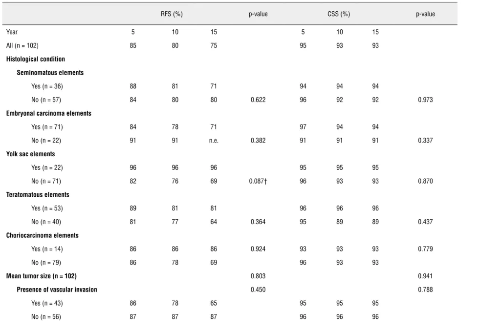
Considering pT-stage, relative risk (OR) for CS > I was significantly elevated with higher pT--stage (p = 0.039). The OR for pT3 compared to pT1 tumors was 8.996 and for pT3 compared to pT2 tumors 8.329. In terms of histological condi-tion, the OR for metastasis was significantly re-duced in cases of teratoma (p = 0.009; OR: 0.428) and YS elements (p = 0.002; OR: 0.262). Other pa-rameters as patients’ age, tumor size or vascular invasion showed no association to the status of metastasis at diagnosis.
Predictive factors for recurrence and survival
IBJU |CHANGES IN NONSEMINOMATOUS TESTICULAR CANCER
12
(0-84). 5-, 10- and 15-year relapse free survival (RFS) was 85%, 80% and 75% respectively. 5-, 10- and 15- year CSS was 95%, 93% and 93% respec-tively. In terms of CSS patients in CS > I showed a reduced CSS compared to patients in CS I (p = 0.055). The histological condition of YS elements tended to be associated to an improved RFS (p = 0.087). The survival data of the patients is listed in Table-3.
DISCUSSION
Patients’ age increased continuously over time. The presence of YS elements gained impor-tance and more pT2 tumors were diagnosed. In terms of adjuvant therapy, RPLND lost importance in favor of chemotherapy. RFS and CSS improved over the study period due to an improvement in patients with disseminated disease. However, RFS in CS I patients declined. The presence of YS and teratoma elements showed a reduction in the re-lative risk (OR) for metastasis at diagnosis where-as higher pT-stage wwhere-as where-associated to the presence of metastasis. Patients with disseminated disease showed a declined CSS compared to patients in CS I. The histological condition of YS elements ten-ded to be associated to an improved RFS.
Although in our study a change of the number of NSGCT patients diagnosed over time was significant, a closer view uncovers that it ac-tually remained stable. This finding is in contra-diction to our recently published results about se-minoma (8). However, it reflects the finding of several authors that the widely reported increase in TC incidence mainly affects seminomas (9). A change in prevalence factors over time, either in terms of differing risk factors for seminoma and NSGCT or of a relation between the intensity of risk factor exposure and the histological type of TC, might account on that (9). Over time, more of our NSGCT patients presented in CSI as reported earlier by other study groups (10,11). The similar results in seminoma favor the hypothesis of an improved awareness of TC as well as an improved accessibility to health care institutions (12). Addi-tionally it, was discussed that NSGCT develops out of seminoma. Thus, a diagnosis of TC in earlier tumor stages would result in an increase of
IBJU |CHANGES IN NONSEMINOMATOUS TESTICULAR CANCER
13
Table 1 - Characteristics of patients with nonseminomatous testicular cancer.
Characteristics Number of contributing patients
Mean patients’ age, years ± SD 147 30.7 ± 8.4
Histological condition, n (%) 129
-Seminomatous elements - 55 (43)
Embryonal carcinoma elements - 101 (78)
Yolk sac elements - 30 (23)
Teratomatous elements - 71 (55)
Choriocarcinoma elements - 21 (16)
Mean tumor size, cm ± SD 130 3.8 ± 2.2
Presence of small vessel invasion, n (%) 140
-Yes - 56 (40)
pT stage, n (%) 142
-pT1 - 85 (60)
pT2 - 43 (30)
pT3 - 14 (10)
pT4 - 0 (0)
Tumor markers 147
-Median AFP, ng/mL (range) 139 69 (1- 37 759)
Median β-HCG, ng/mL (range) 141 22 (0.01- 450 370)
Median LDH, U/l (range) 95 226 (135- 4277)
Clinical stage, n (%) 147
-I - 81 (55)
II - 44 (30)
III - 22 (15)
> I - 66 (45)
IGCCCG classification 64
-Good - 40 (63)
IBJU |CHANGES IN NONSEMINOMATOUS TESTICULAR CANCER
14
Poor - 10 (16)
Adjuvant treatment, n (%) -
-CS I 78
-Surveillance - 15 (19)
Chemotherapy - 33 (42)
RPLND - 29 (37)
Radiation therapy - 1 (1)
CS II 43
-Chemotherapy - 22 (51)
RPLND - 2 (5)
Chemotherapy + RPLND - 19 (44)
CS III 21
-Chemotherapy - 13 (62)
Chemotherapy + surgery* - 8 (38)
Median follow-Up, months (range) 102 80 (0-243)
Median time to relapse, months (range) - 18 (3-199)
Median time to death, months (range) - 10 (0-84)
Relapse, n (%) - 16 (16)
Death, n (%) - 6 (6)
Patients alive, n (%) - 96 (94)
SD = standard deviation; n.e.= not evaluable; AFP = alpha-fetoprotein;β-HCG = human chorionic gonadotropin; LDH = Lactate de-hydrogenase; UPN = upper limit of normal; CS = Clinical stage; IGCCCG = International germ cell cancer consensus group; RPLND = Retroperitoneal lymph node dissection; *surgery includes RPLND and residual tumor resection
IBJU
|
CHANGES IN NONSEMINOMA
T
OUS TESTICULAR CANCER
15
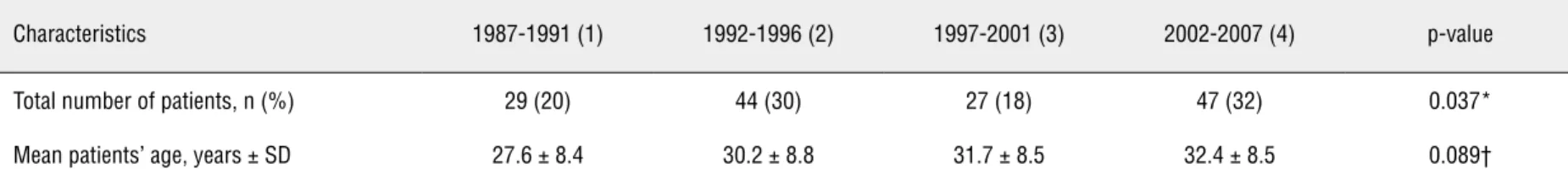
Table 2 - Clinical and pathological patient data and treatment modalities according to the different studied periods.
Characteristics 1987-1991 (1) 1992-1996 (2) 1997-2001 (3) 2002-2007 (4) p-value
Total number of patients, n (%) 29 (20) 44 (30) 27 (18) 47 (32) 0.037*
Mean patients’ age, years ± SD 27.6 ± 8.4 30.2 ± 8.8 31.7 ± 8.5 32.4 ± 8.5 0.089†
(1) vs (3): 0.065† (1) vs (4): 0.015*
Histological condition, n (%)
Seminomatous elements 12 (43) 15 (36) 8 (35) 17 (40) 0.926
Embryonal carcinoma elements 18 (86) 29 (88) 19 (73) 33 (70) 0.204
Yolk sac elements 2 (10) 3 (9) 10 (39) 15 (32) 0.011*
Teratomatous elements 10 (48) 18 (55) 15 (58) 28 (60) 0.827
Choriocarcinoma elements 3 (14) 6 (18) 3 (12) 8 (17) 0.901
Mean tumor size, cm ± SD 4.2 ± 2.8 3.8 ± 1.8 3.0 ± 1.8 3.9 ± 2.2 0.266
(1) vs (3): 0.068† (3) vs (4): 0.090†
Presence of vascular invasion, n (%)
Yes 8 (32) 13 (31) 8 (31) 27 (57) 0.030*
pT stage, n (%) - - - - 0.119
pT1 19 (70) 31 (72) 16 (62) 19 (41) 0.100
pT2 3 (11) 6 (14) 9 (35) 25 (54) < 0.001**
pT3 5 (19) 6 (14) 1 (4) 2 (4) 0.183
IBJU
|
CHANGES IN NONSEMINOMA
T
OUS TESTICULAR CANCER
16
Clinical stage, n (%)
I 12 (41) 25 (57) 16 (59) 28 (59) 0.040*
II 12 (41) 10 (23) 10 (37) 12 (26) 0.948
III 5 (17) 9 (20) 1 (4) 7 (15) 0.095†
> I 17 (59) 19 (43) 11 (41) 19 (40) 0.456
IGCCCG classification 0.292
Good 10 (59) 13 (68) 7 (78) 10 (53)
-Intermediate 4 (24) 3 (16) 1 (44) 6 (32)
-Poor 3 (18) 3 (16) 1 (44) 3 (16)
-Adjuvant treatment n (%)
CS = I
Surveillance 1 (8) 4 (17) 2 (13) 7 (28) 0.432
Chemotherapy 4 (33) 4 (14) 11 (69) 16 (64) 0.004*
RPLND 10 (83) 15 (63) 5 (31) 3 (12) < 0.001**
Radiation therapy 0 (0) 1 (4) 0 (0) 0 (0) 0.530
CS > I
RPLND 1 (6) 0 (0) 0 (0) 1 (5) n.e.
Chemotherapy 5 (31) 5 (26) 10 (91) 15 (79) < 0.001**
RPLND + Chemotherapy 10 (63) 13 (68) 1 (9) 3 (16) < 0.001**
IBJU
|
CHANGES IN NONSEMINOMA
T
OUS TESTICULAR CANCER
17
Table 3 - Relapse free survival (RFS) and cancer specific survival (CSS) according to the clinical and pathological patient data at 5, 10 and 15 years after diagnosis.
RFS (%) p-value CSS (%) p-value
Year 5 10 15 5 10 15
All (n = 102) 85 80 75 95 93 93
Histological condition
Seminomatous elements
Yes (n = 36) 88 81 71 94 94 94
No (n = 57) 84 80 80 0.622 96 92 92 0.973
Embryonal carcinoma elements
Yes (n = 71) 84 78 71 97 94 94
No (n = 22) 91 91 n.e. 0.382 91 91 91 0.337
Yolk sac elements
Yes (n = 22) 96 96 96 95 95 95
No (n = 71) 82 76 69 0.087† 96 93 93 0.870
Teratomatous elements
Yes (n = 53) 89 81 81 96 96 96
No (n = 40) 81 77 64 0.364 95 89 89 0.437
Choriocarcinoma elements
Yes (n = 14) 86 86 86 0.924 93 93 93 0.779
No (n = 79) 86 78 69 96 93 93
Mean tumor size (n = 102) 0.803 0.941
Presence of vascular invasion 0.450 0.788
Yes (n = 43) 86 78 65 95 95 95
IBJU
|
CHANGES IN NONSEMINOMA
T
OUS TESTICULAR CANCER
18
pT stage 0.757
pT1 (n = 56) 85 79 72 96 94 94
pT2 (n = 34) 87 87 n.e. 94 94 n.e.
pT3 (n = 7) 71 71 71 0.790 100 100 100
CS I (n = 57) 87 81 81 98 98 98
CS II (n = 32) 77 72 72 CS I/CSII/ CSIII: 90 84 84 CS I/ CSII/ CSIII:
CS III (n = 13) 92 92 74 0.564 92 92 92 0.127
> CS I (n = 45) 82 78 69 CSI/ CS >
I: 0.503
91 87 87 CSI/ CS >
I: 0.056†
Periods
1987-1991 (n = 15) 67 59 59 73 65 65
1992-1996 (n = 25) 80 75 67 100 100 100
1997-2001 (n = 25) 92 90 n.e. 0.071† 100 100 n.e. < 0.001**
2002-2007 (n = 37) 92 n.e. n.e. 97 n.e. n.e.
CS = I
1987-1991 (n = 5) 100 100 100 100 100 100
1992-1996 (n = 16) 75 66 66 100 100 100
1997-2001 (n = 14) 93 93 n.e. 0.247 100 100 n.e. 0.662
2002-2007 (n = 22) 98 n.e. n.e. 95 n.e. n.e.
CSI > I
1987-1991 (n = 10) 50 40 40 60 48 48
1992-1996 (n = 9) 89 89 74 100 100 100
1997-2001 (n = 11) 91 91 n.e. 0.014* 100 100 n.e. <0.001**
2002-2007 (n = 15) 93 n.e. n.e. 100 n.e. n.e.
RFS= relapse free survival; CSS= cancer specific survival; n= number of patients; CS= clinical stage; n.e.= not evaluable; †= p< 0.100; *= p< 0.050; **= p< 0.001; a p-value of p< 0.050 was
IBJU |CHANGES IN NONSEMINOMATOUS TESTICULAR CANCER
19
88%) (10). An upcoming focus on life quality mi-ght be a possible explanation for such observa-tions. Nevertheless, 62% of performed RPLND is still high compared to our results of 12-31%. Thus, regional as institutional circumstances might also play a role. The latest European guidelines on NSGCT nowadays give explicit recommendations, classifying RPLND as rather second or third treat-ment of choice (18). However, in the EAU guideli-ne of 2001 yet there was a clear recommendation for RPLND besides surveillance and chemotherapy (19). Our data demonstrate that treatment changes in clinical practice took place even earlier. A trend towards more surveillance, the nowadays stan-dard treatment option for low risk NSGCT patients, was not observed in our study collective (18). In our patient collective RFS in disseminated disease continuously improved over time. We believe that this change in RFS is mainly due to a historical
policy of RPLND in CS IIA/B patients in the past that changed to a policy of primary chemotherapy in CS IIA/B NSGCT patients after 1996. The prefer-red treatment regimen in CS IIA/B patients still remains controversial as for both chemotherapy and RPLND survival rates of more than 95% have been described (20,21). Stephenson et al. reported on an increase in RFS in their CS IIA/B patient collective after 1999 and hypothesized that this was due to a more adequate patient selection ac-cording to established parameters of progression as elevated tumor markers or adenopathy larger than 2 cm (22). The current EAU guideline on NSGCT recommend RPLND in CS IIA/B patients only in case of negative markers (18). However, at the same time there was an incline in RFS of CS I patients. Most probably this is due to the higher rate of surveillance regimens after 1991 that will always cause a certain amount of relapses. CSS in our patient collective improved especially since 1992. Sonneveld et al. reported on a significant improvement in the survival rate of their metasta-sized NSGCT patients from 1977-1986 compared to 1987-1996 (23). Data of the Roland-Koch Insti-tute in Germany report on a first incline in survi-val rates in the 1970s due to the introduction of Cisplatin based chemotherapy followed by a fur-ther incline since the 1980s with relative 5 year survival rates increasing from 80% to 95% (24). In our institution the improvement in CSS was main-ly due to a harsh incline in events of death after 1992. We found a reduction in the relative risk for metastases at diagnosis in case of the presence of teratoma and YS elements. This finding is in con-cordance to a study of Klepp et al. of a CSI patient collective, undergoing RPLND. Herein they repor-ted that the absence of teratoma or YS elements was a predictor for retroperitoneal metastases (25). Other studies reported on a higher rate of metasta-tic disease in tumors containing less than 50% of teratoma elements (26). In our study collective hi-gher pT-stage was associated with the presence of metastasis at diagnosis. Nicolai and colleagues created a model for the prediction of nodal metas-tasis at diagnosis. However, t-stage did not serve as a predictor, in contrary VI served as a reliable predictor (27). We found no association between VI and the status of metastasis. We report on a 5-,
Figure 1 - Cancer specific survival curves of metastasized NSGCT patients according to the different studied groups (1987-1991; 1992-1996; 1997-2001; 2002-2007).
Sur
vival probability
Time to death (months) 1,0
0,8
0,6
0,4
0,2
0,0
IBJU |CHANGES IN NONSEMINOMATOUS TESTICULAR CANCER
20
10- and 15- year CSS of 95%, 93% and 93%, res-pectively. This is in concordance to other results reported by Sokoloff and colleagues on their Ame-rican study collective from 1975-2001 with a 96% survival rate. However, no discrimination between NSGCT and seminoma was made (28). Compared to Europe such CSS rates even exceed the upper limit of reported data, ranging for NSGCT from 47% - 90%. Yet this data only comprised an early studied period from 1987-1992 and only an ex-cerpt of European countries was included (29). Sonneveld et al. report on a 10-year CSS of 82% for NSGCT. However, their study period comprises an early time period from 1977 to 1996, too (23). The Roland Koch Institute of Germany delivered latest relative 5-year survival rates of up to 95% (24). Thus, our CSS rates are very good. 16% of our NSGCT patients relapsed and 5-, 10- and 15-year RFS were 85%, 80% and 75%, respectively. For Europe a relapse rate of 12% (0-34%) was re-ported (29). In terms of CSS, patients with metas-tasized disease (CS > I) showed a worse survival compared to patients in CS I. Hence, these results underline the prognostic value of the TNM classi-fication. The histological condition of YS elements was associated with an improved RFS. Other study groups also reported on the absence of YS ele-ments being associated to a reduction in relapse (30). Our study is limited by its retrospective study character that leads to a considerable lack of data concerning histopathology and follow-up data. Furthermore no re-evaluation by application of the recent TNM-classification took place. With TC being a disease of excellent CSS there is a further limitation by the small number of events espe-cially in terms of death. Therefore, a multivariate analysis was not performed. The data here presen-ted are restricpresen-ted to a single-center population. Thus, affirmations above population trends can-not be made.
The remarkable changes in RFS and CSS for both stage I and disseminated disease after the first five year period reflect a change in treatment policies that took place all over Europe. With the excellent survival data provided for NSGCT, pa-tients in early stages of the disease make high de-mands on the form of treatment they will choose. Clinical trials have already proofed the
possibili-ties of surveillance regimens. At the same time the importance of RPLND has declined.
CONCLUSIONS
Detecting patients at high risk of relapse and clearly discriminating them from low-risk otherwise overtreated patients is as urgent as ever. The data of the last decades shows that a further improvement in CSS is possible even after the in-troduction of Cisplatin-based chemotherapy.
ABBREVIATIONS
NSGCT: Nonseminomatous testicular germ cell tumor
CSI: Clinical stage I
YS: Yolk sac
RPLND: Retroperitoneal lymph node dissection
RFS: Relapse free survival
CSS: Cancer specific survival
OR: Odds ratio
CS>I: Disseminated disease
TC: Testicular cancer
CONFLICT OF INTEREST
None declared.
REFERENCES
1. Meeks JJ, Sheinfeld J, Eggener SE: Environmental toxicology of testicular cancer. Urol Oncol. 2012; 30: 212-5.
2. Power DA, Brown RS, Brock CS, Payne HA, Majeed A, Babb P: Trends in testicular carcinoma in England and Wales, 1971-99. BJU Int. 2001; 87: 361-5.
3. Steele GS, Richie JP, Stewart AK, Menck HR: The National Can-cer Data Base report on patterns of care for testicular carcino-ma, 1985-1996. Cancer. 1999; 86: 2171-83.
4. WHO: International histological classification of tumours. 2nd ed. Geneva: WHO; 1981-1999.
5. Sobin LH, Wittekind C: UICC TNM Classification of malignant tumours. 5th ed. New York: Wiley & Sons. 1997.
6. Sobin LH: Wittekind UICC TNM Classification of malignant tu-mours. C. UICC TNM classification of malignant tutu-mours. 6th ed. New York: Wiley & Sons. 2002.
IBJU |CHANGES IN NONSEMINOMATOUS TESTICULAR CANCER
21
8. Heinzelbecker J, Katzmarzik M, Weiss C, Trojan L, Michel MS, Haecker A: Changes of stage, predictive factors and adjuvant treatment modalities in seminomatous testicular cancer from 1987 to 2007 and their impact on the status of metastasis, recurrence-free and overall survival: a single-center analysis. Urol Int. 2011; 87: 282-7.
9. McGlynn KA, Devesa SS, Sigurdson AJ, Brown LM, Tsao L, Tarone RE: Trends in the incidence of testicular germ cell tumors in the United States. Cancer. 2003; 97: 63-70. 10. Cooper DE, L’esperance JO, Christman MS, Auge BK: Testis
cancer: a 20-year epidemiological review of the experience at a regional military medical facility. J Urol. 2008; 180: 577-81; discussion 581-2.
11. Sonneveld DJ, Hoekstra HJ, Van Der Graaf WT, Sluiter WJ, Schraffordt Koops H, Sleijfer DT: The changing distribution of stage in nonseminomatous testicular germ cell tumours, from 1977 to 1996. BJU Int. 1999; 84: 68-74.
12. Powles TB, Bhardwa J, Shamash J, Mandalia S, Oliver T: The changing presentation of germ cell tumours of the tes-tis between 1983 and 2002. BJU Int. 2005; 95: 1197-200. 13. Oliver RT, Leahy M, Ong J: Combined
seminoma/non-sem-inoma should be considered as intermediate grade germ cell cancer (GCC). Eur J Cancer. 1995; 31A: 1392-4. 14. Kratzik C, Höltl W, Albrecht W, Pont J, Zielinski CH, Breindl E,
et al.: Risk adapted management for NSGCT stage 1 - long-term results of a multicenter study. J Urol. 1996; 157: 547A. 15. Muir KR, Parkes SE, Lawson S, Thomas AK, Cameron AH,
Mann JR: Changing incidence and geographical distribu-tion of malignant paediatric germ cell tumours in the West Midlands Health Authority region, 1957-92. Br J Cancer. 1995; 72: 219-23.
16. Birch JM, Marsden HB, Swindell R: Pre-natal factors in the origin of germ cell tumours of childhood. Carcinogenesis. 1982; 3: 75-80.
17. Molina Saera J, Aparicio Urtasun J, Díaz Beveridge R, Palo-mar Abad L, Giménez Ortiz A, Ponce Lorenzo J, et al.: Epi-demiological pattern and time trends in testicular germ-cell tumors: a single institution 20-year experience. Clin Transl Oncol. 2006; 8: 588-93.
18. Albers P, Albrecht W, Algaba F, Bokemeyer C, Cohn-Ceder-mark G, Fizazi K, et al.: EAU guidelines on testicular cancer: 2011 update. Eur Urol. 2011; 60: 304-19.
19. Laguna MP, Pizzocaro G, Klepp O, Algaba F, Kisbenedek L, Leiva O, et al.: EAU guidelines on testicular cancer. Eur Urol. 2001; 40: 102-10.
20. Donohue JP, Thornhill JA, Foster RS, Rowland RG, Bihrle R: Clinical stage B non-seminomatous germ cell testis can-cer: the Indiana University experience (1965-1989) using routine primary retroperitoneal lymph node dissection. Eur J Cancer. 1995; 31A: 1599-604.
21. Weissbach L, Bussar-Maatz R, Flechtner H, Pichlmeier U, Hartmann M, Keller L: RPLND or primary chemotherapy in clinical stage IIA/B nonseminomatous germ cell tumors? Results of a prospective multicenter trial including quality of life assessment. Eur Urol. 2000; 37: 582-94.
22. Stephenson AJ, Bosl GJ, Motzer RJ, Bajorin DF, Stasi JP, Sheinfeld J: Nonrandomized comparison of primary che-motherapy and retroperitoneal lymph node dissection for clinical stage IIA and IIB nonseminomatous germ cell tes-ticular cancer. J Clin Oncol. 2007; 25: 5597-602.
23. Sonneveld DJ, Hoekstra HJ, van der Graaf WT, Sluiter WJ, Mulder NH, Willemse PH, et al.: Improved long term sur-vival of patients with metastatic nonseminomatous testicular germ cell carcinoma in relation to prognostic classification systems during the cisplatin era. Cancer. 2001; 91: 1304-15. 24. Bertz J, Dahm S, Haberland J, Kraywinkel K, Kurth BM,
Wolf U: Verbreitung von Krebserkrankungen in Deutsch-land. Entwicklung der Prävalenzen zwischen 1990 und 2010. Beiträge zur Gesundheitsberichterstattung des Bundes. Berlin. RKI. 2010.
25. Klepp O, Olsson AM, Henrikson H, Aass N, Dahl O, Stenwig AE, et al.: Prognostic factors in clinical stage I nonsemino-matous germ cell tumors of the testis: multivariate analysis of a prospective multicenter study. Swedish-Norwegian Testicular Cancer Group. J Clin Oncol. 1990; 8: 509-18. 26. Guney S, Guney N, Sonmez NC, Ergenekon E: Risk-adapted
management for patients with clinical stage I non-seminomatous germ cell tumour of the testis. Med Oncol. 2009; 26: 136-42. 27. Nicolai N, Miceli R, Artusi R, Piva L, Pizzocaro G,
Salvi-oni R: A simple model for predicting nodal metastasis in patients with clinical stage I nonseminomatous germ cell testicular tumors undergoing retroperitoneal lymph node dissection only. Urol. 2004; 171: 172-6.
28. Sokoloff MH, Joyce GF, Wise M, Urologic Diseases in America Project: Testis cancer. J Urol. 2007; 177: 2030-41. 29. Sant M, Aareleid T, Artioli ME, Berrino F, Coebergh JW,
Colonna M, et al.: Ten-year survival and risk of relapse for testicular cancer: a EUROCARE high resolution study. Eur J Cancer. 2007; 43: 585-92.
30. Freedman LS, Parkinson MC, Jones WG, Oliver RT, Peck-ham MJ, Read G, et al.: Histopathology in the prediction of relapse of patients with stage I testicular teratoma treated by orchidectomy alone. Lancet. 1987; 2: 294-8.
_____________________
Correspondence address: