Early detection strategy and mortality reduction in
severe sepsis
Estratégia de detecção precoce e redução de mortalidade na sepse
grave
INTRODUCTION
Sepsis is a set of sometimes dramatic and catastrophic reactions of hu-man beings in response to invasion by pathogenic microorganisms. It is a clinical syndrome that presents with diferent degrees of severity. If not diagnosed and adequately treated it may worsen over time. Usually, the clinical condition begins with nonspeciic and subtle changes of the vital signs such as tachycardia and tachypnea.(1-4)
Generally speaking, sepsis often goes unnoticed until advanced stages even in hospital settings(4) because its manifestations are not marked by an
ictus as in acute myocardium infraction (AMI) or stroke (S).
Diagnosis of the septic syndrome is clinical, based on changes that com-prise the systemic inlammatory response syndrome SIRS. It was deined in 1991 by the American College of Chest Physicians/Society of Critical Care Medi-cine Consensus Conference Committee as a set of at least two of the following
Glauco Adrieno Westphal1, Janaína
Feijó2, Patrícia Silva de Andrade3,
Louise Trindade4, Cezar Suchard5,
Márcio Andrei Gil Monteiro6, Sheila
Fonseca Martins7, Fernanda Nunes8,
Milton Caldeira Filho9
1. PhD, Preceptor of Intensive Care Medicine Residency, Hospital Municipal São José - HMSJ - Joinville (SC), Brazil. 2. Resident of Intensive Care Medicine, Hospital Municipal São José - HMSJ - Joinville (SC), Brazil.
3. Physician of General Intensive Care Unit, Imperial Hospital de Caridade, Florianópolis (SC), Brazil.
4. Resident of Intensive Care Medicine, Hospital Municipal São José - HMSJ - Joinville (SC), Brazil.
5. Medical Student of Universidade da Região de Joinville, Joinville (SC), Brazil.
6. Medical Student of Universidade da Região de Joinville, Joinville (SC), Brazil.
7. Nurse from the Hospital Infection Control Center, Hospital Municipal São José - HMSJ - Joinville (SC), Brazil. 8. Nurse of the Hospital Infection Control Center, Hospital Municipal São José – HMSJ - Joinville (SC), Brazil. 9. Physician, General Intensive Care Unit and General Coordinator of Intensive Medical Residency, Hospital Municipal São José – HMSJ - Joinville (SC), Brazil.
ABSTRACT
Objective: To evaluate the impact of implementing an institutional pol-icy for detection of severe sepsis and septic shock.
Methods: Study before (stage I), after (stage II) with prospective data collection in a 195 bed public hos-pital.. Stage I: Patients with severe sepsis or septic shock were included consecutively over 15 months and treated according to the Surviving Sepsis Campaign guidelines. Stage II: In the 10 subsequent months, patients with severe sepsis or septic shock were enrolled based on an active search for signs suggesting infection (SSI) in hos-pitalized patients. he two stages were compared for demographic variables, time needed for recognition of at least two signs suggesting infection (SSI-Δt), compliance to the bundles of 6
and 24 hours and mortality.
Results: We identified 124 pa-tients with severe sepsis or septic shock, 68 in stage I and 56 in stage II. The demographic variables were similar in both stages. The Δt-SSI was 34 ± 54 hours in stage I and 7 ± 8.4 hours in stage II (p <0.001). There was no difference in compli-ance to the bundles. In parallel there was significant reduction of mortality rates at 28 days (54.4% versus 30%, p <0.02) and hospital (67.6% versus
41%, p <0.003). Conclusion: The
strategy used helped to identify early risk of sepsis and resulted in decreased mortality associated with severe sepsis and septic shock.
Keywords: Shock, septic /diagno-sis; Shock, septic/therapy; Shock, sep-tic/mortality; Sepsis/diagnosis; Sepsis/ therapy; Sepsis/mortality
Received from Hospital Municipal São José - HMSJ - Joinville (SC), Brazil.
Submitted on March 30, 2009 Accepted on May 12, 2009
Author for correspondence:
Janaina Feijó
Rua Profª Ana Maria Harger, 62 – apt. 201- Bairro Anita Garibaldi
CEP: 89202-020 – Joinville (SC), Brasil.
manifestations: a) fever or hypothermia; b) tachycardia c|) tachypnea, d) leukocytosis or leukopenia. It is an acute condition caused by systemic release of inlam-matory mediators and generalized activation of the en-dothelium, generating break of the endothelial homeo-stasis with impairment and dysfunction of organs distant from the primary focus. It relects the level of organic stress associated to diferent clinical conditions such as: trauma, burns, acute severe pancreatitis, surgery, transfu-sion therapy and infection. When SIRS is secondary to infection, the diagnosis is sepsis. Sepsis is considered se-vere when there is at least one associated organ dysfunc-tion and, if hypotension persists regardless of vigorous administration of water, it is septic shock.(1)
It was proven that adopting the therapeutic strategy proposed by the Serving Sepsis Campaign (SSC) that includes early tissue reperfusion and control of the in-fection focus,(2,5,6) bring about decreased mortality.(7-14) In our hospital, as well as in other Brazilian institutions, notwithstanding adhesion to SSC, mortality rates con-tinue to be unacceptably high.(15-17) Perhaps this was related to delay in diagnosis of sepsis. Failure to iden-tify sepsis delays onset of adequate treatment, causes progress of multiple organ dysfunction and severely jeopardizes prognosis of patients.(16) herefore, ongoing search for detection of signs of SIRS and organ dys-function during routine control of vital signs, might involve identiication of patients at risk of sepsis. In this context we proposed a simple institutional proce-dure to facilitate identiication of severe sepsis or septic shock in our hospital.
his study intended to verify if institutional emphasis to identify risk of sepsis may help early recognition of severe sepsis or septic shock and inluence its prognosis.
METHODS
his is a before/after study (stage 1/stage II) con-ducted from August 2005 to September 2007, in the wards of the emergency department and intensive care unit (ICU) of the Hospital Municipal São José (HMSJ), Joinville, Santa Catarina, Brazil. HMS is a general and public hospital with 195 beds for general admission and 2 ICU with 14 beds. Written consent was not giv-en, as it is an institutional program to attend patients. Patients detected in any sector of the hospital with a diagnosis of severe sepsis or septic shock was included. Terminal disease or shock by other etiologies were con-sidered exclusion criteria.
he study encompasses two distinct periods (stage
I and stage II) that difer according to the screening strategy of patients with risk of sepsis. In stage I (15 months) were consecutively included patients with se-vere sepsis or septic shock, managed according to the SSC recommendations. Diagnosis and treatment strat-egy was divided into three parts, shown in chart 1.(17)
Chart 1 - Strategy for diagnosis and treatment of stage I Screening - According to the following questions for diag-nosis
a. Is there an infectious focus?
b. Are there two or more SSI: temperature > 38oC or < 36oC;
Chills and shivering; heart rate > 90 bpm; respiratory rate > 20 mpm; systolic arterial pressure < 90 or mean arterial pres-sure < 65 mmHg.
c. Airmative reply to two questions results in diagnosis of sepsis
d. Is there organ dysfunction?
(e) Airmative reply to all previous questions results in diag-nosis of severe sepsis
Initial management – Patients with severe sepsis or septic shock must fulfill all of the seven goals below in the first 6 hours after diagnosis, and the sum of these goals is called the “6 hours bundle”.
a. Mesurment of serum lactate
b. Collection of at least two samples of hemoculture from di-ferent sites
c. Onset of adequate antibiotic therapy in the irst hour after diagnosis
d. If there is hypotension or lactate above or equal to 4mmol/l, administer 20 to 30 ml/kg of crystalloid.
e. Initiate vasopressor if MAP of 65 mmHg or more has not been reached after crystalloid infusion.
f. Reach a CVP above 8 mmHg in patients needing this gene-rous infusion of crystalloid
g. Reach a central venous saturation above 70%
24 hours bundle – goals that must be fulilled in the irst 24 hours after onset of treatment
a. administer low dose corticoids according to the ICU policy, if institutional policy is not to use any, record it.
b. administer (according to the policy of each institution) re-combinant human activated protein C. HMS policy regar-ding application of rhPC is not to use the drug.
c. glycemic control with insulin therapy according to institu-tional protocol
d. Keep inspiratory plateau pressure < 30 cmH20 in patients under mechanical ventilation.
At stage II, (10 months) patients with sepsis or sep-tic shock were identiied as from an active search strat-egy for signs suggesting infection (SSI) in all patients admitted to the hospital. A new form was devised for a record of SSI (Appendix 1), grouping vital signs and eventual clinical signs of organ dysfunction of all pa-tients in each ward. Register of at least two SSI in this form were promptly informed to the responsible nurse by the sector that completed the screening form (Ap-pendix 2). A single nursing technician in each ward was in charge of the task. After initial assessment by the responsible nurse and by the sector, the nursing staf of the Hospital Infection Control Committee (HICC) was advised to evaluate and follow-up the case. he on duty physician (internal medicine resident) was imme-diately called when suspicion of sepsis was conirmed (Appendix 2). When diagnosis was deined, therapeutic bundles were started from 6 and 24 hours (Appendix 3 and 4) according to SSC guidelines (Figure 1).
Nurses and resident physicians of intensive care and internal medicine of HMS were trained and supervised by intensivists to ascertain that patients were adequate-ly treated in any ward. In our hospital, as well as many others in Brazil, often a bed is not available in the ICU. hat is why many training sessions were carried out so that all understood severe sepsis/septic shock, stressing the importance of changes in the vital signs.
The groups of patients of stage I and stage II were compared for: age, gender, provenance (ward, ICU, emergency room), time elapsed since first re-cord (medical chart) of at least two SSI, moment of diagnosis of severe sepsis (Δt-SSI), APACHE, Acute Physiology and Chronic Health Evaluation II score, complete compliance to the bundles of 6 and of 24 hours, ICU and hospital stay, mortality at 28th day and intra-hospital.
For statistical analysis of data, the programs NCSS: Statistical Software 2000 & PASS 2000: Power Analy-sisande Sample Size and GraphPad Prism 4 were used. Continuous variables were presented as mean ± stan-dard deviations and compared using the Student’s t test. Categorical variables were expressed in absolute and relative values and compared by the Chi-Square test. A p=0.05 value was considered statistically signiicant.
RESULTS
hree hundred seventy eight patients were consecu-tively assessed. During stage I, 76 patients were identi-ied with severe sepsis or septic shock, of which 8 were excluded for lack of a therapeutic perspective related to the baseline disease. At stage II, 240 patients had two or more signs suggesting infection. Sixty two patients presented severe sepsis criteria (n=26) or septic shock
Only one nursing technician assigned to measure vital signs in each ill person. ↓
Detection of Sign Suggesting Infection by the nursing technician. ↓
Immediate communication to the sector’s nurse. ↓
Nurse evaluates the case and communicates to the HICC the detection of a patients with risk of sepsis. ↓
HICC nurse reevaluates the case and once risk of sepsis is conirmed requests medical evaluation. ↓
Medical evaluation – (1) start the therapeutic bundle if there is severe sepsis based upon clinical data and (2) requests tests (Appendix 2).
↓
Speed tests!!! Maintain the therapeutic bundle if there is severe sepsis based on test results. ↓
Supervision of the entire process by the nursing staf.
(n=30) and 6 were excluded. hat is to say, in the sec-ond stage of the study for each 2.7 patients with at least two signs suggesting infection, one patient presented severe sepsis or septic shock (Figure 2).
he groups of patients of stage I and of stage II were similar regarding age, gender and APACHE II at the time of diagnosis. Compliance to the therapeutic bun-dles of 6 and 24 hours was similar at the two stages, while Δt-SSI was lower at stage II (p < 0.001). At this stage, the number of severe sepsis or septic shock de-tected was signiicantly higher in the wards (p < 0.02). Together with earlier detection, there was a signiicant drop of mortality at the 28th day (p < 0.02) and in hos-pital (p < 0.003). It was further observed that length of stay in the ICU and hospital was not signiicantly diferent between stages (Table 1).
It was noted that dosage of lactate (p < 0.001) and creatinine (p < 0.001), oliguria (p < 0.001) and hy-potension (p < 0.008) were signiicantly more present in stage I patients.
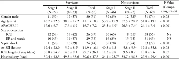
Table 2 shows comparisons among survivors and not survivors in the 2 stages of this study. When comparing the total of survivors to the total of not survivors it was found that age, APACHE II, number of patients in sep-tic shock, number of patients of male gender and time of detection of severe sepsis were signiicantly higher among not survivors. Length of hospital stay was sig-niicantly shorter among not survivors.
APACHE II score was evidently higher among not survivors when compared to survivors in both stages. At stage II, the Δt-SSI was lower among survivors as well as not survivors. Time of detection of survivors was similar in both stages (Table 2).
Figure 2 - Flowchart representing distribution of patients enrolled in each stage of the study.
Table 1- Comparative summary of data observed during the two stages of the Surviving Sepsis Campaign
Variable Stage I
(N=68)
Stage II (N=56)
p Value
Male gender 49 (72) 31 (55.3) NS
Age (years) 51.1 ± 19.5 47 ± 21 NS
APACHE II 21.5 ± 7.3 21.9 ± 8.6 NS
Infectious focus Pulmonary Urinary
Abdominal infection Meningitis
Soft parts Blood low Indeterminate
33 (48.5) 5 (7.3) 11 (16.2) 2 (2.9) 6 (8.8) 2 (2.9) 9 (13.2)
17 (30.4)*
2 (3.6) 11 (19.6) 9 (16)**
6 (10.7) 2 (3.6) 8 (14.2)
< 0.05 NS NS < 0.02
NS NS NS
Septic shock 47 (69.1) 30 (53.6) NS
Site of diagnosis Emergency room Wards
ICU
18 (26.4) 8 (11.7) 42 (61.7)
13 (23.2) 21 (37.5)*
22 (39.3)*
NS < 0.001 < 0.02 Compliance to the 6 h
bundle
11 (17) 11 (19.4) NS
Compliance to the 24 h bundle
20 (30) 17 (31) NS
ICU length of stay (days)
14.3 ± 13.1 11.3 ± 9.4 NS
Length of hospital stay 32.2 ± 32.8 42.3 ± 35.7 NS
Δt-SSI (hours) 33.8 ± 53.9 6.8 ± 8.4** < 0.001
Mortality at 28th day 37 (54.4) 18 (30)* < 0.02
Hospital mortality 46 (67.6) 23 (41)** < 0.003
DISCUSSION
Findings of this study disclosed that the organized search for signs suggesting infection leads to an earlier di-agnosis of sepsis and implies decreased mortality related with this disease.
A series of evidences presented in the last decades clearly point that quick and systematic assistance in clini-cal situations like AMI, stroke and trauma results in an impressive decrease of associated deaths. However, severe sepsis and septic shock related mortality has undergone changes in the last 25 years.(2,18-23) In Brazil it is higher than in other countries, 56% of mortality versus 30% in the developed countries and 45% in other developing coun-tries.(2,24) Possibly these high rates are due to delay in start-ing therapy which greatly contributes to spreadstart-ing of the inlammatory response and development of multiple or-gan dysfunction (MOD). Patients under treatment, even when appropriate, after multiple organ dysfunction have a worse prognosis.(13,14,25-28)
here is evidence that therapeutic intervention with hemodynamic resuscitation and antibiotic therapy are as-sociated to lower mortality rates.(7-12,15) As such, agile and adequate treatment is the “mainstay” for a successful ap-proach to severe sepsis.(18-20)
Goal directed early therapy proposed by Rivers et al.(13), an early hemodynamic resuscitation protocol, pro-vided an evident decrease of mortality in patients with se-vere sepsis and septic shock. he basis of this strategy is to
Table 2 – Summary of the comparison between survivors and not survivors encompassed in the two stages of the Surviving Sepsis Campaign
Survivors Not survivors P Value
(Compa-riing totals) Stage I
(N=22)
Stage II (N=33)
Total (N=55)
Stage I (N=46)
Stage II (N=23)
Total (N=69)
Gender male 11 (50) 19 (57) 30 (54) 39 (85) 12 (52)b 51 (74) < 0.03
Age (years) 45.7 ± 22.5 38.8 ± 17.2 41.1 ± 18.9 53.9 ± 17.9 57.5 ± 20.2# 54.8 ± 19.1 < 0.001
APACHE II 17.6 ± 6.7 17.6 ± 6.9 16.7 ± 7.2 23.5 ± 6.9* 26.5 ± 7.4# 24.1 ± 7.4 < 0.001
Site of detection ICU
ER and wards
12 (54) 10 (45)
14 (42) 19 (57)
26 (47) 29 (53)
30 (65) 16 (35)
8 (35)a
15 (65)
38 (55) 31 (45)
NS NS
Septic shock 11 (50) 13 (39) 24 (44) 36 (78) 17 (74) 53 (77) < 0.001
Δt-SSI (hours) 19.4 ± 22.0 5.9 ± 8.2‡ 11.9 ± 16.4 40.3 ± 6.2 5.8 ± 5.3a 19.8 ± 35.8 < 0.03
ICU length of stay (days) 30.8 ± 54.7 14.5 ± 9.1 29.7 ± 36.4 11.3 ± 9.8 9.6 ± 8.7 10.8 ± 9.6 0.07
Hospital stay (days) 50.4 ± 42.5 49.5 ± 33.6 50.6 ± 37.3 24.1 ± 23.7* 33.7 ± 36.8 27.9 ± 29.4 < 0.001
APACHE II - Acute Physiology and Chronic Health Evaluation II; ICU – intensive care unit; ER – emergency room; Δt-SSI – time elapsed between the irst record (on medical chart) of at least two signs suggesting infection and time of diagnosis of severe sepsis; NS – not signiicant. Results ex-pressed in mean ± standard deviation or N (%). *p < 0.01 for comparison between survivors and not survivors of Stage I. #p < 0.01 for comparison
between survivors and not survivors of Stage II. ‡p < 0.05 for comparison among survivors of Stages I and II. ap < 0.05 and bp < 0.01 for comparison
among not survivors of Stages I and II.
treat overall tissue hypoxia as fast as possible to revert the unbalance between ofer and consumption of oxygen to avoid development of MOD.(13,26-28) Furthermore, control of the infection focus, with broad spectrum antibiotics and/or surgical drainage in the irst hours after diagnosis, also has a major impact on prognosis.(9,10)
All patients cared in the irst stage of this study were treated according to SSC guidelines. hey set forth that management of the patient be grouped in two “bundles” of procedures which should be accomplished until the sixth and 24th hours. Respectively, “6 hours bundle” and 24 hours bundle”.( 5,6) At the irst stage, compliance to these bundles (6 hours = 17%; 24 hours = 30%) was even higher that that observed by SSC worldwide (6 hours= 13%; 24 hours = 15%).(17) Notwithstanding the good performance regard-ing management of severe sepsis., mortality remained unac-ceptably high (67,6%). his rate was higher than Brazilian mortality observed in the PROGRESS study (56%), years before implementation of the SSC.(24)
Probably, the high mortality rate of patients was as-sociated to delayed identiication of the septic condition. he long time period needed to detect sepsis at stage I, if compared to stage II, was remarkable. It is possible that organizational shortcomings associated to the low speci-icity of the systemic signs of infection are the main causes of delay in reaching diagnosis of sepsis, as noted in the irst stage.
stage II. Probably, early detection permitted identiication of patients prior to worsening of lactic acidosis and organ dysfunction such as renal failure, and volume-nonrespon-sive hypotension. Subsequent early intervention brings about more efective reperfusion and interruption of the sepsis “cascade” efect blocking evolution of this dysfunc-tion. Furthermore, am immeasurable aspect must be con-sidered, the motivational factor that resulted in greater collective involvement surrounding the septic patients and better quality of assistance (Hawthorne efect).
It was possible to reproduce indings from other stud-ies showing a decrease in mortality after adoption of the SSC guidelines.(7,8,11-16) At the second stage, even if there had not been a greater compliance to the bundles, mortal-ity decreased considerably, showing that prognosis does not rely on compliance to the therapeutic bundles, but also on the earlier diagnosis.
Unquestionably, subjectivity and subtlety of signs of inlammation delay diagnosis of sepsis in some patients, with no evident focus of infection at the syndrome’s early stages.(1,5,6,29-31) At the same time, international consensus that reviewed SIRS criteria, concluded that: “… these cri-teria are excessively sensitive and not speciic”.(29,30) his makes identiication and dealing with such a common and lethal syndrome even more diicult. In this context, we added to the screening of sepsis protocols besides the most recent leukometry analysis, manifestations that show organ dysfunction and that might be clinically detected. Probably, increase of sensitivity generated by these screen-ing models has facilitated early identiication of physi-ological changes associated to infectious activity.
Although lack of speciicity of the discrete diagnostic signs make earlier recognition of sepsis more diicult, im-plementation of systematic search for signs of SIRS and/or organic dysfunction in all sectors of the hospital redressed operational shortcomings. his correction was based on re-trieval of the importance of care with the patient, the role of each professional involved and importance of vital signs as marker for alert.(31) Changes of the vital signs must be promptly reported by the nursing staf and duly registered
by the physician. To investigate the cause of these changes and assess the need for an aggressive treatment is crucial.
CONCLUSION
To adopt a multidisciplinary institutional strategy fo-cused on early identiication of patients at risk of sepsis, thwarts evolution of the syndrome towards more severe stages and brings about a decreased risk of death associ-ated to severe sepsis and septic shock.
RESUMO
Objetivo: Avaliar o impacto da aplicação de uma política institucional para detecção da sepse grave ou choque séptico.
Métodos: Estudo antes (fase I)/depois (fase II) com coleta prospectiva de dados em hospital público de 195 leitos. Fase I: Pacientes com sepse grave ou choque séptico foram incluídos consecutivamente durante 15 meses e tratados conforme
dire-trizes da Campanha Sobrevivendo à Sepse. Fase II: Nos 10
me-ses subseqüentes, pacientes com sepse grave ou choque séptico foram arrolados a partir da busca ativa de sinais sugestivos de infecção nos pacientes internados. As duas fases foram compa-radas entre si no que diz respeito às variáveis demográicas, tem-po necessário para reconhecimento de pelo menos dois sinais sugestivos de infecção (Δt-SSI), aderência aos pacotes de 6 e 24 horas, e mortalidade.
Resultados: Foram identiicados 124 pacientes com sepse grave ou choque séptico, 68 na fase I e 56 na fase II. As variáveis demográicas foram semelhantes nas fases. O Δt-SSI foi de 34 ± 54 horas na fase I e 7 ± 8,4 horas na fase II (p < 0,001). Não houve diferença na aderência aos pacotes de tratamento. Parale-lamente, observou-se redução signiicativa das taxas de mortali-dade ao 28º dia (54,4% na fase I versus 30% na fase II; p < 0,02) e hospitalar (67,6% na fase I versus 41% na fase II; p < 0,003).
Conclusão: A estratégia utilizada contribuiu para a identii-cação antecipada do risco de sepse e resultou em diminuição da mortalidade associada à sepse grave e ao choque séptico.
Descritores: Choque séptico/diagnóstico; Choque séptico/ terapia; Choque séptico/mortalidade; Sepse/diagnóstico; Sepse/ terapia; Sepse/mortalidade
REFERENCES
1. Bone RC, Balk RA, Cerra FB, Dellinger RP, Fein AM, Knaus
WA, et al. Deinitions for sepsis and organ failure and guide-lines for the use of innovative therapies in sepsis. he ACCP/ SCCM Consensus Conference Committee. American College of Chest Physicians/Society of Critical Care Medicine. Chest. 1992;101(6):1644-55.
2. Teles JM, Silva E, Westphal G, Filho RC, Machado FR.
Survi-ving sepsis campaign in Brazil. Shock. 2008;30 Suppl 1:47-52.
3. Instituto Latino Americano Para Estudos da Sepse. Sepse
ma-nual. 2a ed. Rio de Janeiro: Atheneu; 2006.
5. Dellinger RP, Carlet JM, Masur H, Gerlach H, Calandra T, Cohen J, Gea-Banacloche J, Keh D, Marshall JC, Parker MM, Ramsay G, Zimmerman JL, Vincent JL, Levy MM; Surviving Sepsis Campaign Management Guidelines Committee. Survi-ving Sepsis Campaign guidelines for management of severe sepsis and septic shock. Crit Care Med. 2004;32(3):858-73. Review. .
6. Dellinger RP, Levy MM, Carlet JM, Bion J, Parker MM,
Ja-eschke R, Reinhart K, Angus DC, Brun-Buisson C, Beale R, Calandra T, Dhainaut JF, Gerlach H, Harvey M, Marini JJ, Marshall J, Ranieri M, Ramsay G, Sevransky J, hompson BT, Townsend S, Vender JS, Zimmerman JL, Vincent JL; In-ternational Surviving Sepsis Campaign Guidelines Commit-tee; American Association of Critical-Care Nurses; American College of Chest Physicians; American College of Emergency Physicians; Canadian Critical Care Society; European Society of Clinical Microbiology and Infectious Diseases; European So-ciety of Intensive Care Medicine; European Respiratory Socie-ty; International Sepsis Forum; Japanese Association for Acute Medicine; Japanese Society of Intensive Care Medicine; Society of Critical Care Medicine; Society of Hospital Medicine; Surgi-cal Infection Society; World Federation of Societies of Intensive and Critical Care Medicine. Surviving Sepsis Campaign: inter-national guidelines for management of severe sepsis and septic shock: 2008. Crit Care Med. 2008;36(1):296-327.
7. Kortgen A, Niederprüm P, Bauer M. Implementation of an evidence-based “standard operating procedure” and outcome in septic shock. Crit Care Med. 2006;34(4):943-9.
8. Micek ST, Roubinian N, Heuring T, Bode M, Williams J, Harrison C, et al. Before-after study of a standardized hospital order set for the management of septic shock. Crit Care Med. 2006;34(11):2707-13.
9. Kumar A, Roberts D, Wood KE, Light B, Parrillo JE, Sharma
A, et al. Duration of hypotension before initiation of efective antimicrobial therapy is the critical determinant of survival in human septic shock. Crit Care Med. 2006;34(6):1589-96. 10. Kumar A, Kazmi M, Ronald J, Seleman M, Roberts D, Gurka
D, et al. Rapidity of source control implementation following onset of hypotension is a major determinant of survival in human septic shock: 564. Crit Care Med. 2004;32(12 Suppl):A158. 11. Marshall JC, Maier RV, Jimenez M, Dellinger EP. Source control
in the management of severe sepsis and septic shock: an eviden-ce-based review. Crit Care Med. 2004;32(11 Suppl):S513-26. 12. Otero RM, Nguyen HB, Huang DT, Gaieski DF, Goyal M,
Gunnerson KJ, et al. Early goal-directed therapy in severe sepsis and septic shock revisited: concepts, controversies, and contem-porary indings. Chest. 2006;130(5):1579-95.
13. Rivers E, Nguyen B, Havstad S, Ressler J, Muzzin A, Knobli-ch B, Peterson E, TomlanoviKnobli-ch M; Early Goal-Directed he-rapy Collaborative Group. Early goal-directed thehe-rapy in the treatment of severe sepsis and septic shock. N Engl J Med. 2001;345(19):1368-77.
14. Gao F, Melody T, Daniels DF, Giles S, Fox S. he impact of compliance with 6-hour and 24-hour sepsis bundles on hospi-tal morhospi-tality in patients with severe sepsis: a prospective
obser-vational study. Crit Care. 2005;9(6):R764-70.
15. Fernandes Júnior CJ, Souza AG, Santos GPD, Silva E, Aka-mine N, Lisboa LF. Mortality rate reduction associated with a severe sepsis management protocol implementation. Crit Care 2007; 11(Suppl 3):30.
16. Freitas FG, Salomão R, Tereran N, Mazza BF, Assunção M, Jackiu M, et al. he impact of duration of organ dysfunction on the outcome of patients with severe sepsis and septic shock. Clinics (Sao Paulo). 2008;63(4):483-8.
17. Latin American Sepsis Institute. Campanha sobrevivendo a sepse [Internet].[cited 2009 Jan 12]. Available frim: Available at: http://www.sepsisnet.org/site/conteudo/SSCUH.pdf. 18. Hollenberg SM. Top ten list in myocardial infarction. Chest.
2000;118(5):1477-9.
19. Mullins RJ, Mann NC. Population-based research assessing the efectiveness of trauma systems. J Trauma. 1999;47(3 Suppl):S59-66.
20. Yang Q, Botto LD, Erickson JD, Berry RJ, Sambell C, Jo-hansen H, Friedman JM. Improvement in stroke mortality in Canada and the United States, 1999 to 2002. Circulation. 2006;113(10):1335-43.
21. Friedman G, Silva E, Vincent JL. Has the mortality of septic sho-ck changed with time. Crit Care Med. 1998;26(12):2078-86. 22. Angus DC, Wax RS. Epidemiology of sepsis: an update. Crit
Care Med. 2001;29(7 Suppl):S109-16.
23. Martin GS, Mannino DM, Eaton S, Moss M. he epidemio-logy of sepsis in the United States from 1979 through 2000. N Engl J Med. 2003;348(16):1546-54.
24. Beale R, Reinhart K, Brunkhorst FM, Dobb G, Levy M, Mar-tin G, MarMar-tin C, Ramsey G, Silva E, Vallet B, Vincent JL, Janes JM, Sarwat S, Williams MD; for the PROGRESS Advisory Board. Promoting Global Research Excellence in Severe Sepsis (PROGRESS): Lessons from an International Sepsis Registry. Infection. 2009 Apr 28. [Epub ahead of print]
25. Rivers EP, Kruse JA, Jacobsen G, Shah K, Loomba M, Otero R, Childs EW. he inluence of early hemodynamic optimization on biomarker patterns of severe sepsis and septic shock. Crit Care Med. 2007;35(9):2016-24.
26. Kern JW, Shoemaker WC. Meta-analysis of hemodyna-mic optimization in high-risk patients. Crit Care Med. 2002;30(8):1686-92.
27. Vincent JL, Gerlach H. Fluid resuscitation in severe sepsis and septic shock: an evidence based review. Crit Care Med. 2004;32(11 Suppl):S451-4.
28. Rhodes A, Bennett ED. Early goal-directed therapy: an eviden-ce-based review. Crit Care Med. 2004;32(11 Suppl):S448-50. 29. Giuliano KK. Continuous physiologic monitoring and the
identiication of sepsis: what is the evidence supporting current clinical practice? AACN Adv Crit Care. 2006;17(2):215-23. 30. Gropper MA. Evidence-based management of critically
ill patients: analysis and implementation. Anesth Analg. 2004;99(2):566-72
Appendix 1 – General form for record of vital signs (one sheet for every time of SSI verification Scanning of patients for severe sepsis
Data: Time:
Room /bed
AP
Hypotension - SBP< 90
HR Tachicardia >90 bpm
RR Tachipnea > 20 bpm
Temperature
- Hyperthermia > 38 ºC - Hypotermia < 36 ºC
Oliguria (<0,5 ml/kg/h)
Mental confusion psychosis
Supplementary oxygen
501 – 1 2 3 502
503 – 1 2 504 – 1
2 505 – 1 2 506 – 1 2 507 – 1 2 508 – 1 2 509 – 1 2 510 – 1 2 511 – 1 2 3
AP – arterial pressure; SBP – systolic blood pressure; HR- heart rate; RR – respiratory rate
Appendix 2
Date Hour A. Card for detection of septic patients (Screening)
1. Were two of the items below marked? ( ) Hyperthermia > 38 °C
( ) Hypothermia < 36 °C ( ) Tachipnea > 20 rpm
( ) Need for oxygen supplementation ( ) Tachicardia > 90 bpm
( ) SBP < 90 ou MAP < 65 mmHg
( ) Acute Headache (drowsiness, confusion, agitation, coma) ( ) Oliguria (urinary output < 0.5 ml/kg/h)
2. Is the history suggestive of acute myocardial infarction? ( ) Pneumonia/Empyema
( ) Urinary infection ( ) Intra-abdominal infection ( ) Meningitis
( ) Inlammation of soft parts or skin ( ) Infection of joints or bones ( ) Wound infection
( ) Intravascular catheter infection ( ) Endocarditis
3. If the reply to question 1 and 2 is Yes: suspect infection
( ) Request: blood cultures (1 pair) before antibiotic, with a 15 minute interval. ( ) Request: Blood gas and blood lactate, blood count, glucose, Na, K, Ur, Cr, bilirubin According to the clinic: ( ) Urine test ( ) Chest X-ray, ( ) Amylase, ( ) CT scan
4. Is there some (one is enough) criterion of acute organ dysfunction? ( ) Acute encephalopathy (drowsiness, confusion, agitation, coma) ( ) SBP < 90 or MAP < 65 mmHg
( ) SpO2 < 90% with or without oxygen supplementation ( ) Creatinine > 2.0 mg/dl or urinary output < 0,5 ml/kg/h ( ) Bilirubin > 2 mg/dl
( ) Platelet count < 100,000 ( ) Lactate > 4 mmol/L (36 mg/dl)
5. If one item was marked in question 4 – it is severe sepsis
Urgently start the resuscitation package. Adopt the check-list of Appendix 3
Appendix 3
Date Hour B1. Resuscitation bundle (of the 6 hours)
Lactate and antibiotic therapy (suggestion of ATB – Attachement 4) ( ) Record date and hour of obtaining lactate result
( ) Start broad spectrum antibiotic in time < 1 h (ICU and wards) or < 3 hs (ER) ( ) Drainage or removal (URGENT) of infectious focus if any (abscess, catheter...) Procedures
( ) Arterial catheterization (ATS) ( ) Central venous catheter (CVP) ( ) Bladder catheterization (BC)
( ) After clinical appraisal considered unnecessary ( ) MAP) ( ) CVP ( ) BC Intravenous liquid therapy
( ) Saline solution 0.9% or Ringer lactate IV 20 ml/kg. Give 500 ml every 30 minutes,
Repeat until CVP between 8 -12 mmHg or 12 -15 mmHg in patients under mechanical ventilation
( ) Crystalloid 20 ml/kg to 30 ml/Kg without CVP or ScvO2
Vasopressors
If MAP remains < 65 mmHg even though reaching a CVP of 8 -15 mmHg, start vasopressor therapyrapia therapy. Early use of vasopressors may be needed as an emergency in patients with septic shock
( ) Dopamine title dose until MAP ≥ 65 to 90 mmHg (record time of MAP ≥ 65) ( ) Noradrenaline title dose until MAP ≥ 65 a 90 mmHg (record time of MAP ≥ 65) Assessment of tissue perfusion
( ) Central venous blood gas 60/60 min until ScvO2 ≥ 70 mmHg (record time of ScvO2 ≥ 70)
( ) blood gases
Continuous monitoring of ScvO2 until ≥ 70mmHg (record time of ScvO2 ≥ 70)
Blood product transfusion
If ScvO2 ≤ 70mmHg notwithstanding PVC 8-15mmHg and use of vasopressors, patient must receive
transfusion of packaged blood cells until reaching hematocrit (Ht) > 30% Inotropic therapy
If CVP, MAP and Ht were optimized and SvcO2 < 70%, consider initropic therapy
( ) Dobutamine 2.5 mg/kg/min, title every 30 min until SvcO2 ≥ 70% or 20 mg/kg/min
Date Hour B2. Bundle for management of the septic patient (of the 24 hourss)
Corticosteroids
( ) It is a ICU policy not to administer this drug to septic patients
( ) Vasopressor-dependent patient – Administer hydrocortisone 50 mg IV every 6/6 ( ) Patient has no indication because is not vasopressor-dependent
Glycemic control
( ) Start with catheter obtained capillary or blood glycemias from 2/2 to 4/4 hours ( ) Start continuous infusion of insulin if glycemia > 150 mg/dl.
Drotrecogin alfa activated
( ) It is the policy of the ICU not to administer this drug to septic patients
( ) APACHE II ≥ 25 and with no contraindications - Administer drotrecogin alfa activated. Mechanical ventilation
( ) Inspiratory plateau pressure < 30 cm H2O
( ) Title lowest PEEP needed to prevent lung collapse and warrant SaO2 > 90% Nurse (Sig.):
Physician (Sig.):
Source: Adapted from Micek et al.(8) ) ATB – antibiotics; ER – emergency department; ScvO
2 – central venous oxygen saturation; ICU – intensive
Appendix 4
Empirical antimicrobial therapy
(Must be started within 3 hours in the ER and in 1 hour in the ICU and other sectors) Community acquired pneumonia (CAP - PORT III, IV and V
With no risk factors for pseudomonas ( ) Levoloxacin 750mg/d OR
( ) Azyithromicin 500mg 1x + Amoxi/clavulanate 0.5 to 1g IV 3x OR ( ) Azyt + Ampi/sulbactam 1.5 toa 3g IV 4x OR
( ) Azyt + Ceftriaxone 1 to 2g IV 1x
Risk for pseudomonas ( ) Levoloxacin 750mg/d PLUS( ) Pipe/tazobactam 4.5g IV 4x Bronchiectasis or ICU ( ) Levoloxacin 750mg/d + ( ) Cefepime 1 to 2g IV 2x
Aspiration ( ) Crystalline Penicillin 2 millionUI 6x or ( )Ampi/sulbactam 1.5 to 3g IV 4x HIV ( ) Bactrim (100mg of sulfamehtoxazole/kg/dose) 4x. Assess associations. Nosocomial Pneumonia
< 5 days of admission ( ) Levoloxacin 750mg 1x OR
( ) Ampi/sulbactam 1.5 to 3g IV 4x OR
( ) Ceftriaxone 1 to 2 g 1x (strong resistance inducer) ≥ 5 days of stay
(according to local lora )
( ) Pipe/tazobactam 4.5g IV 4x OR ( ) Cefepime 1 to 2g IV 2x OR
( ) Ceftazidime 1 to 2 g IV 3x (only if culture + for Pseudomonas) OR ( ) Imipenem 1gr IV 3x OR Meropenem 2g IV 3x OR
( ) Aztreonam 2 g IV 3x
Risk for Stailococcus aureus ( ) Vancomycin 1 to 2 g (15 mg/kg) IV 2x OR
( ) Teicoplanin 400 mg (2x in irst 24 hs). After 24hs - 1x/day OR ( ) Linezolid 600 mg IV 2x
Sepsis of unknown origin
Severe communnity sepsis ( ) Ampi/sulbactam 3g IV 4x OR ( ) Cefepime 1 to 2 g 2x OR
( ) Ceftriaxone 1 to 2 g 1x (strong resistance inducer) Severe nosocomial sepsis
(According to local lora)
( ) Pipe/tazobactam 4.5g IV 4x OR ( ) Cefepime 1 to 2g IV 2x OR
( ) Ceftazidime 1 to 2 g IV 3x (only if culture + for Pseudomonas) OR ( ) Imipenem 1 gr IV 3x OR Meropenem 2g IV 3x OR
( ) Aztreonam 2 g IV 3x Risk for Stailococcus aureus resistent to
meticillin, associate:
( ) Vancomicin 1 to2 g (15 mg/kg) IV 2x OR
( ) Teicoplanin 400 mg (2x in the irst 24 hs). After 24hs - 1x/day OR ( ) Linezolid 600 mg IV 2x
Sepsis of abdominal origin
Spontaneous peritonitis ( ) Ampi/sulbactam 3g IV 4x Secondary peritonitis with mild-moderate
manifestation
( ) Ampi/sulbactam 3g IV 4x OR
( ) Cefepime 1 to 2 g 2x + Metronidazol 500 mg 3x OR ( ) Pip/tazobactam 4.5g IV 4x
Secondary peritonitis with severe mani-festation
( ) Imipenem 1 gr IV 3x OR Meropenem 2gr IV 3x PLUS
( ) Vanco OR ( ) Teico OR ( ) Linez if Risk of Enterococcus resistant to vancomyicin or MRSA Necro-hemorrhagic pancreatitis ( ) Imipenem 1gr IV 3x OR Meropenem 2 gr IV 3x
Urinary tract infection
Communnity ( ) Ciproloxacin 400mg IV 2x OR ( ) Ampi/sulbactam 3g IV 4x Nosocomial ( ) Pipe/tazobactam 4.5g IV 4x OR
( ) Cefepime 1 to 2g IV 2x OR
( ) Imipenem 1gr IV 3x or Meropenem 2gr IV 3x Catheter related bloodstream infection
Immunocompetent ( ) Oxacillin 2 g IV 6x (more potent against Stailococcus aureus sensitive to metacillin) or ( ) Vancomycin 1 to 2 g ( 15 mg/kg) IV 2x or ( ) Teico or ( ) Linez
Immunocompromized and/or catheter tunneled
( ) Vancomycion 1 to 2 g (15 mg/kg) IV 2x PLUS ( ) Pipe/tazobactam 4.5g IV 4x or
( ) Ceftazidime 1 to IV 3x or
( ) Imipenem 1gr IV 3x or Meropenem 2 gr IV 3x (according to lora)
* all doses adjusted for creatinine clearance > 75 ml/min. Dose adjustments may be needed after 24 h. Always take heed of risk of fungal infection. Desassign ATB after results of culture. Function of ATB is restricted without urgent removal of infection focus.