Endoscopic endonasal transsphenoidal
approach for pituitary adenomas
Technical aspects and report of casuistic
Américo Rubens Leite dos Santos1, Roberto Monteiro Fonseca Neto1, José Carlos Esteves Veiga1, José Viana Jr.2, Nilza Maria Scaliassi2, Carmen Lúcia Penteado Lancellotti3, Paulo Roberto Lazarini4
ABSTRACT
Objective: Analyse technical aspects, effectiveness and morbidity of the endoscopic endonasal transphenoidal approach for pituitary adenomas. Method: From January 2005 to September 2008, 30 consecutive patients underwent endoscopic endonasal resection of pituitary adenomas with a follow up from 3 to 36 months. Their medical charts were retrospectively analysed. Results: There were 18 women and 12 men, mean age 44 years (range 17-65 yr). Among the 30 patients, 23 had macroadenomas and 7 microadenomas. Twelve patients had non-functioning tumors, 9 had ACTH-secreting tumors, 8 had GH-secreting tumors and 1 prolactinoma. Complete resection and hormonal control was achieved in all microadenomas. Macroadenomas were completely removed in 6 patients, subtotal resection in 6 and partial resection in 11. Three patients had diabetes insipidus and 5 had CSF leaks treated with lumbar drainage. Conclusion: The endonasal endoscopic approach for pituitary tumors is effective and has low morbidity.
Key words: endoscopy, pituitary, nasal, surgery.
Abordagem endoscópica endonasal para adenomas de hipófise: aspectos técnicos e relato de casuística
RESUMO
Objetivo: Analisar aspectos técnicos, eficácia e morbidade do acesso transesfenoidal endonasal endoscópico para adenomas hipofisários. Método: Estudo retrospectivo de trinta pacientes consecutivos submetidos à ressecção endoscópica endonasal de adenomas hipofisários, entre janeiro de 2005 e setembro de 2008, com seguimento pós-operatório entre três e 36 meses. Resultados: Foram operados 18 mulheres e 12 homens com idades variando entre 17 e 65 anos (média 44 anos). Entre os 30 casos operados, 23 eram macroadenomas e sete microadenomas. Doze pacientes apresentavam adenomas não-funcionantes, nove tumores secretores de ACTH, oito tumores secretores de GH e um prolactinoma. Ressecção macroscópica completa e controle endócrino foram conseguidos em todos microadenomas. Ressecção dos macroadenomas foi completa em seis pacientes, subtotal em seis e parcial em seis casos. Três pacientes desenvolveram diabetes insipidus e cinco tiveram fístula liqüórica pós-operatória controlada com drenagem lombar. Conclusão: A abordagem endoscópica endonasal para adenomas de hipófise é eficaz e apresenta baixa morbidade.
Palavras-chave: endoscopia, hipófise, nasal, cirurgia.
Correspondence
Américo Rubens Leite dos Santos Rua Martinico Prado 131 / 5 01224-010 São Paulo SP - Brasil E-mail: [email protected]
Received 25 November 2009 Received in final form 25 February 2010 Accepted 4 March 2010
Faculdade de Ciências Médicas da Santa Casa de São Paulo, São Paulo SP, Brazil: 1Discipline of Neurosurgery,
Department of Surgery; 2Discipline of Endocrinology, Department of Medicine; 3Department of Pathology; 4Department of
Otorhinolaryngology.
he irst transnasal resection of a pi-tuitary tumor was performed by Schlof-fer in 19071. Cushing2 systematically
ap-plied a transsphenoidal approach for
sell-ar lesions. his technique was posterior
re-ined and popularized by Guiot3 and
Har-dy4 with the introduction of the operative
he development of endoscopic techniques for sur-gery of paranasal sinuses5 awoke the possibility of an
en-doscopic approach for the pituitary gland. Jankowski et al.6 described the endoscopic endonasal removal of
pitu-itary adenomas in 3 patients. Jho and Carraufurther
de-veloped the pure endonasal endoscopic surgery of pitu-itary tumors7-15.
here is still controversy regarding the beneits of pi-tuitary endoscopic surgery. We started endonasal endo-scopic skull base surgery in 2005 as part of a minimally invasive concept in neurosurgery. Our results and the en-doscopic technique for pituitary adenomas concerning its efectiveness and morbidity are analyzed.
METHOD
Retrospective analysis of 30 patients submitted to en-doscopic endonasal removal of pituitary adenomas be-tween January 2005 and September 2008 with a follow up from 3 to 36 months. Patients with previous pituitary surgery and lesions other than pituitary adenomas were excluded from this study.
he sample consisted of 18 women and 12 men, age range 17-65 years, median age 44 year-old.
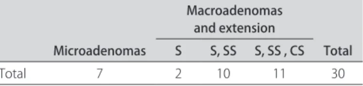
he tumors were divided in microadenomas (<1 cm) and macroadenomas (>1 cm). he macroadenomas were further classiied according to their extensions in intra-sellar (S), supraintra-sellar (SS) and invasion of cavernous si-nus (CS) (Table 1).
he following parameters were evaluated preoperative and 3 months after the operation: symptoms, endocrino-logical assessment (measurement of cortisol, corticotro-phin (ACTH), free thyroxin, thyrotropin (TSH), prolactin (PRL),growth hormone (GH), luteinizing hormone (LH), follicle-stimulating hormone (FSH), insulin-like growth factor-1, testosterone, estradiol, progesterone), neurora-diologic imaging and ophthalmologic examination in pa-tients with clinic or radiologic evidence of chiasmal com-pression. Histology and immunohistochemistry of the le-sions were investigated in all cases.
he degree of surgical resection was deined by the postoperative magnetic resonance imaging (MRI) as gross macroscopical resection (100%), subtotal resection (more than 80%) or partial resection (less than 80%).
Surgical procedure
Our surgical technique was based on the publications of Jho e Carrauwith minor modiications7-15.
he patients were operated on general anesthesia and orotracheal intubation, in supine position with the head ixed in a standard 3-pin-holder, slightly lexed and turned 10 degrees toward the surgeon. he video monitor was positioned behind the patient’s head in front of the sur-geon. he patient’s face, nasal and oral cavities were
pre-pared and draped in an aseptic manner. A gauze roll was packed into the oropharynx in order to prevent aspiration of stagnant blood at the time of extubation. ceftriaxone and clyndamicin were given 30 minutes before the sur-gery and kept 3 days after it. Both nostrils were washed with a topical vasoconstrictor. No intraoperative image guidance (frameless stereotatic navigation or C-arm lu-oroscopy) was necessary for patients with normally pneu-matized sphenoid sinus. All procedures were performed solely guided by anatomical landmarks. A 0-degree en-doscope with lens diameter of 4 mm connected to a suc-tion/irrigation device (Richard Wolf, Knittlingen, Germa-ny) was used in all cases.
A binostril approach was always performed. he sur-gery was divided in nasal and sellar phases.
Nasal phase: A “two-hand technique” was used. Dur-ing this step the surgeon held the endoscope with his left hand and an instrument with his right hand. he middle turbinate was lateralized and the sphenoid ostia located bilaterally. A wide sphenoidotomy followed by removal of inter and intrasinusal septa and exposure of clivus, sell-ar loor, csell-arotid prominences, opticocsell-arotid recesses and planum sphenoidale were done (Fig 1).
Sellar phase: A “three-hand” technique was used. An assistant held the endoscope in order to give the surgeon the possibility of working with 2 instruments and perform a bimanual dissection. he sellar loor was removed using a high-speed drill. he dura mater was opened and sellar content exposed (Fig 2).
All attempts were made to preserve normal pituitary tissue. he sellar tumor was removed with standard neu-rosurgical technique. he endoscope was then introduced inside the sella in patients with macroadenomas and a careful search for tumor remnants was made. In the case of macroadenomas with supraselar extensions the endo-scope was further directed to the suprasellar region and the tumor removed (Fig 3).
In case of signiicant perioperative cerebrospinal luid (CSF) leak, a multilayer sellar reconstruction was made using fat graft, fascia lata, Gel foam (Upjohn, Kalamazoo) and ibrin glue. When no perioperative CSF leak was seem only Gel foam and ibrin glue were used to close the sella.
All procedures were performed with informed con-sent of the patients.
his study was approved by the Research Ethics
Com-Table 1. Case distribution by tumor size and extension.
Microadenomas
Macroadenomas and extension
Total S S, SS S, SS , CS
Total 7 2 10 11 30
mittee of “Santa Casa de Misericórdia de São Paulo” (proj-ect number 427/08).
RESULTS
here were 12 (40%) non-functioning pituitary ad-enomas, 9 (30%) ACTH-secreting adad-enomas, 8 (26%) GH-secreting adenomas and one (3%) prolactinoma con-irmed with histology and immunohistochemistry.
Non-functioning adenomas
here were 6 macroadenomas with sellar and supra-sellar extensions. Another 6 macroadenomas had addi-tionally cavernous sinus invasion. he tumor was totally removed in 3 cases, sub totally (more than 80 %) in 3 cas-es and partially (lcas-ess than 80 %) in 6 cascas-es.
he immunohistochemistry of the lesions revealed 6 null cell tumors and 6 LH and FSH secreting-adenomas.
ACTH-secreting adenomas
here were 9 patients with conirmed Cushing’s dis-ease based on clinical endocrinological and immuno-histochemical evaluation, 5 macroadenomas and 4 mi-croadenomas. Complete tumor resection and biochemi-cal control of the disease (deined as postoperative plas-matic cortisol levels lower than 7 mg/dl) were achieved in all microadenomas and in one case of macroadenoma re-stricted to the sella. Two patients with sellar and suprase-lar lesions had subtotal and partial resection of their le-sions and should be submitted to reoperation. Two pa-tients had lesions with extensions to the cavernous sinus and their tumors were partially removed. One of then was submitted to a supraorbital craniotomy and complete re-moval of the lesion followed by hormonal control. he other one was referred to radiosurgery but remains with high cortisol levels.
GH-secreting adenomas
There were 8 acromegalic patients with confirmed GH-secreting adenomas. Hormonal control was deined as GH <1.0 ng/ml and not higher than 0.4 ng/ml after glu-cose load and IGF-1 levels normal for age.
Gross macroscopical tumor removal and hormonal control was obtained in 3 cases of microadenomas, in 1 case of macroadenoma restricted to the sella and in 1 case of sellar and supraselar macroadenoma. No addi-tional therapy was necessary for these patients.
One sellar/supraselar macroadenoma and two cases with cavernous sinus invasion were sub totally removed and therapy with Octreotide was started. he patient with a remnant tumor sellar/supraselar was reoperated and gross macroscopical removal was achieved.
One case of cavernous sinus invasion was submitted to radiosurgery due to lack of control with octreotide.
Fig 1. Endoscopic view of the sphenoidal sinus with the right side partially covered by an intrasinusal septum [Spt]. [C] clivus, [S] sell-ar loor, [PS] planum sphenoidale, [CP] csell-arotid prominence, [OC] opticocarotid recess.
Fig 2. Endoscopic view of the sellar content in a patient with mac-roadenoma after opening the dura mater. [T] tumor.
Prolactin-secreting adenomas
One patient had a giant prolactinoma that caused sudden visual deterioration and oculomotor nerve palsy. his tumor was partially removed and carbegoline thera-py started. Prolactin levels were normalized and the neu-rological deicits improved.
Table 2 summarizes the overall amount of tumor re-moval compared with tumor size and extension.
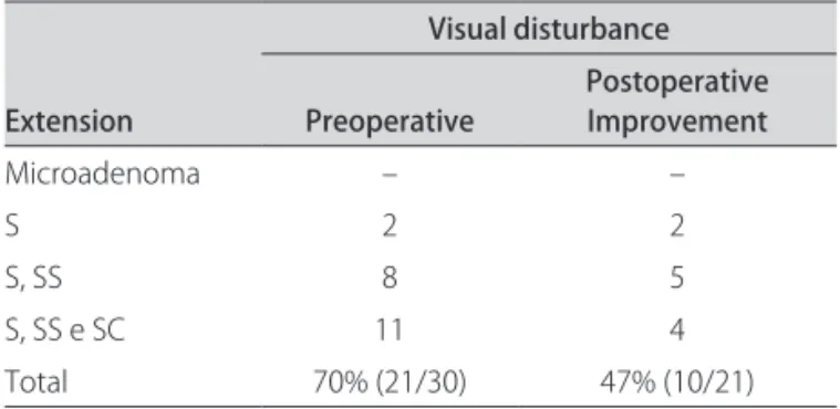
Table 3 summarizes pre and post operative visual dis-turbances compared with tumor size extension.
Complications
Five patients developed postoperative cerebrospinal luid (CSF) leaks treated successfully with 3 days of lum-bar drainage. None of the patients developed meningitis. One patient had communicating hydrocephalus and re-quired a ventriculo-peritoneal shunt. hree patients had diabetes insipidus, two of them permanent. here was no postoperative nasal bleeding or other signiicant na-sal disturbances.
here were no neurological disturbances or casualties related to the operations in this series.
DISCUSSION
Any new surgical technique must show beneits that overcome the previous surgical options and more impor-tant than that; it must be safe.
he widespread use of endoscopes in all surgical disci-plines has reached neurosurgical procedures in a delayed way. A great step related with the development of endo-scopic neurosurgery was the team work with otorhino-laryngologists in skull base approaches. he previous oto-rhinolaryngologic experience with endoscopic surgery for paranasal sinuses5 was the milestone for development of
endoscopic endonasal pituitary surgery.
The endoscopic approach has obvious advantages when compared with the traditional sublabial transsep-tal microscopic surgery. It avoids sublabial incision, dis-section of septal mucosa and fracture of nasal septum re-ducing postoperative morbidity16. here is no need of
na-sal speculum which narrows the surgical corridor limit-ing visualization and manipulation of instruments. he endoscopes ofer a wide angle of view that provides bet-ter visualization of anatomical landmarks. Based on an-atomical landmarks it is possible to avoid intraoperative luoroscopy, commonly used in traditional surgery, pre-venting patients and surgical team to unnecessary expo-sure to radiation.
Another advantage of endoscopic surgery is the pos-sibility of introduction of the endoscope inside the sella turcica and the supraselar region for the search of tumor remnants not accessible with microsurgery.
here is still controversy regarding the superiority of endoscopic pituitary surgery compared with transphenoi-dal microsurgery17-19 but the beneits of endoscopy should
no longer be questioned.
he literature of pituitary surgery has signiicant dif-ferent rates of tumor removal that relects distinct surgi-cal techniques and stages of learning curve11,16-22. Our
re-sults relect our learning process and the preponderance of patients with advanced disease. Tumors with sellar/su-praselar and cavernous sinus extensions were the most common in our series (36%) and gross macroscopical re-section was achieved in only 1 case (9%). In the other hand, it was possible to completely remove all function-ing microadenomas (23%) resultfunction-ing in a better endocri-nological outcome for those patients. Our rate of
visu-Table 2. Tumor extension and removal.
Extension No patients
Tumor removal
Partial
Total Subtotal
Microadenoma
Macroadenoma 7 100% (7) _ _
S 2 100% (2) _ _
S, SS 10 30% (3) 40% (4) 30% (3)
S, SS e SC 11 9% (1) 18% (2) 73% (8)
Total 30 43% (13) 20% (6) 37% (11)
S: sellar; SS: suprasellar; CS: cavernous sinus.
Table 3. Visual improvement and tumor extension.
Extension
Visual disturbance
Preoperative Postoperative Improvement
Microadenoma – –
S 2 2
S, SS 8 5
S, SS e SC 11 4
Total 70% (21/30) 47% (10/21)
al improvement after surgery (47%), based on visual ield campimetry, is comparable to other series18-22.
Complications common to microscopic or endo-scopic transsphenoidal approach include CSF leaks and meningitis19,23-25. Five of our patients had CSF leaks but no
one developed meningitis. his is probably due to lack of routine reconstruction of sella turcica. We only recon-structed the sella when perioperative CSF leak was visual-ized. Additionally, the free fat grafts that are normally used may not be the best material to prevent the leaks. Recent-ly the use of nasal vascularized laps has proved superiori-ty to prevent CSF leaks26; therefore we believe that
recon-struction of sella turcica using this technique is advisable. Based in the results of our series and the reports of literature we found that the endoscopic endonasal ap-proach for pituitary adenomas is efective and has low morbidity.
REFERENCES
Landolt AM. History of transsphenoidal pituitary surgery. In Landolt AM, 1.
Vance ML, Reilly PL (Eds). Pituitary adenomas. London: Churchill Livingstone 1996:307-314.
Rosegay H. Cushing’s legacy to transsphenoidal surgery. J Neurosurg 1981;54: 2.
448-454.
Guiot A. Transsphenoidal approach in surgical treatment of pituitary ade-3.
nomas: general principles and indications in nonfunctioning adenomas. In: Kohler PO, Ross GT (Eds). Diagnosis and treatment of pituitary tumors. Amster-dam: Excerpta Medica, International Congress Series No 303, 1973:159-178. Hardy J. Transphenoidal microsurgery of the normal and pathological pitu-4.
itary. Clin Neurosurg 1969;16:185-217.
Stammberger H. Endoscopic endonasal surgery: concepts in treatment of 5.
recurring rhinosinusitis. Part II. Surgical technique. Otolaryngol Head Neck Surg 1986;94:147-156.
Jankowski R, Auque J, Simon C, Marchal JC, Hepner H, Wayof M. Endoscop-6.
ic pituitary tumor surgery. Laryngoscope 1992;102:198-202.
Carrau RL, Jho HD, Ko Y. Transnasal-transsphenoidal endoscopic surgery of 7.
the pituitary gland. Laryngoscope 1996;106:914-918.
Jho HD. Endoscopic endonasal pituitary surgery: technical aspects. Contem-8.
porary Neurosurgery 1997;19:1-7.
Jho HD, Carrau RL, Ko Y. Endoscopic pituitary surgery. In: Wilkins RH, Ren-9.
gachary SS (Eds). Neurosurgical operative atlas. Baltimore: Williams&Wilkins 1996;5:1-12.
Jho HD, Carrau RL. Endoscopic assisted transsphenoidal surgery for pituitary 10.
adenoma. Technical note. Acta Neurochir (Wien) 1996;138:1416-1425. Jho HD, Carrau RL. Endoscopic endonasal transsphenoidal surgery: experi-11.
ence with 50 patients. J Neurosurg 1997;87:44-51.
Jho HD, Carrau RL, Ko Y, Daly M. Endoscopic pituitary surgery: an early expe-12.
rience. Surg Neurol 1997;47:213-223.
Jho HD.Endoscopic pituitary surgery. Pituitary 1999;2:139-154. 13.
Jho HD, Alieri A. Endoscopic endonasal pituitary surgery: evolution of sur-14.
gical technique and equipment in 150 operations. Minim Invasive Neuro-surg 2001;44:1-12.
Jho HD, Alieri A. Endoscopic transsphenoidal pituitary surgery: various surgi-15.
cal techniques and recommended steps for procedural transition. Br J Neu-rosurg 2000;14:432-440.
Santos Rde P, Zymberg ST, Abucham Filho JZ, Gregório LC, Weckx LL. En-16.
doscopic transnasal approach to sellar tumors. Braz J Otorhinolaryngol 2007;73:463-475.
Sheehan MT, Atkinson JL, Kasperbauer JL, Erickson BJ, Nippoldt TB. Prelimi-17.
nary comparison of the endoscopic transnasal vs. the sublabial transseptal approach for clinically nonfunctioning pituitary macroadenomas. Mayo Clin Proc 1999;74:661-670.
Frank G, Pasquini E, Farneti G, et al. The endoscopic versus the traditional ap-18.
proach in pituitary surgery. Neuroendocrinology 2006;83:240-248. Dehdashti AR, Ganna A, Karabatsou K, Gentili F. Pure endoscopic endonasal ap-19.
proach for pituitary adenomas: early surgical results in 200 patients and com-parison with previous microsurgical series. Neurosurgery 2008;62:1006-1017. Cappabianca P, Cavallo LM, Colao A, et al. Endoscopic endonasal transsphe-20.
noidal approach: outcome analysis of 100 consecutive procedures. Minim Invasive Neurosurg 2002;45:193-200.
Ferrante L, Trillò G, Ramundo E, et al. Surgical treatment of pituitary tu-21.
mors in the elderly: clinical outcome and long-term follow-up. J Neuroon-col 2002;60:185-191.
Mortini P, Losa M, Barzaghi R, Boari N, Giovanelli M. Results of transsphenoi-22.
dal surgery in a large series of patients with pituitary adenoma. Neurosur-gery 2005;56:1222-1233.
Cappabianca P, Cavallo LM, Colao A, de Divitiis E. Surgical complications as-23.
sociated with the endoscopic endonasal transsphenoidal approach for pi-tuitary adenomas. J Neurosurg 2002;97:293-298.
Black PM, Zervas NT, Candia GL. Incidence and management of complica-24.
tions of transsphenoidal operation for pituitary adenomas. Neurosurgery 1987;20:920-924.
Ciric I, Ragin A, Baumgartner C, Pierce D. Complications of transsphenoidal 25.
surgery: results of a national survey, review of the literature, and personal ex-perience. Neurosurgery 1997;40:225-237.
Hadad G, Bassagasteguy L, Carrau RL, et al. A novel reconstructive technique 26.
![Fig 1. Endoscopic view of the sphenoidal sinus with the right side partially covered by an intrasinusal septum [Spt]](https://thumb-eu.123doks.com/thumbv2/123dok_br/15432852.595125/3.955.60.438.94.372/endoscopic-sphenoidal-sinus-right-partially-covered-intrasinusal-septum.webp)