The efect of breath physiotherapeutic
maneuvers on cerebral hemodynamics
A clinical trial
Manoel Luiz de Cerqueira-Neto1, Álvaro Vieira Moura2,
Rosana Herminia Scola3, Esperidião Elias Aquim4, Álvaro Rea-Neto5, Mirela Cristine Oliveira6, Telma Cristina Fontes Cerqueira7
ABSTRACT
Objective: To observe the repercussion of respiratory physiotherapy techniques on the mean arterial pressure (MBP), intracranial pressure (ICP), cerebral perfusion pressure (CPP), jugular venous oxygen pressure (PjvO2) and jugular venous oxygen saturation (SjvO2).
Method: The sample consisted of 20 patients with head trauma. The protocol consisted of physiotherapy techniques application of vibrocompression (VBC), expiratory flow increase (EFI) and suction. Results: The results show the maintenance on variables of cerebral hemodynamics during the techniques of VBC and EFI. However, in relation to suction, there was an increase of MBP, ICP, with maintenance of CPP, PjvO2 and SjvO2 and return to baseline of MBP and ICP 10 minutes after the end of suction. Conclusion: The respiratory physiotherapy techniques (VBC, EFI) do not promote cerebral hemodynamic repercussion, unlike suction, in severe head injury patients, mechanically ventilated, sedated and paralyzed. Key words: physical therapy, respiratory, brain.
Efeitos das manobras de fisioterapia respiratória na hemodinâmica cerebral: um ensaio clínico
RESUMO
Objetivo: Observar a repercussão das técnicas de fisioterapia respiratória na pressão arterial média (PAM), pressão intracraniana (PIC), pressão de perfusão cerebral (PPC), pressão venosa jugular de oxigênio (PjO2) e saturação venosa jugular de oxigênio (SjO2).
Método: Foram incluídos no estudo 20 pacientes com traumatismo cranioencefálico. O protocolo consistiu na aplicação das manobras fisioterapêuticas de vibrocompressão (VBC), aumento de fluxo expiratório (AFE) e aspiração (ASP). Resultados: Os resultados mostraram a manutenção das variáveis da hemodinâmica cerebral durante as manobras de VBC e AFE. Porém, em relação à ASP, houve uma elevação da PAM e PIC, com manutenção da PPC, PjO2 e SjO2 e retorno aos valores basais da PAM e PIC dez minutos após o final da aspiração. Conclusão: As manobras de fisioterapia respiratória (VBC, AFE) não promovem alterações sobre a hemodinâmica cerebral, ao contrário da ASP traqueal, em pacientes com traumatismo cranioencefálico grave, em ventilação mecânica, sedados e curarizados. Palavras-chave: fisioterapia, respiratória, cérebro.
Correspondence
Manoel Luiz de Cerqueira-Neto Rua Mario Jorge Menezes Vieira 635 49035-660 Aracaju SE - Brasil E-mail: [email protected]
Received 21 June 2009
Received in final form 10 February 2010 Accepted 17 February 2010
University Federal of Paraná (UFPR), Curitiba PR, Brazil: 1Ms, Departament Medicine, UFPR; 2Ph.D., Montreal Heart Institute; 3Ph.D., Neuromuscular/Neurology Division, Internal Medicine Department, Hospital de Clínicas, UFPR; 4Dr, University of Buenos Aires, Argentina; 5Msc, Departament Medicine, UFPR; 6Especialist, AMIB; 7Msc, Departament Medicine, University Federal of Sergipe, Brazil.
Head injury (HI) is the largest cause of incapacity and death among Western na-tions1. he total number of deaths per year
resulting directly or indirectly from HI are estimated in 500.0002.
in-volving motor and cardio-respiratory care oriented to aspects like possible neurological sequels, bed immobi-lization and lung secretion accumulation. Without that speciic treatment modality, most of the patients devel-op lung infection associated to mechanical ventilation or even acute respiratory distress syndrome rising up mor-bidity and mortality indexes3.
In most of the developed nations’ hospitals, physio-therapy constitutes an integral part of the intensive care unit (ICU) management procedures. Among the several techniques used by ICU physiotherapists, the most com-mon are postural drainage, mobilization, vibration, per-cussion, manual hyperinsulation, aspiration (ASP) and many types of breathing exercises. hese techniques, rou-tinely combined, focus the patient’s underlying physiopa-thology condition intending to avoid lung complications4.
he physiotherapeutic maneuvers of vibrocompres-sion, expiratory low increase (EFI) and aspiration act in the intracranial compartment’s pressure through venous stream, in the same way as the positive end expiration pressure. By that, the central venous pressure can be in-creased, elevating intrathoracic pressure levels, obstruct-ing and delayobstruct-ing the superior and inferior vena cava ve-nous return. In addition, positive end expiration pressure can decrease cardiac output and mean blood pressure (MBP), reducing cerebral perfusion pressure (CPP) and increasing intracranial pressure (ICP)5.
his research was designed to verify the inluence of breathing physiotherapy maneuvers of vibrocompression (VBC), EFI and aspiration (ASP) in order to analyze their efects in the variables; MBP, ICP, CPP, jugular venous partial pressure in oxygen (PjvO2) and internal jugular
venous oxygen saturation (SjvO2) in patients with HI
ad-mitted to ICU.
METHOD
Design of study
his descriptive longitudinal study, allowing a quan-titative approach, is a prospective non-randomized clini-cal trial approved by the ethics and research committee of ‘Hospital de Clinicas da Universidade Federal do Paraná’- 065EXT019/2002-11.
Setting
The clinical trial was accomplished in the ICU for adult patients of ‘Hospital do Trabalhador de Curitiba’.
Participants
The sample consisted of 20 consecutive adult pa-tients of both genders randomly chosen on admission to ICU. he parameters of choice were; presence of severe HI (Glasgow Coma Scale score ≤8-eight) in the irst 48 hours of admission, absence of lung radiological
altera-tions, propofol sedation (Ramsay Scale score 6-six, enclo-sure 3), curarization with pancuronium bromide inhibit-ing cough relex, intubation with mechanical ventilation (Neumovent Graph) assisted and controlled by volume estimated at 8 ml/Kg/ body weight and positive end expi-ration pressure of 5 mmHg with an oxygen inspired frac-tion of 40%. Hemodynamic unstable patients presenting MBP levels <60 mmHg with pulmonary contusion and costal arch fractures were excluded from the trial.
Description
After reading and signing Free and Informed Consent Form, all patients underwent intra-arterial catheterization for monitoring of MBP levels as well as placement of an intra-ventricular catheter for ICP gauging. he catheters then were connected to a HP multi parametric monitor (Hewlett Packard). In addiction, a catheter was placed in the jugular bulb to collect PjvO2 and SjvO2 gasometry data.
Interventions
he patients were positioned in dorsal decubitus with headboard at 30º. All hemodynamic variables were col-lected: MBP, ICP (by multi-parametric monitor) and CPP as well as PjvO2 and SjvO2 gasometry data and the results
obtained were established as basal values.
he VBC and EFI physiotherapeutic maneuvers were performed in sequence; 5 minutes on each hemithorax, each maneuver lasting for 10 minutes. he irst applied maneuver was VBC in order to modify mucous’ physi-cal properties, decreasing its viscosity by the provided thixotropism. After that, it was applied the EFI maneu-ver responsible for mobilize, displace and eliminate pe-ripheral secretions towards the windpipe utilizing the in-creased expiratory low. Next, ASP was undertaken pre-ceded by 5 ml physiological saline solution (0.9%) instil-lation and three hyperinsuinstil-lations and hyperoxygenations with a manual resuscitation ambu connected to an oxy-gen source (10 l/s low). he chosen sterile/dischargeable valve probe was number 12. he ASP procedure total time was about 10 to 15 seconds and accomplished just once. Between each maneuver it was given a 5 minutes rest period in order to prevent relections of the irst maneu-ver to the second and from the second to the third one, trying this way to avoid cumulative efect of one maneu-ver into the other.
Statistical analysis
repeated analysis of variance measures. For the null hy-pothesis rejection, Newman-Keuls test was used, eval-uating all pairwise comparisons. When comparing only two moments it was considered the Student’s t test for dependent samples. Data’s normal condition was evalu-ated by Kolmogorov-Smirnov test (p<0.05 indicevalu-ated sta-tistical signiicance).
RESULTS
Sample characterization
Twenty patients were included in the study, 18 male (90%) and 2 female (10%), age mean average of 33.5 (±11.94), Apache 26 (±4) and Ramsay 6.
All patients presented HI (100%) and admitted to ICU (‘Hospital do Trabalhador de Curitiba’) in the period of November 2002 to May 2004.
Analysis results
In relation to VBC, there were no signiicant behav-ioral changes in MBP, ICP and CPP values during the ma-neuver 10 minutes execution period (Table 1).
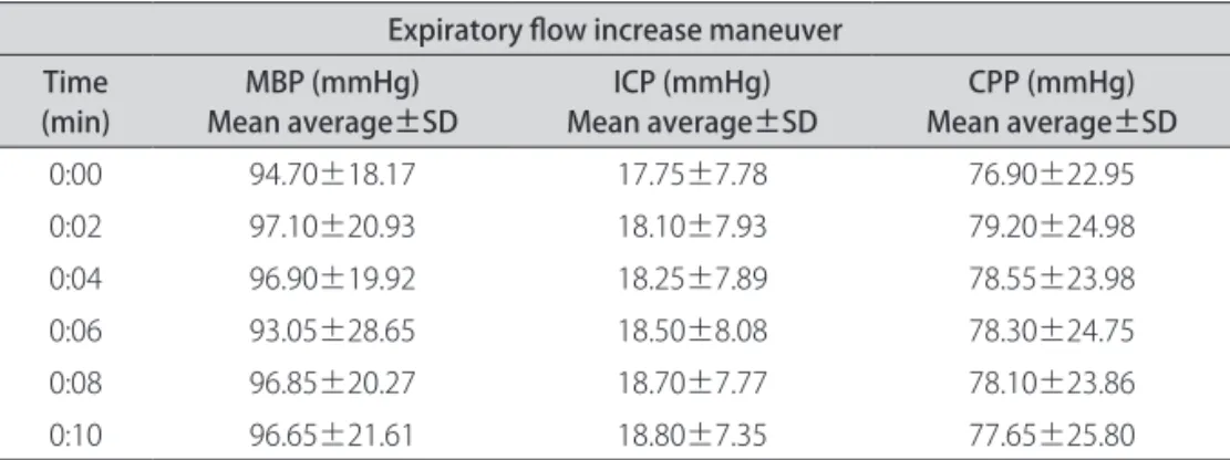
here were no signiicant changes related to MBP, ICP and CPP behavior during the EFI maneuver 10 minutes execution period as shown by additional results in Table 2.
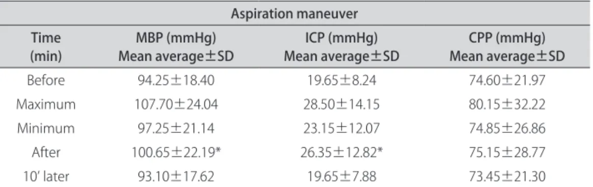
Table 3 depicts the mean variance behavior of MBP, ICP and CPP during ASP maneuver. Comparing the mo-ments before and after ASP, the variables MBP (p=0.0111) and ICP (p=0.0004) presented a signiicant change. How-ever, by comparing the moments before ASP against 10 minutes after the inal procedure, there was no statistical relevancy related to MBP and ICP, showing that the vari-ables returned to their basal values. CPP kept unchanged during the whole protocol.
he variables PjvO2 and SjO2 did not show signiicant
changes during MBP, ICP, CPP and 10 minutes after ASP procedures compared to basal moment (Table 4).
DISCUSSION
Stiller4 reviewing selected articles in MEDLINE and
CINAHL (Cumulative Index to Nursing and Allied Health Literature), as well as Dean and Ross6, veriied the
impor-tance of ICP, CPP and hemodynamics monitoring proce-dures during physiotherapy interventions.
According to hiesen et al.7 physiotherapy maneuvers,
when applied upon the patient’s thoracic box, increase in-trathoracic pressure with adverse efect in the cerebral ve-nous return by rising the right ventricle and large vessels pressure levels, including superior vena cava.
Table 1. MBP, ICP and CPP standard deviation/mean average results for each VBC moment. Vibrocompression maneuver
Time
(min) Mean average±SDMBP (mmHg) Mean average±SDICP (mmHg) Mean average±SDCPP (mmHg)
0:00 94.00±18.78 16.95±7.25 77.25±22.45
0:02 94.50±20.46 17.65±7.73 76.85±23.92
0:04 95.95±23.30 17.75±7.86 78.20±26.36
0:06 97.20±24.18 17.55±7.96 79.65±27.59
0:08 94.60±20.46 17.15±8.09 77.45±24.27
0:10 93.00±17.54 16.80±7.68 76.15±22.38
SD: standard deviation; MBP: mean blood pressure; ICP: increasing intracranial pressure; CPP: cerebral perfusion pressure; VBC: vebrocompression.
Table 2. MBP, ICP and CPP standard deviation/mean average results for each EFI moment. Expiratory low increase maneuver
Time
(min) Mean average±SDMBP (mmHg) Mean average±SDICP (mmHg) Mean average±SDCPP (mmHg)
0:00 94.70±18.17 17.75±7.78 76.90±22.95
0:02 97.10±20.93 18.10±7.93 79.20±24.98
0:04 96.90±19.92 18.25±7.89 78.55±23.98
0:06 93.05±28.65 18.50±8.08 78.30±24.75
0:08 96.85±20.27 18.70±7.77 78.10±23.86
0:10 96.65±21.61 18.80±7.35 77.65±25.80
his study tried to observe the repercussion of respi-ratory physiotherapy maneuvers into cerebral hemody-namic variables. In relation to isolated VBC and EFI ma-neuvers, the variables MBP, ICP, CPP, PjvO2 and SjvO2
did not show any significant change, remaining stable during the whole 10 minutes procedure period and even after its conclusion.
he results corroborate with Ciesla apud Irwin and Teclin8 who found out that percussion and vibration do
not cause any adverse efect in ICP.
hiessen et al.7, in a study with severe HI patients, did
not observe changes in MBP, ICP and CPP when costal manual vibration and manual expiratory pressure were applied, corroborating with this study results.
In terms of ASP procedure, this study observed a sig-niicant increase related to variables MBP and ICP but did not ind changes in CPP, PjvO2 and SjvO2. he ICP
increased level was gathered by an increase of MBP. his can be justiied by the probable condition of limited or absent cerebral self-regulation mechanism observed in patients with severe HI.
In a study quoted by Stiller4, when evaluating several
physiotherapeutic maneuvers including ASP, the observed increase in ICP was less than 10 mmHg. In the studies in-volving MBP, ICP and CPP measures, the increase of ICP
values was accompanied by an increase of MBP, resulting this way in a less than 10 mmHg CPP elevation.
Gemma et al.9 observed that ASP caused increases in
ICP, CPP and SjvO2 levels without ischaemic evidences
in well-sedated patients as well as decreases of CPP and SjvO2 levels in patients presenting cough relex.
he observed ICP increase is justiied by Fortune et al.10, Kerr et al.11, Knobel12 and Carvalho13 as a result of
the intrathoracic pressure elevation, due to carinal stim-uli, leading to venous return impairment what elevates cerebral blood low (CBF) with a secondary increase of the arterial pressure.
Ersson et al.14 found more ICP variations in the group
where cough relex was not inhibited and lesser ICP in-creases associated to groups where the relex was abol-ished. he largest pressure levels variation were veriied in the irst minute after procedures conclusion. At the end of the ifteenth minute, pressure levels presented stable with values similar to basal.
he care procedures involving airways, performed in the previous study, can increase ICP values at least by two diferent manners. In ASP and bagsqueezing the intratho-racic pressure is momentarily elevated by cough and lung insulation respectively. Consequently, there will be an increase in central venous pressure level, decreasing ce-rebral venous drainage and elevating ICP. his sequence occurs during the procedures’ initial period.
Another mechanism explains the ICP variation as a result of CPP changes due to the CBF reduction derived from sympathetic branches vagal activity due to tracheal stimuli. he rise of ICP can be a consequence of an increased cerebral blood volume due to a CPP de-creased level below certain limits (70-80 mm Hg) when vasodilatation is induced by the CBF self-regulation mechanism.
Applying the indings of Ersson et al.14 in this study,
increased ICP levels can occur derived from an elevation of intrathoracic pressure due to pulmonary hyper-insuf-lation (prior to ASP) as well as through carinal stimuli. In addiction, the likely presence of pain during the
pro-Table 3. MBP mean average/standard deviation results for each moment of aspiration. Aspiration maneuver
Time
(min) Mean average±SDMBP (mmHg) Mean average±SDICP (mmHg) Mean average±SDCPP (mmHg)
Before 94.25±18.40 19.65±8.24 74.60±21.97
Maximum 107.70±24.04 28.50±14.15 80.15±32.22
Minimum 97.25±21.14 23.15±12.07 74.85±26.86
After 100.65±22.19* 26.35±12.82* 75.15±28.77
10’ later 93.10±17.62 19.65±7.88 73.45±21.30
SD: standard deviation; *p<0. 05; MBP: mean blood pressure; ICP: intracranial pressure; CPP: cerebral perfusion pressure.
Table 4. SjO2 mean average/standard deviation results in each one of the moments.
Maneuvers
Moments Mean average±SDPjvO2 (mm Hg) Mean average±SDSjO2 (mm Hg)
Basal 45.95±8.64 77.93±7.22
VBC 45.79±13.51 76.50±8.85
EFI 44.22±12.59 75.04±8.61
ASP 45.43±12.24 75.99±8.13
10’ later 45.44±11.25 75.71±7.97
SD: standard deviation; SjO2: jugular venous oxygen saturation; PjvO2:
cedure may contribute to the rising of ICP since the pa-tients were sedated merely by a hypnotic drug.
In order to investigate the early administration eicacy of neuromuscular blocking agents (NMB), related to ICP management, in patients with severe HI by trauma, Hsiang et al.15 and White et al.16 concluded that the use of NMB
must be administrated only in cases presenting intracra-nial hypertension. Other HI patients may be managed by sedation once the use of NMB do not show beneits. On the contrary, it delays hospitalization period and increas-es extra-cranial adverse efects associated to pharmaco-logical paralysis as: accidental disconnection, pulmonary complications linked to the ventilation/perfusion relation-ship, atelectasis, embolism and cardiovascular side efects. he present study agrees with White et al.16 assertion
that isolated NMB administration do not provide neces-sary protection since it was not able to inhibit MBP rises. In addiction, in the event of absent cerebral self-regula-tion mechanism, NMB can also induce increases in ICP. he quoted authors suggest that NMB combined with a topic anesthetics could bring beneits, preventing ICP and MBP risings associated to endotracheal ASP.
Kerr et al.11 reported ICP elevations in patients not
treated with NMB during hyperinsulation, hyper-oxy-genation and in the course of the endotracheal ASP cath-eter insertion procedure. he increases were concomitant with MBP elevations. Otherwise, patients managed with NMB did not present increased ICP during the ASP cath-eter insertion; however, NMB was unable to prevent ICP rising during hyperinsulation and hyper-oxygenation.
Unni et al., Werba et al. (1991) and Klezl et al. (1991)
apud Kerr et al.11 suggest that NMB, by inducing
vaso-motor tonus changes as well as intercostals muscles and diaphragmatic paralysis, can mitigate the expected rising in ICP during endotracheal ASP.
Venous pressure elevations caused by hyperinsula-tion combined with arterial pressure rising, both related to CBF augments, can hinder the ICP return to basal lev-els. his mechanism may explain the reason why it takes 10 minutes or more for ICP to return at basal values.
Oertel et al.17, Oertel et al.18 and Pinaud el al.19
corrob-orate with Knobel12 asserting that, in terms of HI patients,
relaxation and curarization decrease ICP levels, maintain-ing arterial pressure stable.
Rudy et al.20 showed that patients with severe and
closed HI (Glasgow ≤8) show the risk of presenting in-tracranial hypertension peaks during endotracheal ASP procedure. In this type of patient, endotracheal ASP is necessary but raises potential adverse efects. To prevent intracranial hypertension setup, the ASP maneuver time must be lower than 10 seconds and limited to one or at maximum two procedures. Due to ICP rising, observed later than 10 minutes after ASP conclusion (recognized to
provoke mean ICP and MBP increased values), this rest period must be prolonged.
Opposing to the latter, this study observed the return of ICP basal values, as well as CPP levels maintenance, on the tenth minute after ASP conclusion with no signs of cerebral ischemia or hyperemia (evaluated by PjvO2 and
SjvO2 data). Even by that, we are unable to assure the
ab-sent of cerebral vasodilatation and CBF levels risings (the most probable causes of increased ICP during endotra-cheal ASP). In order to conirm this hypothesis, it is nec-essary to carry on transcranial Doppler evaluation.
Cruz21 reported that ASP increased MBP and ICP
lev-els along with SjO2 elevation but with no observed risings
in systemic arterial and venous oxygenation. his study suggests that the elevations induced by ASP in MBP also increased CBF values. Fortune et al.10 reported MBP, CPP
and SjvO2 increasings accompanied by a decrease of the
oxygen extraction associated to ICP augmentation. he study also assures that the observed risings, induced by ASP, were resulted from CBF elevation due to vasodila-tation accompanied by PjvO2 maintenance.
Fortune et al.10 found dramatic increases in SjvO
2
as-sociated to ASP while Kerr et al.22 did not observe
Sj-vO2 luctuations once, as in this study, some aspects of
the ASP protocol, more likely to inluence ICP elevations and/or cerebral oxygenation changes, were controlled (e.g.: hyper-oxygenation, ASP period duration, FiO2 and
catheter type).
Although hypoxemia induced by ASP was quoted to justify MBP risings, there were clear evidences pointing to the absence of hypoxemia in these patients with elevat-ed MBP levels as it was found in this study and in Kerr et al.23, Rudy et al.20, Kerr et al.22,23. In all the quoted studies,
the ASP induced hypoxemia in HI patients was prevented by hyper-oxygenation, previous to ASP (well documented by its beneic efects in other acute injured patients’ sam-ples). hese studies point to the fact that in the absence of hypoxemia, ICP elevations appear synchronically with MBP elevations. In this study, we found ICP and MBP ris-ings while the hypoxemia, ASP induced, was controlled by previous hyper-oxygenation as recommended in Skel-ley et al. and Urban and Weitzner apud Keer et al.23.
Kerr et al.23 depicted that ASP, preceded by
hyper-ox-ygenation, was associated to blood low velocity and ar-terial pressure risings. Signiicant decrease in the pulsa-tility index suggests that vasodilatation plays important part as a mechanism to maintain cerebral oxygenation in the presence of ICP raisings.
herefore, it can be concluded that transient ICP and MBP elevations, associated with ASP procedure preced-ed by cerebral oxygenation, do not seem to provoke haz-ardous efects in cerebral oxygenation once PjvO2, SjvO2
By preserving pulmonary alveoli function, one can guarantee optimized ventilation eicacy. Losses of gas trade zones compromise PaCO2 controlling, raising
crit-ical features to the treatment mainly in the event of cere-bral hyperemia. Consequently, in terms of beneicial ef-fects, respiratory physiotherapy maneuvers must be man-aged in HI patients.
hus, the respiratory physiotherapy, through VBC, EFI and ASP maneuvers, seems to be a required condi-tion since attencondi-tion is given to ICP. In efort to aloud opti-mal tracheobronquial hygiene, physiotherapy procedures must be performed in patients submitted to maximum sedation, always avoiding any other unnecessary stimu-lus. By not performing the procedure, secretions accumu-lation may lead to hypoventiaccumu-lation.
In summary, VBC and EFI respiratory physiothera-py maneuvers do not cause alterations in cerebral hemo-dynamics related to the MBP, ICP, CPP, PjvO2 and SjvO2
variables. Consequently, the procedures can be safely ac-complished in sedated and curarized patients presenting HI and submitted to mechanical ventilation.
he tracheal ASP procedure provokes changes in ce-rebral hemodynamics leading to MBP risings with con-comitant ICP elevation and maintenance of CPP, PjvO2
and SjvO2 levels. By this way, ischaemic events due to
ASP can be avoided since MBP and ICP return to its basal values after 10 minutes of tracheal ASP conclusion.
ACknOwlEDgMEnTS– he authors would like to thank Dr. Francis-co M.B. Germiniani for assisting in the translation of this manuscript.
REFERENCES
Marik PE, Varon J, Trask T. Management of head trauma. Chest 2002;122:699-711. 1.
David CM. Medicina intensiva. Rio de Janeiro: Revinter, 2004. 2.
Gosselink R, Bott J, Johnson M, et al. Physiotherapy for adult patients with 3.
critical illness: recommendations of the European Respiratory Society and Eu-ropean Society of Intensive Care Medicine Task Force on Physiotherapy for Critically Ill Patients. Intensive Care Med 2008;34:1188-1199.
Stiller K. Physiotherapy in intensive care: towards an evidence-based prac-4.
tice. Chest 2000;118:1801-1813.
Yilmazlar S, Kocaeli H. Monitoring and controlling of intracranial pressure in 5.
severe head injury. Ulus Travma Derg 2001;7:151-157.
Dean E, Ross J. Discordance between cardiopulmonary physiology and phys-6.
ical therapy. Chest 1992;101:1694-1698.
Thiesen RA, Dragosavac D, Roquejane AC, et al. Inluência da isioterapia 7.
respiratória na pressão intracraniana em pacientes com traumatismo cran-ioencefálico grave. Arq Neuropsiquiatr 2005;63:110-113.
Irwin S, Tecklin, JS. Fisioterapia cardiopulmonar. 2
8. nd Ed. São Paulo: Manole,
1994:353-354.
Gemma M, Tommasino C, Cerri M, et al. Intracranial efects of endotrache-9.
al suctioning in the acute phase of head injury. J Neurosurg Anesthesiol 2002;14:50-54.
Fortune J, Feustel PJ, Weigle CG, et al. Continuous measurement of jugular 10.
venous oxygen saturation in response to transient elevations of blood pres-sure in head-injured patients. J Neurosurg 1994;80:461-468.
Kerr ME, Sereika SM, Orndof P, et al. Efect of neuromuscular blockers and 11.
opiates on the cerebrovascular response to endotracheal suctioning in adults with severe head injuries. Am J Crit Care 1998;7:205-217.
Knobel E. Condutas no paciente grave. 2
12. nd Ed. São Paulo: Atheneu, 2000.
Carvalho CRR. Ventilação mecânica. São Paulo: Atheneu, 2000. 13.
Ersson U, Carlson H, Mellström A, et al. Observations on intracranial dynam-14.
ics during respiratory physiotherapy in unconscious neurosurgical patients. Acta Anaesthesiol Scand 1990;34:99-103.
Hsiang JK, Chesnut RM, Crisp CB, et al. Early, routine paralysis for intracra-15.
nial pressure control in severe head injury: is it necessary? Crit Care Med 1994;22: 1471-1476.
White PF, Schlobohm RM, Pitts LH, et al. A randomized study of drugs for 16.
preventing increases in intracranial pressure during endotracheal suction-ing. Anesthesiology 1982;57:242-244.
Oertel M, Kelly DF, Lee JH, et al. Eicacy of hyperventilation, blood pressure 17.
elevation, and metabolic supresión therapy in controlling intracranial pres-sure after head injury. J Neurosurg 2002;97:1045-1053.
Oertel M, Kelly DF, Lee JH, et al. Metabolic suppressive therapy as a treat-18.
ment for intracranial hypertension: why it works and when it fails. Acta Neu-rochir 2002;81(Suppl):S69-S70.
Pinaud M, Lelausque JN, Chetanneau A, et al. Efects of diprivan on cerebral 19.
blood low, intracranial pressure and cerebral metabolism in head injured patients. Ann Fr Anesth Reanim 1991;10:2-9.
Rudy EB, Turner BS, Baun M, et al. Endotracheal suctioning in adults with 20.
head injury. Heart Lung 1991;20:667-674.
Cruz J. Combined continuous monitoring of systemic and cerebral oxygen-21.
ation in acute brain injury: preliminary observations. Crit Care Med 1993;21: 1225-1232.
Kerr ME, Weber BB, Sereika SM, et al. Efect of endotracheal suctioning on ce-22.
rebral oxygenation in traumatic brain-injured patients. Crit Care Med 1999; 27:2776-2781.
Kerr ME, Rudy EB, Weber BB, et al. Efect of short-duration hyperventilation 23.