Arq Neuropsiquiatr 2008;66(3-A):575-577
575 Clinical / Scientiic note
CHOREOATHETOSIS SECONDARY TO LEAD TOXICITY
Mariana Spitz
1, Leandro Tavares Lucato
2, Mônica Santoro Haddad
1, Egberto Reis Barbosa
1COREOATETOSE SECUNDÁRIA A INTOXICAÇÃO POR CHUMBO
Departments of Neurology1 and Radiology2, University of São Paulo Medical School, São Paulo SP, Brazil.
Received 29 February 2008, received in inal form 8 May 2008. Accepted 30 May 2008.
Dra. Mariana Spitz – Rua Paulo César de Andrade 200/402 - 22221-090 Rio de Janeiro RJ - Brasil. E-mail: marianaspitz@gmail.com
Firearm projectiles have been described, albeit rarely, as a cause of lead toxicity. The usual route of lead expo-sure is oral ingestion, but toxicity secondary to retained bullet fragments has been well documented1. Switz et
al. described the irst case in 1976 – a bullet was lodged in a patient’s left ankle and gastrointestinal symptoms emerged forty years after the wound2. Since then
sever-al reports followed, including one by Goodheart et sever-al.3,
in 1999, in which the authors reported 25 patients with lead intoxication secondary to intentional petrol snifing, among whom 8 presented with chorea, though the most common neurological complication was altered mental state, observed in all subjects. The major clinical manifes-tations of lead toxicity are gastrointestinal, hematologi-cal and neurologihematologi-cal, namely abdominal cramps, anorex-ia, nausea, vomiting, constipation, anemanorex-ia, headache, pe-ripheral neuropathy and encephalopathy4. This is
charac-terized by delirium, seizures, somnolence, and even coma. Joint and muscle pain and nephropathy may also be part of the clinical picture5.
We herein describe an interesting case, where choreo-athetosis was attributed to lead toxicity due to retained bullet fragments.
CASE
A 42-year-old right-handed man was shot in 1987 at the age of 23. He had been a drug addict since the age of 14 – he had used marijuana and inhaled cocaine, but denied intravenous drug use. He was shot in the left side of the abdomen, left shoulder and right thigh. At that time, he had undergone emergency sur-gery – there was no injury to internal organs, but one of the bullets could not be successfully removed because its location in the lumbar spinal column – in close proximity to the spinal cord – was considered too risky. The patient was discharged a week after the surgery.
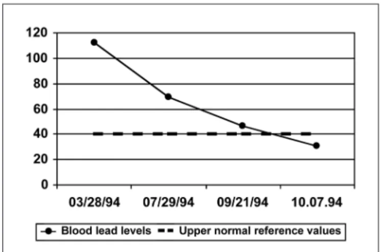
He remained asymptomatic until February 1994, when he started complaining of frequent abdominal cramps, nausea, vomiting and constipation. On that occasion he was referred to the Orthopedics Clinic of our hospital. On physical examination, he was pale, the abdomen was diffusely tender to palpation, but there were no signs of peritonitis. Neurological examination was normal. His blood exams showed normochromic normocyt-ic anemia and blood lead levels were in the toxnormocyt-ic range (112 µg/ dL), which was attributed to the retained bullet. He was diag-nosed with lead toxicity and underwent chelation with dimer-caprol and EDTA (ethylenediaminetetracetic acid). One month later, since the blood lead levels remained high (65 µg/dL),
Arq Neuropsiquiatr 2008;66(3-A)
576
Choreoathetosis: lead toxicity Spitz et al.
lation with EDTA was administered again, followed by an unsuc-cessful attempt to surgically remove the bullet from the spine. There was improvement of the abdominal cramps, but within a few weeks of the procedure, the patient noticed involuntary movements of the hands, trunk and feet. He was then admitted to the Neurologic Clinic for diagnostic evaluation.
The patient is the irst child of non-consanguineous par-ents. His family history is negative for neurological diseases. On neurological examination, there were generalized, partial-ly-suppressible choreoathetoid movements involving the face and proximal limbs, in addition to the toes and ingers. He had a dystonic posture on the right upper limb. His muscle strength, deep tendon relexes and cognitive function were normal. There was rigidity in the four limbs and plantar relexes were lexor.
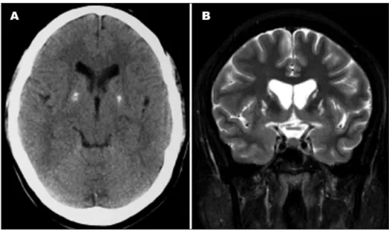
Blood exams were unremarkable, including liver and thyroid function and rheumatologic tests. HIV antibody testing was neg-ative, as well as hepatitis serology. Serum and urinary (24 hour collection) copper and serum cerulopasmin were within normal limits. Huntington disease genetic testing was negative. Electro-neuromyography of the four limbs was normal. Cerebrospinal luid (CSF) analysis revealed normal protein and glucose levels, negative VDRL, no red blood cells and 3 leucocytes. Computed tomography performed in 1996 showed bilateral and symmetric pallidal calciications and a slight hypoattenuation in both pu-tamen and caudate nuclei. He also underwent brain MRI, which demonstrated some volumetric reduction in both caudate and lentiform nuclei, associated with bilateral and symmetric T2 hy-perintensity and T1 hypointensity of these structures (Fig 1). Blood lead levels were still increased (69 µg/dL) and another course of EDTA was prescribed.
The patient was given chelation therapy with EDTA for four months and blood lead levels eventually returned to normal (Fig 2). Abnormal movements persisted and were only controlled with neuroleptics. Patient is currently taking haloperidol 7 mg daily. Since the beginning of its administration, the patient tried to withdraw the drug on several occasions, resulting in signii-cant worsening of the choreoathetosis.
DISCUSSION
Although it is not common in clinical practice, move-ment disorders can occur following exposure to several chemical substances, such as heavy metals, pesticides and organic solvents. Goldings et al. in 1982 described the case of a 15 year-old boy who inhaled gasoline and developed chorea, myoclonus and ataxia due to lead encephalopa-thy6. Goodheart et al. in 1999 also reported patients with
petrol snifing history, lead toxicity and chorea3, but to
our knowledge no cases of choreoathetosis secondary to lead toxicity because of a retained bullet have been pub-lished to date.
In the case herein described neurological symptoms consisted of choreoathetosis and were ascribed to lead toxicity due to a retained bullet lodged in the spinal col-umn for the past 7 years. But could the patient develop lead toxicity after such a long interval from the gunshot wound? It has been demonstrated that blood lead levels tend to increase with time after injury in patients with projectile retention7.Onset of symptoms from the initial
gunshot wound has been reported from 2 days up to 52 years1.
The pathophysiology of lead absorption from retained bullets is still unclear, but there seems to be a correlation between bullet location and lead toxicity7. Synovial and
bony interfaces and contact with pleural and CSF have been associated with elevated absorption rates1. As the
onset of symptoms may be insidious and the asymptom-atic interval may be prolonged, a high index of suspicion is required to diagnose lead toxicity8.
In 1997 Centers for Disease Control and Prevention deined elevated blood lead levels for children as exceed-ing 10 µg/dL9. In adults, severe lead toxicity is associated
with blood lead concentrations of 100 µg/dLl or more10.
This patient had lead levels of up to 112 µg/dL, consistent with severe toxicity.
CT in this case disclosed pallidal calciications, bilat-eral and symmetric, which is a relatively common inding in patients over age 4011. However, the patient was only 32
years old by the time the irst CT was performed, which raises the possibility that these calciications could be as-sociated to lead intoxication. Calciications are commonly described in this condition, especially in the chronic form (as is the case described here). They are usually linear in the subcortical area and they can be seen also in the cer-ebellum and basal ganglia12.
MR imaging showed in detail striatal lesions which are probably the substrate for the choreoathetoid move-ments. There was a clear decrease in volume of the cau-date nuclei and also of the putamen, associated to signal abnormalities that are unspeciic, but associated to the
Arq Neuropsiquiatr 2008;66(3-A)
577
Choreoathetosis: lead toxicity Spitz et al.
atrophy of these structures, can be related to gliosis and end-stage changes. The irreversibility of the clinical pic-ture corroborates the imaging aspects, although revers-ibility can sometimes be appreciated in this entity, mainly in acute forms13. MR indings in lead toxicity are variable,
including white matter, basal ganglia, insula and thalamus involvement13,14 – usually presenting T2-hyperintensity.
Strictly from an imaging standpoint, differential diagnosis in this case includes diseases with basal ganglia involve-ment, especially Wilson disease, which was excluded after other diagnostic tests.
We cannot certify that movement disorders and in-tracranial lesions were due to the lead toxicity, but the temporal course certainly suggests it. There remains a possibility that the neurological symptoms were not the direct effect of the bullet, but we believe that lead poi-soning was due to the bullet lodged in the spinal cord and direct penetration into the CSF compartment. We came to the conclusion that the cause of choreoathetosis was lead toxicity considering that other common causes were excluded.
In this case there was no regression of the involuntary movements despite normalization of blood lead levels. After prolonged exposure to lead, effects do not appear to be reversible even if blood lead levels are lowered with chelation15.
Lead toxicity is a very rare cause of choreoathetosis. There must be surveillance for people with retained
bul-lets concerning the possible development of lead toxicity, since this is a dificult diagnosis.
REfERENCES
1. Coon T, Miller M, Shirazi F, Sullivan J. Lead toxicity in a 14-year-old fe-male with retained bullet fragments. Pediatrics 2006; 117: 227-230. 2. Switz DM, Elmorshidy ME, Deyerle WM. Bullets, joints, and lead
in-toxication: a remarkable and instructive case. Arch Intern Med 1976; 136: 939-941.
3. Goodheart RS, Dunne JW. Petrol sniffer’s encephalopathy: a study of 25 patients. Med J Aust 1994; 160: 178-181.
4. Brodkin E, Copes R, Mattman A, Kennedy J, Kling R, Yassi A. Lead and mercury exposures: interpretation and action. CMAJ 2007; 176: 59-63. 5. Ford MD. Clinical toxicology. Philadelphia: Saunders, 2001.
6. Goldings AS, Stewart RM. Organic lead encephalopathy: behavioral change and movement disorder following gasoline inhalation. J Clin Psychiatry 1982; 43: 70-72.
7. Ibrahim D, Froberg B, Wolf A, Rusyniak DE. Heavy metal poisoning: clinical presentations and pathophysiology. Clin Lab Med 2006; 26: 67-97, viii.
8. McQuirter JL, Rothenberg SJ, Dinkins GA, Manalo M, Kondrashov V, Todd AC. The effects of retained lead bullets on body lead burden. J Trauma 2001; 50: 892-899.
9. Centers for Disease Control and Prevention. Preventing lead poison-ing in young children. Atlanta; CDC, 2005.
10. Gracia RC, Snodgrass WR. Lead toxicity and chelation therapy. Am J Health Syst Pharm 2007; 64: 45-53.
11. Teo JG, Goh KY, Ahuja A, et al. Intracranial vascular calcifications, glioblastoma multiforme, and lead poisoning. AJNR Am J Neuroradi-ol 1997; 18: 576-579.
12. Reyes PF, Gonzalez CF, Zalewska MK, Besarab A. Intracranial calciica -tion in adults with chronic lead exposure. AJR Am J Roentgenol 1986; 146: 267-270.
13. Atre AL, Shinde PR, Shinde SN, et al. Pre- and posttreatment MR
im-aging indings in lead encephalopathy. AJNR Am J Neuroradiol 2006;
27: 902-903.
14. Tuzun M, Tuzun D, Salan A, Hekimoglu B. Lead encephalopathy: CT
and MR indings. J Comput Assist Tomogr 2002; 26: 479-481.