Lead Toxicity Risks in Gunshot Victims
Gabriel Costa Serrão de Araújo1*, Natália Teixeira Mourão2, Igor Natário Pinheiro1,
Analúcia Rampazzo Xavier1, Vinicius Schott Gameiro1
1Hospital Universitário Antônio Pedro, Faculdade de Medicina, Universidade Federal Fluminense, Niterói, RJ, Brazil,2Hospital Central da Polícia Militar, Rio de Janeiro, RJ, Brazil
Abstract
Background
Gunshot wounds require surgeons to decide whether to remove or leave bullet fragments in the body. Surgeons also decide how to follow up with patients who have lead fragments retained in their body. Current literature recommends to remove only intra-articular frag-ments without the need for a follow-up for patients with the metal retained. Therefore, this study investigates chronic lead toxicity for gunshot wounds.
Methods
The study was performed in the metropolitan area of Rio de Janeiro/Brazil, between 2013 and 2015. It was a case-control study that included 45 victims of gunshot lesions with metal-lic fragments retained for more than 6 months. The 45 controls were matched for gender, age, and race. We compared the lead blood levels and frequency of symptoms.
Results
The control group had average blood lead levels of 2.17μg/dL (95% Confidence Interval [CI]; 1.71–2.63) and median 2.1μg/dL. The case group had average values of 9.01μg/dL (CI; 6.07–11.96) and median values of 6.5μg/dL with p-values<= 0.001. The case group reported the following more frequently: irritancy, bad mood, headache, memory losses, day-light drowsiness, myalgia, weakness, abdominal pain, joint pain, trembling, tingling limbs. There was statistical significance for the differences of symptoms frequencies and for odds ratio between groups.
Conclusions
Although the mean lead levels found were lower than the current laboratory references, low levels have been associated with both rising morbidity and mortality. The WHO stated:
“There is no known level of lead exposure that is considered safe”. In conclusion, this work showed that bullets retained in the body are not innocuous. There are impacts in the blood lead levels and symptoms related to it, even with few fragments, extra-articular located or existing with low blood lead levels.
OPEN ACCESS
Citation:Araújo GCSd, Mourão NT, Pinheiro IN, Xavier AR, Gameiro VS (2015) Lead Toxicity Risks in Gunshot Victims. PLoS ONE 10(10): e0140220. doi:10.1371/journal.pone.0140220
Editor:David O. Carpenter, Institute for Health & the Environment, UNITED STATES
Received:June 9, 2015
Accepted:September 23, 2015
Published:October 28, 2015
Copyright:© 2015 Araújo et al. This is an open access article distributed under the terms of the Creative Commons Attribution License, which permits unrestricted use, distribution, and reproduction in any medium, provided the original author and source are credited.
Data Availability Statement:All relevant data are within the paper and its Supporting Information files. Exams and questionnaires were archived but were not publicly available in order to preserve anonymity. Interested researchers could obtain the dataset pending ethical approval by the Comitê de Ética em Pesquisa of Universidade Federal Fluminense (http:// www.cep.uff.br). The corresponding author can be contacted to assist in this procedure.
Introduction
The high incidence of gunshot wounds has required surgeons to be skilled in treating the wide variety of lesions caused by bullets. Treatment requires the decision to remove or leave the metallic fragments retained in the body.[1] Sometimes, removal demands difficult surgical approaches, with extensive tissue dissection and high morbidity in a patient that is debilitated by the trauma.
When the metal stays retained in soft tissues, there is a foreign body reaction that forms a fibrous capsule that contains it and blocks the contact to blood and the lead (Pb) input. How-ever, when the fragments are retained in a joint, the synovial fluid blocks the foreign body reac-tion, stimulates the metal dissolureac-tion, and facilitates absorption.[2,3]
The literature has few case reports of patients that present clinical signs of lead poisoning caused by gunshot wounds. Blood lead levels are not routinely monitored, but the few studies that were proposed to investigate it showed increases in the blood lead levels.[4–7] The main issue is that lead chronic exposure may cause symptoms that are rarely associated with poison-ing by doctors, such as psychological alterations, headache, cramps, or weakness.[8] There are clinical manifestations on a number of systems, including the hematological system[9] such as anemia, basophilic stippling, or porphyria; the digestive system such as anorexia, vomiting, constipation, abdominal pain, or cramps; the cardiovascular system such as arterial hyperten-sion; and the neurological system such as peripheral neuropathy or encephalopathy;[10] which may lead to death.[11]
This work, thus, was designed as a case-control study to compare blood lead levels of gun-shot victims to unexposed age, gender, and ethnicity matched controls.
Materials and Methods
The study was performed in the metropolitan area of Rio de Janeiro, Brazil, between November 2013 and May 2015. Two hospitals were enrolled in the study: the Antônio Pedro University Hospital of Fluminense Federal University and the Central Hospital of Military Police of the Rio de Janeiro State. Two groups of volunteers were compared. We used a convenience sample of patients with records in the two participant institutions. The case group had volunteers that suffered gunshot wounds and had metal fragments in the limbs, spine, or pelvic ring. Inclusion criteria included that the projectiles must be retained for at least 6 months, and they must be more than 18 years of age and be lucid and able to answer the questionnaire. The control group was paired by gender, age, and ethnicity. It included patients attended at the Orthopedic Department, older than 18 years, who had previously given permission to collect preoperative blood exams for elective procedures not related to gunshot wounds. Exclusion criteria was the same for both groups: professions associated to lead exposure, for example, metallurgic work-ers, battery factory workwork-ers, and shooting instructors. However, no one met these conditions and has to be excluded.
All doctors of the Orthopedic Departments in both institutions were informed about the study. Even though they attend all different conditions in orthopedics, they were invited to refer all gunshot victims to the researchers. Volunteers were also enrolled after having been attended to in the emergency. In those cases, they were invited to return after 6 months when they matched the inclusion criteria.
The control group was matched and enrolled after their pairs in the cases. Every time one patient was interned in the orthopedic nursery to treat any condition or had pre-operative exams scheduled, they were checked to see if they match the criteria to be paired. Ethnicity was divided into the following categories: Asian, black, brown, Indian or white. The criteria for age was a 5-year difference between the case pair. All volunteers were informed of the research and
study design, data collection and analysis, decision to publish, or preparation of the manuscript.
consented. The research project was submitted to and approved by an independent ethical committee, respecting the institutions guidelines and the international agreements for scientific experiments with human tissues, including the Declaration of Helsinki (1964) and their follow-ing recommendations of Fortaleza/Brazil (2013). The Ethics Research Committee of the Flumi-nense Federal University gave their approval for the study. They authorized the informed consent procedure, which was written obtained. Detailed information is documented under the approval/registration number 20597413.6.0000.5243.
Blood samples were collected by upper limb venipuncture. Four milliliters were stored in a plastic, lead free, vacuum tube with heparin. Lead dosage in the whole blood was determined by spectrometry, Agilent 7700 ICP-MS system, in a laboratory certified by The College of American Pathologists and The International Certification Network (IQNET and FCAV).
The questionnaire was filled out by the researchers during anamnesis. It collected data about demographic information, clinical conditions, medicine usage, lead poisoning symptoms and professional history. We directly asked about 22 symptoms that are known to be associated with lead exposure. They included the following: abdominal pain, mental confusion, headache, loss of appetite, weakness, tingling limbs, decreased libido, eyesight losses, metallic taste, vomit-ing, irritancy, bad mood, myalgia, tremblvomit-ing, limbs sensitivity alterations, daylight drowsiness, trouble sleeping, constipation, seizure, memory losses, joints pain, unstable gait.
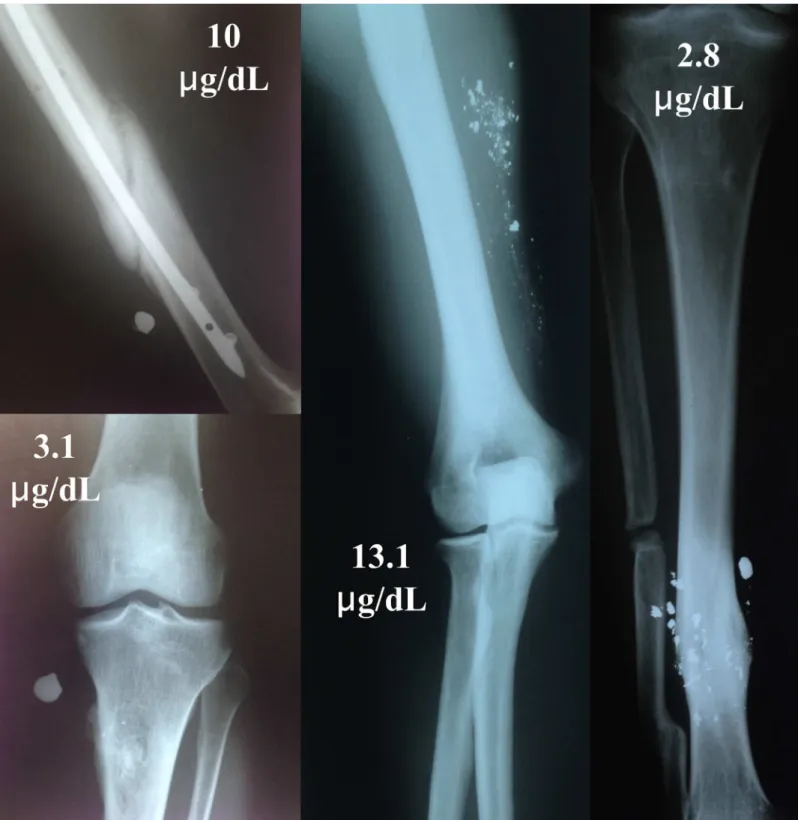
The case group was submitted to plain radiographic examination to confirm the metal frag-ments retained and to collect information about number of fragfrag-ments and location. For this group, we also collected information about the gunshot trauma as well as the date and hospital-ization history. The influence of fragment dispersion was analyzed and separated into 3 groups: volunteers with 1 fragment retained; those with 2 to 10 fragments; and those with more than 10 fragments. The fragments were counted by the researchers in the plain radiographs.
Descriptive statistics were used to evaluate the data. The inferential analysis for qualitative var-iables was investigated by chi-squared test. When the test was inconclusive, we alternatively used the Fisher’s exact test. Inferential analysis for quantitative variables,“lead levels”, and“time since trauma”was performed by non-parametric tests of Mann-Whitney to compare 2 groups and Kruskall-Wallis test for multiple groups comparison. The non-parametric tests were used because the Shapiro-Wilk test rejects the hypothesis of normal distribution for those samples. Both groups compared were treated as independent samples for statistical analysis. The matching procedure was used to homogenize the groups for confounding factors. Statistical significance was defined as p-value<0.05. All data analysis was performed using SPSS software version 22.0.
Results
45 male subjects were enrolled in each group. There was no woman assessed as volunteer. The demographic distribution for ethnicity was the following: 5 black, 14 pardo (brown), and 26 white. The matching method resulted in mean age of 38 (range from 18 to 69) years in the case group and 39 (18–66) in controls. Statistical analysis for lead blood levels resulted in significant differences between case and control groups. The average blood lead levels were 2.17μg/dL
(95% CI; 1.71–2.63) with median 2.1μg/dL for the control group, and the average levels were
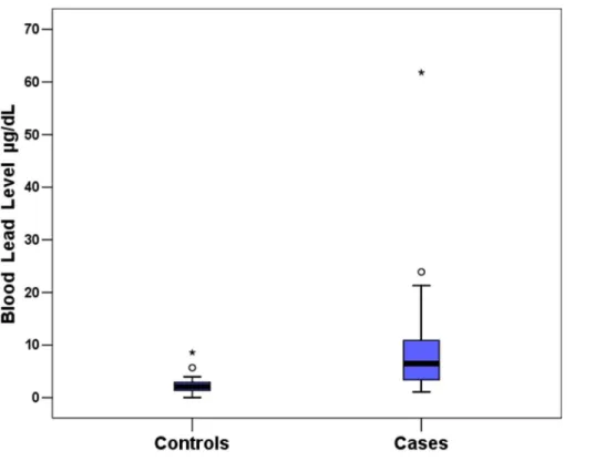
9.01μg/dL (CI; 6.07–11.96) with median 6.5μg/dL for the cases with p value<0.001.Fig 1is a
box plot for lead levels, where it is possible to see 2 outliers samples. The case group had a vol-unteer with 61.8μg/dL who had a multi-fragmentary knee fracture with bullets retained
intra-articular. The control outlier had a blood lead level of 8.6μg/dL, but we did not found any
bio-logical explanation for it. We tested the mean differences without the discrepant samples and verified that groups were also significantly different (p<0.001).Table 1presents descriptive
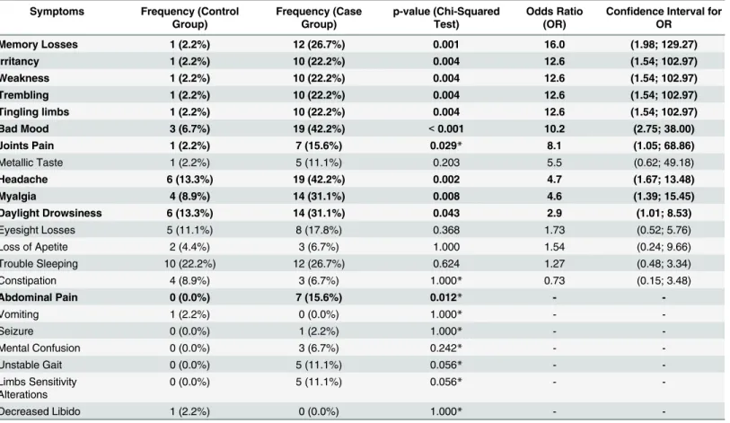
The frequencies of symptoms were compared between the controls and the cases and for some symptoms, a significant difference was found. The strength of those associations are described inTable 2.
We found no association between the number of symptoms in each volunteer and the time that the bullets were retained. The Spearman correlation between the variables was 0.209 (p-value = 0.167). We also searched for an association between the number of symptoms in each volunteer and the lead levels; the Spearman correlation was weak (0.26) but significant (p = 0.012).
The analysis for correlation between lead levels and time of bullet retention resulted in a Spearman correlation of -0.38 (p = 0.01). Therefore, blood lead levels are weakly correlated to bullet retention time.
Fig 1. A box plot to represent the samples distribution for blood lead levels. doi:10.1371/journal.pone.0140220.g001
Table 1. Descriptive statistics for blood lead levels (μg/dL).
Leads Level Statistics Control
Group
Case Group
Size of Group (n) 45 (50%) 45 (50%)
Mean 2.17 9.01
Confidence Interval for Mean (95% level) (1.71; 2.63) (6.07; 11.96)
Median 2.10 6.50
Standard Deviation (SD) 1.53 9.80
Mimimum 0.0 1.1
Maximum 8.6 61.8
Variation Coefficient (SD/Mean) 0.70 1.09
Significance from Shapiro Wilk Test <0.001 <0.001
Significance from Mann-Whitney Test (comparing Control and Case Groups)
The dispersion pattern effect of retained bullet fragments was analyzed using the Kruskall-Wallis test to compare the lead levels between 3 groups: volunteers with 1 fragment retained (12 cases); with 2 to 10 fragments (24 cases); and with more than 10 fragments (9 cases). The mean lead levels from subgroup of volunteers with 1 fragment retained was 9.2 (± 16.7)μg/dL
with median 3.55μg/dL; for the subgroup of volunteers with 2 to 10 fragments, the mean level
was 8.8 (± 6.2)μg/dL with median 7.6μg/dL; and from subgroup of volunteers with more than
10 fragments, the mean lead level was 9.3 (± 5.9)μg/dL with median 7.4μg/dL.Fig 2shows
examples of the different number of fragments retained and their respective lead levels. The statistical test resulted in a p-value of 0.233, which suggests that the lead level is not associated to the number of bullet fragments in the body.
While investigating the sites of the retained bullets, we found that the fragments were often widely distributed in multiple tissues (i.e., muscles, bones, subcutaneous, joints and spinal canal). No relations were found between lead levels and the sites of the retained bullets. A larger sample would be necessary for this analysis.
Discussion
The industrial application of lead has impacted the blood levels of the general population. Sur-veys in the USA reported that in the 1960s blood levels were higher than 20μg/dL and lowered
to 13μg/dL in that late 1970s until reaching 3.5μg/dL in the last decade.[12] A Brazilian
popu-lation study published this year[13] reported an average of 1.97μg/dL (95% CI; 1.9–2.04μg/
Table 2. Associations of symptoms and the presence of bullets retained.
Symptoms Frequency (Control Group)
Frequency (Case Group)
p-value (Chi-Squared Test)
Odds Ratio (OR)
Confidence Interval for OR
Memory Losses 1 (2.2%) 12 (26.7%) 0.001 16.0 (1.98; 129.27) Irritancy 1 (2.2%) 10 (22.2%) 0.004 12.6 (1.54; 102.97) Weakness 1 (2.2%) 10 (22.2%) 0.004 12.6 (1.54; 102.97) Trembling 1 (2.2%) 10 (22.2%) 0.004 12.6 (1.54; 102.97) Tingling limbs 1 (2.2%) 10 (22.2%) 0.004 12.6 (1.54; 102.97) Bad Mood 3 (6.7%) 19 (42.2%) <0.001 10.2 (2.75; 38.00) Joints Pain 1 (2.2%) 7 (15.6%) 0.029* 8.1 (1.05; 68.86)
Metallic Taste 1 (2.2%) 5 (11.1%) 0.203 5.5 (0.62; 49.18)
Headache 6 (13.3%) 19 (42.2%) 0.002 4.7 (1.67; 13.48)
Myalgia 4 (8.9%) 14 (31.1%) 0.008 4.6 (1.39; 15.45)
Daylight Drowsiness 6 (13.3%) 14 (31.1%) 0.043 2.9 (1.01; 8.53)
Eyesight Losses 5 (11.1%) 8 (17.8%) 0.368 1.73 (0.52; 5.76)
Loss of Apetite 2 (4.4%) 3 (6.7%) 1.000 1.54 (0.24; 9.66)
Trouble Sleeping 10 (22.2%) 12 (26.7%) 0.624 1.27 (0.48; 3.34)
Constipation 4 (8.9%) 3 (6.7%) 1.000* 0.73 (0.15; 3.48)
Abdominal Pain 0 (0.0%) 7 (15.6%) 0.012* -
-Vomiting 1 (2.2%) 0 (0.0%) 1.000* -
-Seizure 0 (0.0%) 1 (2.2%) 1.000* -
-Mental Confusion 0 (0.0%) 3 (6.7%) 0.242* -
-Unstable Gait 0 (0.0%) 5 (11.1%) 0.056* -
-Limbs Sensitivity Alterations
0 (0.0%) 5 (11.1%) 0.056* -
-Decreased Libido 1 (2.2%) 0 (0.0%) 1.000* -
-*Fisher exact test.
dL). Both studies also identified gender, race and age as risk factors for blood lead levels. In agreement with the literature, our control group presented a mean level of 2.17 (Standard Devi-ation; ± 1.53)μg/dL.
The first study that was designed to describe the association between gunshot wounds and blood lead levels, was performed by Farrell and col[5] in 1999. They compared two groups of 15 patients in a matched case-control study. They found a mean (± SD) of 17 (± 9.78)μg/dL
for the cases and 7 (± 3.77)μg/dL for the controls; these were found to be statistically
signifi-cantly different. Both means were higher than in our study, indicating a higher environmental exposure. It is important to highlight that: their data was collected during the 90’s, the location of the study was not given, and the professions of half of the volunteers were not recorded. These factors could contribute to the differences in our mean values.
McQuirter et al.[6] prospectively studied a volunteer sample of 451 subjects with retained bullets. There was a tendency of blood lead levels' elevation during the first 6 months after the trauma and stabilization after that time. For that reason, in our transversal study we included only volunteers with more than 6 months after trauma. They considered that three factors were as predictive to elevated blood levels: number of fragments, torso bone fracture and bullet fragments in the humerus. In our study, neither the number of bullet fragments nor their loca-tion were correlated to intoxicaloca-tion. They recommended, and we agree, that patients should have the lead blood levels recorded at hospital admission and be advised to obtain yearly blood lead determinations thereafter. Despite the methodological differences between the prospec-tive/transversal studies, they found a prevalence at 12 months after trauma of 20,1% of individ-uals with blood lead levels above 10μg/dL and 2,6% for those above 20μg/dL. We found,
respectively, 31,11% and 6,66%. Our controls did not values above those limits.
A case-control study[7] with 120 patients with extra-articular retained missiles reported blood lead mean values (95% CI) of 6.71 (5.68–7.74)μg/dL and 3.16 (2.79–3.53)μg/dL in case
and control groups, respectively, with significant differences verified using a matched pairs t-test (p = 0.0001). They also demonstrated an association between recent fractures and elevated lead levels. The authors suggested that the periodically blood lead levels screening may benefit those patients, especially after episodes of increased metabolic stress. This work was the most similar to our study, including methods and results. They also did not find relationships between the number of fragments and the blood lead elevation. However, in contrast to our study, they did not verify more symptoms in their cases.
In 2014, Moazeni et al.[14] reported a comparison of 25 patients with some retained lead pellets in their bodies and the same number of volunteers without similar lead exposure. The results showed lead levels of 29 (± 12.8)μg/dL and 25.3 (± 6.4)μg/dL in case and control
groups, respectively, without any significant difference (p = 0.3). But, they confirmed the previ-ous studies that found that there is no association between long time retention of bullet frag-ments in the body and rising lead levels. Although, they found that patients who had more lead pellets in their bodies had higher blood lead levels (p = 0.025). Our study found whole blood lead levels in the case group to be between 1.1μg/dL and 61.8μg/dL, with mean and standard
deviation of 9.01 ±9.8μg/dL and median 6.5μg/dL. The control group mean was 2.1 (±1.53) μg/dL with median 2.1μg/dL. The statistical analysis showed significance differences in the
lead levels between the two groups (p<0.001). These differences suggest that their controls
were more exposed to lead than ours controls. Their discrepant results could perhaps be explained by an environmental or occupational exposure that was not well controlled.
literature can be explained by pathophysiology. Initially, the metal retained induce the inflam-matory process, the foreign body reaction and it is transported to the bone stocks. After that, there is a stabilization between the absorption rates, blood distribution, renal elimination, bone storage and stocks mobilization. That is why, theoretically, after long periods, the dispersion of fragments do not influence the blood lead levels.
The Brazilian regulatory standard number 7,[15] which was published by the Ministry of Work and Employment, is the official laboratory reference for lead exposure. It established 40μg/dL as the accepted lead blood level for unexposed individuals. This guideline considers
60μg/dL as the maximum allowed biological index for workers exposure. Although cases
pre-sented mean levels were lower than the laboratory references, low levels have been associated with rising both morbidity and mortality. Along this line, the World Health Organization stated:“There is no known level of lead exposure that is considered safe”.
Sanders et al.[16] and Florea at al.[17] published wide reviews of the literature regarding the neurotoxic effects of lead exposure. This metal can exist throughout almost every organ and system. The toxicity in target organs is widely variable but is especially important in the central nervous system. Lead (Pb2+) can mimic calcium (Ca2+) functions and modifies their pre- and post-synaptic effects. There are concerns about the toxic impact on cognition and mortality in chronic exposures. Coon et al.[18] studied chronic Pb exposure and confirmed that it is a risk factor for Parkinson’s disease. Khalil et al.[19] followed 533 women aged 65–87 years in a 12-years prospective cohort study. They compared individuals with blood lead concentrations above and below 8μg/dL. A three-fold risk in coronary heart disease mortality and 73% higher
risk for total mortality risk were found. The odds ratio of diastolic hypertension was 8.1 com-paring women with blood lead levels of 4.0–31.1μg/dL to lower ones 0.5–1.6μg/dL. A
prospec-tive cohort study[20] found that men with one standard deviation increase in blood lead level were associated with a 1.27 (CI; 1.01–1.59)-fold greater risk for ischemic heart disease. Fang et al[21], in their case-control study, found an increased risk for amyotrophic lateral sclerosis, even in extremely low blood lead levels. Their means levels were 1.76μg/dL (range, 0.32–6.90)
among controls and 2.41μg/dL (range, 0.72–7.58) among cases. An editorial[22] at the
periodi-cal Circulation alerted:“Low-level environmental exposure to lead unmasked as silent killer”. It discussed that levels below 10μg/dL can lead to a higher risk of death. They also observed
that levels as low as 2.07μg/dL, in adolescents, is a public health hazard.
The U.S. Centers for Disease Control and Prevention considers 10μg/dL as the limit for
children exposure, but Gilbert and Weiss[23] published their perspective to lower the limit to 2μg/dL. The developing brain is very sensitive to lead toxicity. There are several studies that
relate lower blood lead levels with a rising risk of poor reading ability, arithmetic and others academic performances.[24] Concerns about lead exposure during pregnancy and reduced intellectual development in children with prenatal lead exposure have been reported.[25] Although we did not have any women in our study, the feminine presence in the security forces is increasing worldwide. Women soldiers in the battle fronts are an especially susceptible popu-lation, because bullets retained can theoretically jeopardize their children. Another reason for this concern is that lead in the skeleton is mobilized during pregnancy, as demonstrated by Gulson an cols.[26] The blood lead elevation is expected during pregnancy and is also a poten-tially harmful factor for the fetus.
joint pain, trembling, and tingling limbs. These symptoms can influence the social life and the quality of life of these individuals.
The main limitation of this study is inherent to all case-controls. We would like to follow-up a coorte with all patients received at the emergency since they arrived at the day of trauma. It would allow us to establish causative relations. However, in our region, gunshot victims are difficult to follow up with because usually they have security issues such as having their identity revealed. The case group included individuals who voluntarily participated. For that reason, it was not possible to evaluate gunshot victims who did not have personal concerns about their own health. Socioeconomic status is a common concern for studies on lead poisoning. Although, we work in a public hospital that offers free health assistance and, consequently, we attend to mainly the low-income population.
In conclusion, this work showed that bullets retained in the body are not innocuous. There are impacts in the blood lead levels and symptoms related to it, even with few fragments, extra-articular located or existing with low blood lead levels. These findings contribute to a new per-spective on treating patients as well as recommendations to test blood lead and lead toxicity investigations. We also think that it is important to keep in mind the possibility to remove frag-ments. Nowadays, while the benefits and risks involved in removing fragments are not clear, the patients should be aware of the issues and take part in these decisions.
Supporting Information
S1 Table. Raw data table file with all information used for statistical analysis. (XLSX)
Acknowledgments
The authors are grateful for Professor Salim Kanaan M.D., who provided the laboratory sup-port and research project design, and Professor Doctor Keila Cassiano, who performed the sta-tistical analysis. It is also important to thanks the American Manuscript Editors team for the English revision.
Author Contributions
Conceived and designed the experiments: GCSA ARX VSG. Performed the experiments: GCSA NTM INP. Analyzed the data: GCSA ARX. Contributed reagents/materials/analysis tools: GCSA NTM INP ARX. Wrote the paper: GCSA ARX VSG.
References
1. Ganocy K 2nd, Lindsey RW. The management of civilian intra-articular gunshot wounds: treatment con-siderations and proposal of a classification system. Injury. 1998; 29 Suppl 1:SA1–6. PMID:9764222. 2. Leonard MH. The solution of lead by synovial fluid. Clinical orthopaedics and related research. 1969;
64:255–61. PMID:5793011.
3. Bolanos AA, Demizio JP Jr., Vigorita VJ, Bryk E. Lead poisoning from an intra-articular shotgun pellet in the knee treated with arthroscopic extraction and chelation therapy. A case report. The Journal of bone and joint surgery American volume. 1996; 78(3):422–6. PMID:8613450.
4. Roux P, Pocock F. Blood lead concentration in children after gunshot injuries. South African medical journal = Suid-Afrikaanse tydskrif vir geneeskunde. 1988; 73(10):580–2. PMID:3375904.
5. Farrell SE, Vandevander P, Schoffstall JM, Lee DC. Blood lead levels in emergency department patients with retained lead bullets and shrapnel. Academic emergency medicine: official journal of the Society for Academic Emergency Medicine. 1999; 6(3):208–12. PMID:10192672.
7. Nguyen A, Schaider JJ, Manzanares M, Hanaki R, Rydman RJ, Bokhari F. Elevation of blood lead lev-els in emergency department patients with extra-articular retained missiles. The Journal of trauma. 2005; 58(2):289–99. PMID:15706190.
8. Capitani EMD. Diagnóstico e tratamento da intoxicação por chumbo em crianças e adultos. Medicina (Ribeirão Preto). 2009; 42(3):319–29.
9. Souza AM, Tavares CFF. Chumbo e anemia. Medicina (Ribeirão Preto). 2009; 42(3):337–40. 10. Bartlett CS. Clinical update: gunshot wound ballistics. Clinical orthopaedics and related research.
2003;(408: ):28–57. PMID:12616039.
11. DiMaio VJ, DiMaio SM, Garriott JC, Simpson P. A fatal case of lead poisoning due to a retained bullet. The American journal of forensic medicine and pathology. 1983; 4(2):165–9. PMID:6859004. 12. Theppeang K, Glass TA, Bandeen-Roche K, Todd AC, Rohde CA, Schwartz BS. Gender and
race/eth-nicity differences in lead dose biomarkers. American journal of public health. 2008; 98(7):1248–55. doi:
10.2105/AJPH.2007.118505PMID:18511728; PubMed Central PMCID: PMC2424096.
13. de Almeida Lopes AC, Navas-Acien A, Zamoiski R, Silbergeld EK, Carvalho Mde F, Buzzo ML, et al. Risk factors for lead exposure in adult population in southern Brazil. J Toxicol Environ Health A. 2015; 78(2):92–108. doi:10.1080/15287394.2014.942125PMID:25424618.
14. Moazeni M, Mohammad Alibeigi F, Sayadi M, Poorya Mofrad E, Kheiri S, Darvishi M. The Serum Lead level in Patients With Retained Lead Pellets. Arch Trauma Res. 2014; 3(2):e18950. doi:10.5812/atr. 18950PMID:25147780; PubMed Central PMCID: PMC4139699.
15. Programa de Controle Médico de Saúde Ocupacional., NR 7 (1994).
16. Sanders T, Liu Y, Buchner V, Tchounwou PB. Neurotoxic effects and biomarkers of lead exposure: a review. Reviews on environmental health. 2009; 24(1):15–45. PMID:19476290; PubMed Central PMCID: PMC2858639.
17. Florea A- M, Taban J, Varghese E, Alost BT, Moreno S, Büsselberg D. Lead (Pb2+) neurotoxicity: Ion-mimicry with calcium (Ca2+) impairs synaptic transmission. A review with animated illustrations of the pre- and post-synaptic effects of lead. Journal of Local and Global Health Science. 2013: 4. doi:10. 5339/jlghs.2013.4
18. Coon S, Stark A, Peterson E, Gloi A, Kortsha G, Pounds J, et al. Whole-body lifetime occupational lead exposure and risk of Parkinson's disease. Environmental health perspectives. 2006; 114(12):1872–6. PMID:17185278; PubMed Central PMCID: PMC1764163.
19. Khalil N, Wilson JW, Talbott EO, Morrow LA, Hochberg MC, Hillier TA, et al. Association of blood lead concentrations with mortality in older women: a prospective cohort study. Environmental health: a global access science source. 2009; 8:15. doi:10.1186/1476-069X-8-15PMID:19344498; PubMed Central PMCID: PMC2670287.
20. Jain NB, Potula V, Schwartz J, Vokonas PS, Sparrow D, Wright RO, et al. Lead levels and ischemic heart disease in a prospective study of middle-aged and elderly men: the VA Normative Aging Study. Environmental health perspectives. 2007; 115(6):871–5. doi:10.1289/ehp.9629PMID:17589593; PubMed Central PMCID: PMC1892138.
21. Fang F, Kwee LC, Allen KD, Umbach DM, Ye W, Watson M, et al. Association Between Blood Lead and the Risk of Amyotrophic Lateral Sclerosis. American journal of epidemiology. 2010; 171(10):1126–
33. doi:10.1093/aje/kwq063PMID:20406759
22. Nawrot TS, Staessen JA. Low-level environmental exposure to lead unmasked as silent killer. Circula-tion. 2006; 114(13):1347–9. doi:10.1161/CIRCULATIONAHA.106.650440PMID:17000919. 23. Gilbert SG, Weiss B. A rationale for lowering the blood lead action level from 10 to 2 microg/dL.
Neuro-toxicology. 2006; 27(5):693–701. doi:10.1016/j.neuro.2006.06.008PMID:16889836; PubMed Central PMCID: PMCPMC2212280.
24. Marturano EM, Elias LCS. Efeitos cognitivos, neuropsico- lógicos e comportamentais da exposição a baixas concentrações de chumbo na infância. Medicina (Ribeirão Preto). 2009; 42(3):291–5. 25. Schnaas L, Rothenberg SJ, Flores MF, Martinez S, Hernandez C, Osorio E, et al. Reduced intellectual
development in children with prenatal lead exposure. Environmental health perspectives. 2006; 114 (5):791–7. PMID:16675439; PubMed Central PMCID: PMC1459938.
26. Gulson BL, Mahaffeya KR, Jamesona CW, Mizona KJ, Korscha MJ, Camerona MA, et al. Mobilization of lead from the skeleton during the postnatal period is larger than during pregnancy. Journal of Labora-tory and Clinical Medicine. 1998; 131(4):324–9. doi:http://dx.doi.org/10.1016/S0022-2143(98)90182-2
PMID:9579385