DOI: 10.1590/0004-282X20150002
ARTICLE
Visual evoked potentials show strong positive
association with intracranial pressure in
patients with cryptococcal meningitis
Potenciais evocados visuais mostram correlação positiva forte com a pressão
intracraniana em pacientes com meningite criptocócica
Marcelo Adriano da Cunha Silva Vieira1, Maria do Amparo Salmito Cavalcanti1, Dorcas Lamounier Costa2, Kelsen
Dantas Eulálio1, Otoni Cardoso do Vale3, Chrystiany Placido de Brito Vieira4, Carlos Henrique Nery Costa2
Intracranial hypertension (ICH) is responsible for both early mortality and the auditory, visual, and cognitive se-quelae of meningitis caused by Cryptococcus neoformans1,2,3.
More than half of the patients with cryptococcal meningitis (CM) exhibit ICH1,3,4,5. Approximately 600,000 deaths
associ-ated with CM occur annually worldwide6.
Cryptococcus neoformans is the main cause of meningi-tis among immunocompromised individuals7. he absence of
host immune response and the particularities inherent to the
genesis of increased cerebrospinal luid (CSF) pressure make clinical and radiological manifestations weak predictors of
ICH in neurocryptococcosis1,2,3,4,5,8,9. he Infectious Diseases
1Instituto de Doenças Tropicais Natan Portella, Departamento de Neurologia, Teresina PI, Brazil; 2Universidade Federal do Piauí, Departamento de Medicina Especializada, Teresina PI, Brazil;
3Universidade Federal do Ceará, Departamento de Medicina Clínica, Hospital Universitário Walter Cantídio, Serviço de Neurofisiologia, Fortaleza CE, Brazil; 4Universidade Federal do Piauí, Departamento de Enfermagem, Teresina PI, Brazil.
Correspondence: Marcelo Adriano Cunha Silva Vieira; Departamento de Neurologia, Instituto de Doenças Tropicais Natan Portella; Rua Anfrísio Lobão, 1235; 64049-280 Teresina PI, Brasil; E-mail: [email protected]
Conflict of interest: There is no conflict of interest to declare. Support: Instituto de Doenças Tropicais Natan Portella.
Received 06 August 2014; Received in final form 14 November 2014; Accepted 03 December 2014.
ABSTRACT
Objective: To verify the relationship between intracranial pressure and flash visual evoked potentials (F-VEP) in patients with cryptococcal meningitis. Method: The sample included adults diagnosed with cryptococcal meningitis admitted at a reference hospital for infectious diseases. The patients were subjected to F-VEP tests shortly before lumbar puncture. The Pearson’s linear correlation coefficient was calculated and the linear regression analysis was performed. Results: Eighteen individuals were subjected to a total of 69 lumbar punctures preceded by F-VEP tests. At the first lumbar puncture performed in each patient, N2 latency exhibited a strong positive correlation with intracranial pressure (r = 0.83; CI = 0.60 - 0.94; p < 0.0001). The direction of this relationship was maintained in subsequent punctures.
Conclusion: The intracranial pressure measured by spinal tap manometry showed strong positive association with the N2 latency F-VEP in patients with cryptococcal meningitis.
Keywords: visual evoked potentials, intracranial pressure, cryptococcal meningitis.
RESUMO
Objetivo: Verificar a relação entre pressão intracraniana e potencial evocado visual por flash (PEV-F) em pacientes com meningite criptocócica. Método: A amostra incluiu pacientes admitidos em um hospital de referência para doenças infecciosas. Realizou-se PEV-F antes de cada punção lombar. Calculou-se o coeficiente de correlação de Pearson e a equação de regressão linear entre as variáveis latência N2 e pressão intracraniana inferida através de raquimanometria. Resultados: Dezoito pacientes foram submetidos a um total de 69 punções lombares. A latência N2 mostrou correlação positiva forte com a pressão de abertura verificada na primeira punção lombar a que cada paciente foi submetido (r = 0,83; IC = 0,60 – 0,94; p < 0,0001). A positividade da correlação foi mantida nas aferições subsequentes. Conclusão: Houve associação positiva forte entre a latência N2 do PEV-F e pressão intracraniana em pacientes com meningite criptocócica.
Society of America recommends measuring the CSF pressure
of patients with CM using a manometer for lumbar puncture at initial and follow-up assessment. ICH requires the removal
of CSF using lumbar puncture on a daily basis and can even
require shunts in refractory cases10.
he mechanical efects of ICH on the central visual pro
-jection pathways, together with the repercussions on brain perfusion, may slow neuronal conduction, which could be
demonstrated through evoked potential testing. Although the technique to acquire visual evoked potentials using lashes of difuse light exhibits more variability compared
to techniques that apply pattern-reversal stimulation, it of-fers the advantage that it may be performed in the supine position and does not require strict cooperation and visual
ixation, allowing this procedure to be performed even in
comatose patients11,12.
A relationship has been observed between a
prolonga-tion of the latency of waves from the difuse light lash vi
-sual evoked potential (F-VEP) test and increased intracrani
-al pressure (ICP) in patients with hydroceph-alus, traumatic
brain injury or drug-related cerebral oedema13,14,15,16,17,18,19. he
N2 component exhibits the greatest stability and is the most
widely investigated parameter of the F-VEP.
To date, no study has investigated the correlation
be-tween F-VEP latencies and ICP in patients with CM. he demonstration of a correlation between ICP and the latency of the N2 wave of the F-VEP in CM may support the use of this test to infer ICP and guide the use of lumbar punctures or CSF shunts for relief. he aim of the present study was to verify the correlation between ICP and the peak latency of the N2 component of the F-VEP in patients with CM.
METHOD
he sample included adults who were hospitalized at the Natan Portella Institute of Tropical Diseases, Teresina,
Brazil, between May 2011 and February 2012 with a diagno
-sis of CM, which was conirmed by CSF examination with
India ink. he study excluded patients with macular or reti
-nal lesions, as revealed by an ophthalmologic examination, and patients with structural impairment of the central visual pathways, as revealed by imaging techniques (computed to-mography or magnetic resonance). Hepatic encephalopathy, uremia, pneumocystosis or tuberculosis as co-morbidities also led to exclusion of patients.
he ICP was inferred using lumbar puncture manometry
following the recommendations of the American Academy of Neurology20. he procedures were performed and repeated
only when the attending physician established their clinical indication, the patients consented and a minimum 24-hour
interval had passed between measurements. he opening pressure was measured using the MVD 300 digital manometer
(Globalmed, Brazil) and the pressure around which stable os
-cillations occurred up to two minutes after the device was connected to the puncture needle was recorded.
he patients were subjected to F-VEP tests shortly before
each lumbar puncture. Whenever the opening pressure was high (> 20 cm H2O), the test was repeated 20 minutes after
the needle was removed. he standards established by the
American Clinical Neurophysiology Society11 were followed
and the Neuro-MEP.micro (Neurosoft, Brazil) device was used. he electrodes were placed at the sites standardized by the International System 10-20 on Oz (active), A1 (reference),
and A2 (ground).
he stimulation by lashes of difuse light was chosen be
-cause the subjects with CSF hypertension were uncoopera
-tive to reliable ixate on a pattern stimulus. Simultaneous
binocular stimulation was performed using light lashes pro
-jected through glasses with light emitting diodes on the
in-ternal surface of the goggles. he bilateral simultaneous vi
-sual stimulation aimed to minimize the magniication of any
direct harmful efects of fungi on the optic nerves individual
-ly. he stimulation frequency was 1.9 Hz and the stimulus du
-ration was 100 µs. he input impedance was < 5 kΩ, and 1- to 100-Hz ilters and the average of the signals resulting from 60 stimuli were used. hree series were performed to verify the
reproducibility of the curves representing the evoked poten
-tial. he identiication of the peaks of the F-VEP is illustrated in Figure 1.
he parameter selected for measurement and analy
-sis was the peak latency of the second negative delection of the F-VEP recording. his wave is referred to as the “III”
peak by American Clinical Neurophysiology Society11 or “N2”
peak by International Society for Clinical Electrophysiology
of Vision12. he last is the most widely used in clinical studies
involving F-VEP and ICP.
he normality of distribution was investigated using the
D’’Agostino K-squared test. Pearson’s linear correlation co
-eicient was calculated and linear regression analysis was performed using the variables measured at the irst lumbar puncture performed after the CM diagnosis was conirmed. he linear coeicients of correlation were calculated for the
average values of the opening pressure and the N2 latency as-sessed in consecutive lumbar punctures, together with their
corresponding 95% conidence intervals. Student’s t-test for
paired variables was used to investigate whether the N2
la-tency decreased after the ICP normalized.
he level of signiicance was established at 5% with a sta
-tistical power of 80% for inferential analysis. he data were processed using BioStat 2009 software (AnalysSoft Inc., Brazil). he study was approved by the Research Ethics Committee of the Federal University of Piauí and all of the patients (or their
legal guardians) signed an informed consent form.
RESULTS
Eighteen patients, aged 20 to 56 years old (mean = 35.4, standard deviation (SD) = 9.4), were analyzed. Only two of
the patients (2/18) were female, and almost all of the patients
(17/18) were infected by the human immunodeiciency virus (HIV). No other cause of immunosuppression was identiied in the HIV-negative patient.
Sixty-nine lumbar punctures were performed during the
study, and each patient was subjected to this procedure
be-tween one and nine times. As positive India ink examination
was a criterion for inclusion in the study, the measurements were performed after patients have already undergone at
di-agnostic lumbar puncture. Although the irst lumbar punc
-ture performed within the scope of the study exhibited a high opening pressure (> 20 cm H2O) in only eight (8/18) of the patients, most of the patients (13/18) presented ICH at some point during evaluation, resulting in 58% (40/69) of the
mea-surements indicating high CSF pressure. All the sixty-nine
lumbar punctures were preceded by F-VEP test. In the for
-ty procedures in which the opening pressure was high, the
F-VEP test was repeated 20 minutes after CSF drainage. he opening pressure and F-VEP N2 latency variables
exhibited a strong positive correlationat the irst assess
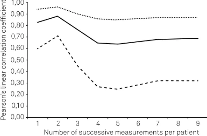
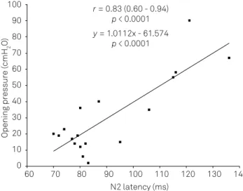
-ment in each patient, whit Pearson’s coeicient (r) equal to 0.83 (CI = 0.60-0.94; p < 0.0001). Linear regression analysis showed that the equation y = 1.01x - 61.57 describes ICP as a function of N 2 latency (p < 0.0001) (Figure 2). he magnitude of Pearson’s linear coeicient of correlation (r) decreased, and the range of it conidence interval increased, when an
increasing number of measurements were included in the
av-erage values for each patient (Figure 2). he N2 latency did not decrease (t = 0.79; p = 0.21) when F-VEP was retested 20 minutes after the reduction of ICP by CSF drainage.
DISCUSSION
he frequency of ICH detected at irst assessment or
during follow-up in the CM patients was similar to those reported in other case series1,3,4. he predominance of
male and HIV seropositive individuals in the investigat
-ed population relects the current epidemiological proile
of neurocryptococcosis1,6,21,22,23.
he positive correlation between ICP and F-VEP wave la
-tency observed in studies of patients with hydrocephalus sug-gested this method as a mean to provide an early detection of shunt dysfunction13,14,16,17,18,19. Changes in the F-VEP latencies
of patients with ICH secondary to cerebral edema have also
been observed, and the use of F-VEP has been suggested to
monitor ICP in head trauma victims14,16,17,18. However, F-VEP
in head trauma is biased by other variables as arterial pO2, sagittal sinus venous pO2 and regional cerebral low and is
not an adequate substitute for direct ICP measures17,19.
A linear correlation between the latency of the F-VEP N2 wave and medium to high ICP values was observed under
several other clinical conditions24. he present study showed similar relationship between F-VEP and ICP in patients with CM (Figure 3).
High levels of CSF pressure contribute to the appearance and irreversibility of visual, auditory, and cognitive deicits in
patients with CM1,25,26. he impact of CSF hypertension on the
morbidity and mortality associated with this disease supports the current recommendations to perform routine and frequent
measurement of ICP using spinal tap manometry. When the
opening pressure is ≥ 25 cm H2O, daily spinal taps and CSF
drainage suicient to reduce it to 20 cm H2O or 50% of the initial
value are recommended. When the opening pressure is normal on two consecutive days, the procedure should be performed
once a week until the patient is discharged from the hospital or
at any moment when the relapse of ICH is suspected10. Number of successive measurements per patient
Pe
arso
n’s
linear correlation coefficient
1 0,00
0,10 0,20 0,30 0,40 0,50 0,60 0,70 0,80 0,90 1,00
2 3 4 5 6 7 8 9
As ICH does not systematically translate into their clas-sical clinical manifestations or ventricular dilation on cra-nial tomography1,2,27, the time of relapse of ICH cannot be recognized. Accordingly, the detection of high ICP through
an alternative of clinical and tomographic methods would
be useful in monitoring of patients in order to indicate CSF
drainage by spinal tap. While failure to address raised intra-cranial pressure could result in more residual neurological damage and death1, the unneeded repetition of spinal taps increases the risk of adverse events, including hemorrhage, bacterial infection, CSF hypotension, radicular involvement,
epidermoid tumor, and brain herniation28. herefore, to avoid
unneeded lumbar punctures is desirable through the
devel-opment of a reliable, non-invasive method to measure ICP
that is easy to perform at the bedside and can identify the
pa-tients who must be subjected to spinal tap. he simple linear
regression equation depicted in Figure 3 show that it is fea
-sible the inference of ICP values from the F-VEP N2 latency. he N2 latency did not decrease when the F-VEP test was repeated 20 minutes after reduction of the ICP. It is possible that a 20-minute interval was insuicient for the chemical and physical efects of ICH on the visual pathways to regress. In addition, some of these efects may be deinitive, and other
mechanisms, such as arachnoiditis, may be involved in the visual impairment of patients25,26,27. Such phenomena could
explain the slight reduction in the strength of the correla-tion between the N2 latency and the opening pressure as a function of the subsequent spinal taps to which the patients
were subjected (Figure 2). Prediction and interpretation of
changes in the amplitude of the potentials after CSF drain
-age would be even more complex. he CSF pressure adjacent to the optic nerves tends to reduce the responses, but difuse
brain dysfunction next to it has the opposite efect11,19.
Despite the reproducibility of the curves representing
the evoked potential has been veriied in every measure
-ment in this study, the intra-individual variability of N2 la-tency on successive tests in healthy subjects advises caution
in interpreting F-VEP changes in clinical work29. To conirm
the clinical usefulness of the F-VEP test in the management
of neurocryptococcosis, studies assessing the reversibili-ty of the visual disorders exhibited by patients are needed.
Such studies must include control groups to assess the ran
-dom intra-individual variability of the N2 latency during the follow-up period and partial correlation analysis to verify if arachnoiditis adjacent to the optic nerves, hypoxia, acidosis,
uremia, liver failure or level of arousal constitute lurking vari
-ables. To date, the only F-VEP abnormality established by the
American Academy of Clinical Neurophysiology as having
deinite clinical value is the absence of potentials11.
Finally, as the results of this study indicate that the N2 la
-tency and CSF pressure exhibit a strong positive correlation, it is suggested that the F-VEP test should be explored as an indirect, non-invasive method in the diagnosis of high ICP in order to indicate the need for CSF drainage in patients with
cryptococcal meningitis.
References
1. Graybill JR, Sobel J, Saag M, van Der Horst C, Powderly W, Cloud G et al. Diagnosis and management of increased intracranial pressure in patients with AIDS and cryptococcal meningitis. Clin Infect Dis. 2000;30(1):47-54. http://dx.doi.org/10.1086/313603
2. Malessa R, Krams M, Hengge U, Weiller C, Reinhardt V, Volbracht L et al. Elevation of intracranial pressure in acute AIDS-related cryptococcal meningitis. Clin Investig 1994;72(12):1020-6. http://dx.doi.org/10.1007/BF00577748
3. Sun HY, Hung CC, Chang SC. Management of cryptococcal meningitis with extremely high intracranial pressure in HIV-infected patients. Clin Infect Dis. 2004;38(12):1790-2. http://dx.doi.org/10.1086/421272
4. Bicanic T, Brouwer AE, Meintjes G, Rebe K, Limmathurotsakul D, Chierakul W et al. Relationship of cerebrospinal fluid pressure, fungal burden and outcome in patients with cryptococcal meningitis undergoing serial lumbar punctures. AIDS. 2009;23(6):701-6. http://dx.doi.org/10.1097/QAD.0b013e32832605fe
5. Denning DW, Armstrong RW, Lewis BH, Stevens DA. Elevated cerebrospinal fluid pressures in patients with cryptococcal meningitis and acquired immunodeficiency syndrome. Am J Med. 1991;91(3):267-72. http://dx.doi.org/10.1016/0002-9343(91)90126-I
6. Park BJ, Wannemuehler KA, Marston BJ, Govender N, Pappas PG, Chiller TM. Estimation of the current global burden of cryptococcal meningitis among persons living with HIV/AIDS. AIDS. 2008;23(4):525. http://dx.doi.org/10.1097/QAD.0b013e328322ffac
7. Hajjeh RA, Conn LA, Stephens DS, Baughman W, Hamill R, Graviss E et al. Cryptococcosis: population-based multistate active surveillance and risk factors in human immunodeficiency virus-infected persons. J Infect Dis. 1999;179(2):449-54. http://dx.doi.org/10.1086/314606
8. Charlier C, Dromer F, Lévêque C, Chartier L, Cordoliani YS, Fontanet A et al. Cryptococcal neuroradiological lesions correlate with severity during cryptococcal meningoencephalitis in HIV-positive patients in the HAART Era. PLoS ONE. 2008;3(4):e1950. http://dx.doi.org/10.1371/journal.pone.0001950
N2 latency (ms)
r = 0.83 (0.60 - 0.94)
p < 0.0001
y = 1.0112x - 61.574
p < 0.0001
Opening pressure (cmH
2
O)
60 0 10 20 30 40 50 60 70 80 90 100
70 80 90 100 110 120 130 140
9. Darzé C, Lucena R, Gomes I, Melo A. Prognostic factors in cryptococcal meningoencephalitis. Arq Neuropsiquiatr. 1999;57(3A):649-52.
http://dx.doi.org/10.1590/S0004-282X1999000400018
10. Perfect JR, Dismukes WE, Dromer F, Goldman DL, Graybill JR, Hamill RJ et al. Clinical practice guidelines for the management of cryptococcal disease: 2010 update by the Infectious Diseases Society of America. Clin Infect Dis. 2010;50(3):291-322. http://dx.doi. org/10.1086/649858
11. American Clinical Neurophysiology Society. Guideline 9B: guidelines on evoked potentials. J Clin Neurophysiol. 2006;23(2):138-56.
12. Odom VJ, Bach M, Brigell M, Holder GE, McCulloch DL, Tormene AP, et al. ISCEV standard for clinical visual evoked potentials (2009 update). Doc Ophthalmol. 2010;120(1):111-9. http://dx.doi.org/10.1007/s10633-009-9195-4
13. Coupland SG, Cochrane DD. Visual evoked potentials, intracranial pressure and ventricular size in hydrocephalus. Doc Ophthalmol. 1987;66(4):321-9. http://dx.doi.org/10.1007/BF00213660
14. Desch LW. Longitudinal stability of visual evoked potentials in children and adolescents with hydrocephalus. Dev Med Child Neurol. 2001;43(2):113-7. http://dx.doi.org/10.1017/S0012162201000196
15. Gumerlock MK, York D, Durkis D. Visual evoked responses as a monitor of intracranial pressure during hyperosmolar Blood-Brain Barrier Disruption. Acta Neurochir Suppl (Wien). 1994;60:132-5.
16. Sjöström A, Uvebrant P, Roos A. The light-flash-evoked response as a possible indicator of increased intracranial pressure in hydrocephalus. Childs Nerv Syst.1991;11(7):381-7. http://dx.doi.org/10.1007/BF00717400
17. Stone JL, Ghaly RF, Hughes JR. Evoked potentials in head injury and states of increased intracranial pressure. J Clin Neurophysiol. 1988;5(2):135-60. http://dx.doi.org/10.1097/00004691-198804000-00002
18. York D, Legan M, Benner S, Watts C. Further studies with a noninvasive method of intracranial pressure estimation. Neurosurgery. 1984;14(4):456-61. http://dx.doi.org/10.1227/00006123-198404000-00011
19. York DH, Pulliam MW, Rosenfeld JG, Watts C. Relationship between visual evoked potentials and intracranial pressure. J Neurosurg. 1981;55(6):909-16. http://dx.doi.org/10.3171/jns.1981.55.6.0909
20. American Academy of Neurology. Practice parameters: lumbar puncture (summary statement). Report of the Quality Standards Subcommittee of the American Academy of Neurology. Neurology. 1993;43(3 Pt 1):625-7.
21. Mirza SA, Phelan M, Rimland D, Graviss E, Hamill R, Brandt ME et al. The changing epidemiology of cryptococcosis: an update from population-based active surveillance in 2 large metropolitan areas, 1992-2000. Clin Infect Dis. 2003;36(6):789-94. http://dx.doi.org/10.1086/368091
22. Mora DJ, Colombo ERC, Ferreira-Paim K, Andrade-Silva LE, Nascentes GA, Silva-Vergara ML. Clinical, epidemiological and outcome features of patients with cryptococcosis in Uberaba, Minas Gerais, Brazil. Mycopathologia. 2012;173(5-6):321-7. http://dx.doi.org/10.1007/s11046-011-9504-9
23. Pappalardo MC, Melhem MS. Cryptococcosis: a review of the Brazilian experience for the disease. Rev Inst Med Trop S Paulo. 2003;45(6):299-305. http://dx.doi.org/10.1590/S0036-46652003000600001
24. Zhao YL, Zhou JY, Zhu GH. Clinical experience with the noninvasive ICP monitoring system. Acta Neurochir Suppl (Wien). 2005;95:351-5. http://dx.doi.org/10.1007/3-211-32318-X_72
25. Johnston SR, Corbett EL, Foster O, Ash S, Cohen J. Raised intracranial pressure and visual complications in AIDS patients with cryptococcal meningitis. J Infect. 1992;24(2):185-9. http://dx.doi.org/10.1016/0163-4453(92)92954-H
26. Lipson BK, Freeman WR, Beniz J, Goldbaum MH, Hesselink JR, Weinreb RN et al. Optic neuropathy associated with cryptococcal arachnoiditis in AIDS patients. Am J Ophthalmol. 1989;107(5):523-7. http://dx.doi.org/10.1016/0002-9394(89)90498-4
27. Tan CT. Intracranial hypertension causing visual failure in cryptococcus meningitis. J Neurol Neurosurg Psychiatry. 1988;51(7):944-6. http://dx.doi.org/10.1136/jnnp.51.7.944
28. Ellenby MS, Tegtmeyer K, Lai S, Braner DA. Lumbar puncture. N Engl J Med. 2006;355(13):e12. http://dx.doi.org/10.1056/ NEJMvcm054952