Arquivos Brasileiros de Cardiologia - Volume 83, Nº 6, Dezembro 2004
466
Original Article
Changes in the Parameters of Left Ventricular
Diastolic Function According to Age on Tissue
Doppler Imaging
Márcia Duarte Pedone, Iran castro, Domingos Hatem, José Carlos Haertel, Flávia Feier,
Fernando Pandolfo
Porto Alegre, RS - Brazil
Instituto de Cardiologia do RS / Fundação Universitária de Cardiologia Mailing address: Iran Castro - Instituto de Cardiologia do RS Av. Princesa Isabel, 395 - Cep 90620-001 - Porto Alegre, RS, Brazil E-mail: [email protected]
Received for publication: 09/03/2003 Accepted for publication: 02/18/2004 English version by Stela Maris Costalonga
Objective
To determine the correlation between diastolic velocities on tissue Doppler imaging and age in a sample of healthy adults and to correlate age with the velocities of transmitral and pul-monary vein flows.
Methods
Echocardiographic assessment of 51 healthy individuals, whose ages ranged from 21 to 69 years. The diastolic myocardial velocities were recorded on tissue Doppler imaging. The velocities of the transmitral and pulmonary vein flows were also determined.
Results
The initial basal septal and basal lateral diastolic myocardial velocities showed an inverse correlation with age [r = - 0.40 (P = 0.004), and r = - 0.60 (P = 0.0001), respectively]. The atriogenic velocities of tissue Doppler imaging correlated directly with age [r = 0.56 (P = 0.0001) in the basal septal segment, and r = 0.50 (P = 0.0001) in the basal lateral segment]. The velocities of transmitral and pulmonary vein flows also correlated with age.
Conclusion
Age correlates with the tissue Doppler diastolic myocardial velocities and with the velocities of transmitral and pulmonary vein flows. In healthy individuals, the parameters of left ventricular diastolic function vary with the natural evolution of age.
Key words
echocardiography, tissue Doppler imaging, left ventricular dias-tolic function
The understanding of left ventricular diastolic function has stimulated medical research, because congestive heart failure is a severe public health problem associated with significant morbidity and mortality. A significant proportion (50%) of patients 1 with
congestive heart failure has preserved systolic function. Patients with diastolic heart failure have a mortality risk 5 to 8 times greater than that of the healthy population 2.
Of the methods for diagnosing diastolic dysfunction, Doppler echocardiography has evolved greatly in recent years. The often-used conventional echocardiographic techniques (transmitral Dop-pler and pulmonary vein DopDop-pler), although very useful, have flaws and limitations. However, tissue Doppler, with its capacity for measuring myocardial velocities, and because it does not undergo significant alterations with afterload changes, has played an im-portant role in determining diastolic dysfunction 3-9.
The distinction between normal physiology and pathological processes presupposes an appropriate knowledge of the normality pattern in the population studied. Myocardial compliance and re-laxation are known to change with age, and different patterns of diastolic filling are expected for distinct age groups. However, several studies on diastolic function and diastolic heart failure have used only young individuals as the control group 10. Thus,
the assessment of variations in left ventricular diastolic myocardial velocities in healthy individuals according to age by use of tissue Doppler imaging will contribute to a better understanding and evaluation of the diastolic function.
The major objective of this study was to determine the corre-lation between the regional myocardial diastolic velocities on tissue Doppler imaging and age in a sample of healthy individuals. The secondary objective was to demonstrate the correlation of age with the velocities of conventional Doppler (Doppler of the trans-mitral and pulmonary vein flows).
Methods
This study assessed 51 healthy, active volunteers (23 men and 28 women), whose ages ranged from 21 to 69 years. All participants had normal heart rate and blood pressure levels. Nei-ther smoking, nor use of medication, nor history of diabetes mellitus or cardiovascular disease was reported. All participants had normal electrocardiograms and transthoracic echocardiograms, and none of them was an athlete.
Arquivos Brasileiros de Cardiologia - Volume 83, Nº 6, Dezembro 2004
467
Changes in the Parameters of Left Ventricular Diastolic Function According to Age on Tissue Doppler ImagingAll participants underwent complete transthoracic echocar-diographic study with analysis focused on the parameters of left ventricular diastolic function.
The examinations were performed in the echocardiography laboratory at our institution, with an HP Sonus 2500 echocardio-graphic device equipped with multifrequency transducer. A native tissue Doppler device was also available. All images were obtained by the same observer with a concomitant electrocardiographic tracing. All examinations were analyzed while being performed, recorded on a VHS tape, later being reassessed by a second observer. The dimensions of the cardiac cavities were measured by use of M mode and recorded in all participants. The criteria adopted were those recommended by the American Society of Echocardiography11.
The myocardial diastolic velocities (fig. 1) of the mitral ring and of the left ventricular basal septal and basal lateral segments12
obtained on the apical 4-chamber view were recorded on tissue Doppler. In each segment, the initial (E’) and atriogenic (A’) dias-tolic velocities were determined (fig.1). Each value recorded re-sulted from the arithmetic mean of 8 to 10 complexes.
The transmitral flow velocity was measured on the apical 4-chamber view, with a sample volume positioned at the extremity of the mitral valve leaflets. The initial (E wave) and atriogenic (A wave) diastolic velocities, and the time for deceleration (TD) were recorded. The velocities of pulmonary vein flow were determined in the same echocardiographic view, with a sample volume posi-tioned 1 cm below the orifice of the right superior pulmonary vein in the left atrium. The following velocities were recorded: systolic, diastolic, and the reverse atrial pulmonary vein flow (PVa) 13.
The continuous variables were presented as means and standard deviations.
The Pearson correlation was adopted for assessing the asso-ciation between age and diastolic variables. Diagrams of dispersion were used for the graphic demonstration of these relations.
Age was categorized into 2 groups (< 40 years and > 40 years), and the diastolic measures were compared by using the Studentt test for independent samples.
The 8.0 version of the statistical program SPSS was used for data analysis.
The alpha level adopted for the comparisons was 0.05.
Results
The means and respective standard deviations of the myocardial diastolic velocities obtained in the basal septal and basal lateral segments are shown in table I, as are the velocities of transmitral flow and pulmonary vein flow. Data were divided into 2 groups (above and below 40 years of age).
The myocardial diastolic velocities were observed to correlate with age (fig. 2 and 3).
The initial basal septal and basal lateral myocardial diastolic velocities were inversely correlated with age [r = -0.40, P = 0.004, in the basal septal segment (fig. 2-A), and r = -0.60, P = 0.0001, in the basal lateral segment (fig. 2-B)].
The atriogenic velocities were directly correlated with age [r = 0.56, P = 0.0001, in the basal septal segment (fig. 3-A), and r = 0.50, P = 0.0001, in the basal lateral segment (fig. 3-B)].
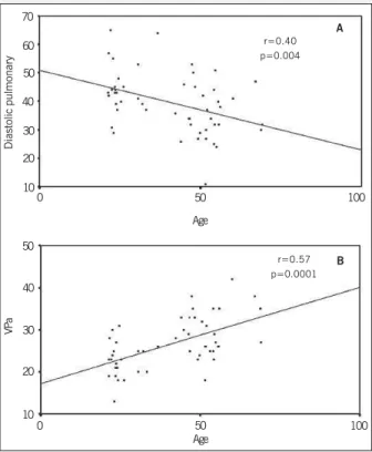
The velocities of transmitral flow also correlated with age (fig. 4). Figure 4-A shows an inverse correlation of age with initial diastolic velocity (r=-0.36; P=0.009), and figure 4-B shows a di-rect correlation of age with atriogenic velocity (r = 0.40; P = 0.004). When assessing the pulmonary venous flow, an inverse correla-tion with age was observed [r= -0.40; P= 0.004 (fig.5-A)], and a direct correlation with systolic velocity was observed (r= 0.33; P= 0.02). The amplitude of the reverse pulmonary A wave also had a direct correlation with age [r= 0.57; P=0.0001 (fig.5-B)].
Discussion
Tissue Doppler imaging has been extensively studied and has proved, among other applications, to be useful in the following situations: the differential diagnosis between constrictive pericarditis and restrictive cardiomyopathy, in the follow-up of heart trans-plantation, in assessing the segmentary ventricular function, and in the diagnostic differentiation between pathological and physio-logical ventricular hypertrophy3,14-16. The method has also been
studied for assessing left and right ventricular systolic function17-20.
Fig. 1 - Tissue Doppler imaging: myocardial diastolic velocities in the basal lateral segment.
Table I - Tissue Doppler velocities: transmitral flow and pulmonary venous flow in healthy individuals
Parameters < 40 years (n = 22) > 40 years (n = 29) P
E’S (cm/s) 9.8 ± 1.9 8.1 ± 1.8 0.002 E’L (cm/s) 13 ± 2.8 9.4 ± 2.3 0.0001 A’S (cm/s) 5.5 ± 1.2 7.2 ± 1.6 0.0001 A’L (cm/s) 5.8 ± 1.6 7.3 ± 1.5 0.001 E mitral (cm/s) 93 ± 31 74 ± 23 0.014 A mitral (cm/s) 49 ± 14 61 ± 17 0.008 E/A ratio 1.9 ± 0.4 1.2 ± 0.3 0.0001 TD (ms) 142 ± 13 154 ± 29 0.06 SP (cm/s) 42 ± 9 50 ± 19 0.08 DP (cm/s) 45 ± 9 35 ± 9 0.001 S/D ratio 1.0 ± 0.2 1.4 ± 0.5 0.0001 PVa (cm/s) 23 ± 4 30 ± 5 0.0001 E/E’L ratio 7.5 ± 3 8.1 ± 2.6 0.412 E/E’S ratio 9.8 ± 3.5 9.3 ± 2.8 0.553
Arquivos Brasileiros de Cardiologia - Volume 83, Nº 6, Dezembro 2004
468
Changes in the Parameters of Left Ventricular Diastolic Function According to Age on Tissue Doppler Imaging
As age advances, a gradual reduction in the myocardial rela-xation rate occurs, as well as in the elastic shortening, which results in a delayed decline in left ventricular pressure and a delayed ventricular filling 21-23.
Our results showed that the initial myocardial diastolic
velo-E’ SEPT
A
L
14
12
10
8
6
4
Age
0 50 100
A
B
r=0.40 p=0.004
E
’ L
A
TERAL
r=-0.60 p=0.0001
Age 20
18 16 14 12 10 8 6 4
0 50 100
Fig. 2 - Correlations between the initial myocardial diastolic velocity (in cm/s) and age (in years). A: initial basal septal myocardial diastolic velocity (E’SEPTAL). B: initial basal lateral myocardial diastolic velocity (E’LATERAL).
A
’ SEPT
A
L
14
12
10
8
2
Age
0 50 100
A
B
r=0.56 p=0.0001
A
’ L
A
TERAL
r=0.50 p=0001
Age 12
10
8
6
4
2
0 50 100
Fig. 3 - Correlations between the atriogenic myocardial velocity (in cm/s) and age (in years). A: basal septal myocardial atriogenic velocity (A’SEPTAL). B: basal lateral myocardial atriogenic velocity (A’LATERAL).
E MITRAL
180 160 140 120
0
Age 50
A
B
r=0.36 p=0.009
A MITRAL
r=0.40 p=0.004
Age 120
100
80
60
40
20
0 50 100
Fig. 4 - Correlations between the transmitral flow velocities (in cm/s) and age (in years). A: initial diastolic transmitral velocity (E MITRAL). B: atriogenic transmitral velocity (A MITRAL).
100
100 80
60 40 20
Diastolic pulmonary
70
60
50
40
0
Age 50
A
B
r=0.40 p=0.004
VP
a
r=0.57 p=0.0001
Age 50
40
30
20
10
0 50 100
Fig. 5 - Correlations between the pulmonary venous flow velocities (in cm/s) and age (in years). A: diastolic pulmonary flow velocity correlated with age. B: reverse atrial pulmonary venous flow velocity correlated with age.
30
100 20
10
Arquivos Brasileiros de Cardiologia - Volume 83, Nº 6, Dezembro 2004
469
Changes in the Parameters of Left Ventricular Diastolic Function According to Age on Tissue Doppler Imaging1. Redfield MM. Heart Failure – An epidemic of uncertain proportions. N Engl J Med 2002; 347:1442-44.
2. Zile MR, Brutsaert DL. New Concepts in Diastolic Dysfunction and Diastolic Heart Failure: Part I. Circulation 2002; 105: 1387-93.
3. Garcia MJ, Thomas JD, Klein AL. New Doppler echocardiographic applications for the study of diastolic function. J Am Coll Cardiol 1998; 32: 865-75.
4. Sohn D, Chai I, Lee D et al. Assessment of Mitral Annulus Velocity by Doppler tissue imaging in the evaluation of left ventricular diastolic function. J Am Coll Cardiol 1997; 30: 474-80.
5. Waggoner AD, Bierig SM. Tissue Doppler Imaging: a useful echocardiographic me-thod for the cardiac sonographer to assess systolic and diastolic ventricular func-tion. J Am Soc Echocardiogr 2001;14:1143-52.
6. Isaaz K. Tissue Doppler imaging for the assessment of left ventricular systolic and diastolic function. Curr Opin Cardiol 2002;17:431-442
7. Ommen SR. Echocardiographic assessment of diastolic function. Curr Opin Cardiol 2001;16: 240-45.
8. Poelaert J. Diagnosis of diastolic dysfunction: importance of spectral Doppler ima-ging. Anesth Analg 2002; 94(4):1043-44.
9. Nagueh SF, Kopelen HA, Middleton KJ, et al. Doppler tissue imaging: a noninva-sive technique for evaluation of left ventricular relaxation and estimation of filling pressures. J Am Coll Cardiol 1997; 30:1527-33.
10. Henein M, Lindqvist P, Francis D, et al. Tissue Doppler analisys of age-dependency in diastolic ventricular behaviour and filling. Eur Heart J 2002; 23:162-171. 11. American Society of Echocardiography Committtee on Standards.
Recommenda-tions for quantification of the left ventricle by two-dimensional echocardiography. J Am Soc Echocardiogr 1989; 2: 358-367.
12. Cerqueira MD, Weissman NJ, Dilsizian V, et al. Standardized for tomographic imaging of the heart. Circulation 2002; 105: 539-42
13. Appleton CP, Jensen JL, Hatle LK, et al. Doppler evaluation of left and right ventri-cular diastolic function: a technical guide for obtaining optimal flow velocity recor-dings. J Am Echocardiogr 1997; 10: 271-292.
14. Silva CE, Ferreira LD, Peixoto LB et al. Estudo das velocidades de contração e rela-xamento miocárdico pela ecocardiografia com Doppler tissular. Nova alternativa na avaliação da função ventricular segmentar. Arq Bras Cardiol 2002; 78(2): 200-205. 15. Garcia MJ, Rodriguez L, Ares M, et al. Differentiation of constrictive pericarditis from
References
restrictive cardiomyopathy: assessment of left ventricular diastolic velocities in lon-gitudinal axis by Doppler tissue imaging. J Am Coll Cardiol 1996; 27:108-114. 16. Vinereanu D, Florescu N, Sculthorpe N, et al. Differentiation between pathologic
and physiologic left ventricular hypertrophy by tissue Doppler assessment of long axis function in patients with hypertrophic cardiomyopathy or systemic hyperten-sion and in athletes. J Am Coll Cardiol 2001; 88: 53-58
17. Meluzin J, Bakala J, Toman J, et al. Pulsed Doppler tissue imaging of the velocity of tricusp annular systolic motion. A new, rapid, and non-invasive method of eva-luating right ventricular systolic function. Eur Heart J 2001; 22: 340-348. 18. Palka P, Lange A, Sutherland GR et al. Doppler tissue imaging: myocardial wall
mo-tion velocities in normal subjects. J Am Soc Echocardiogr 1995; 8:659-68. 19. Isaaz K. Tissue Doppler imaging for the assessment of left ventricular systolic and
diastolic function. Curr Opin Cardiol 2002; 17:431-442.
20. Wang K, Ho SY, Gibson DG, Anderson RH. Architecture of atrial musculature in hu-mans. Br Heart J 1995; 73: 559-65.
21. Greenbaum RA, Ho SY, Gibson DG, et al. Left ventricular fibre architecture in man. Br Heart J 1981; 45: 248-63.
22. Oh JK, Appleton CP, Hatle LK, et al. The noninvasive assessment of left ventricu-lar diastolic function with two-dimensional and Doppler echocardiography. J Am Soc Echocardiogr 1997; 10: 246-270
23. Appleton CP, Hatle LK. The natural history of left ventricular filling abnormalities: assessment by bidimensional and Doppler echocardiography. Echocardiography 1992;9:437-457
24. Gardin JM, Rohan MK, Davidson DM et al. Doppler transmitral flow velocity pa-rameters. Relationship between age, body surface area, blood pressure, and gender in normal subjects. Am Noninv Cardiol 1987; 1: 3-10.
25. Masuyama T, Lee JM, Tamai M et al. Pulmonary venous flow patterns as assessed with transthoracic pulsed Doppler echocardiography in subjects without cardiac disease. Am J Cardiol 1991;67:1936.
26. Klein AL, Tajik AJ. Doppler assessment of pulmonary venous flow in healthy sub-jects and in patients with heart disease. J Am Soc Echocardiogr 1991; 4: 379. 27. Ommen SR, Nishmura RA, Appleton CP, et al. Clinical utility of Doppler
echocardio-graphy and tissue Doppler imaging in the estimation of left ventricular filling pres-sures: a comparative simultaneous Doppler catheterization study. Circulation 2000;102:1788-1794.
in the left ventricular initial diastolic filling, with a compensatory increase in the contribution of atrial systole to maintain an appropriate ventricular filling volume.
Our indices show a correlation of moderate magnitude (qualita-tive assessment by use of the Pearson coefficient), which is relevant in this context, because we are comparing the correlation between 2 distinct elements (age and velocities of cardiac muscle motion).
As previously reported in the literature 24-26, the Doppler
velo-cities of the transmitral flow and pulmonary venous flow in our study also correlate with age. The most evident association was expressed by the direct correlation between age and the velocity of reverse atrial pulmonary venous flow.
Dividing the participants of the study into 2 groups according to age, above and below 40 years, a significant difference in the absolute values is observed between the groups, both in regard to the myocardial velocities determined on tissue Doppler, and in regard to the velocities of transmitral Doppler and the velocities of the diastolic and atrial reverse waves of pulmonary venous flow. All these manifestations may be related to alterations in the left ventricular diastolic performance associated with the natural aging process.
The E/E’ ratio showed no difference between the groups. This may be because that ratio assesses 2 parameters that in isolation vary with age in the same direction (both decrease with age),
which means that, as those velocities vary independently with age, their ratio remains constant.
According to the study by Ommen et al 27, the E/E’ ratio plays
an important role in estimating the ventricular filling pressures as follows: patients with E/E’ > 15 had elevated ventricular filling pressures, and those with E/E’ < 8 had normal or low ventricular filling pressures.
Thus, this study emphasizes the usefulness of that ratio for assessing the ventricular diastolic function, because, in healthy individuals, the E/E’ ratio does not vary with age.
The results of this study could be amplified if the number of participants > 60 years was greater, stratifying the sample in narrower age groups, allowing a more detailed study of a group with more advanced age.
In conclusion, according to our data, the myocardial diastolic velocities of tissue Doppler and age correlate, which is shown by the variation in the parameters of left ventricular diastolic function with aging.