O
RIGINALA
RTICLE Revista Brasileira de FisioterapiaBreathing pattern and thoracoabdominal
motion in healthy individuals: influence of age
and sex
Padrão respiratório e movimento toracoabdominal em indivíduos saudáveis:
influência da idade e do sexo
Verônica F. Parreira1, Carolina J. Bueno2,Danielle C. França3, Danielle S. R. Vieira3, Dirceu R. Pereira2, Raquel R. Britto1
Abstract
Objective: To describe the breathing pattern and thoracoabdominal motion of healthy individuals, taking age and sex into consideration. Methods: The study included 104 individuals aged 20 to 39, 40 to 59, and 60 to 80 years (41 males and 63 females), with normal body mass index and spirometric values. Participants were evaluated at rest in the supine position, by means of respiratory inductive plethysmography. The following variables were measured: tidal volume (Vt), respiratory frequency (f), minute ventilation (VE), inspiratory duty cycle (Ti/Ttot), mean inspiratory flow (Vt/Ti), rib cage motion (%RC), inspiratory phase relation (PhRIB), expiratory phase relation (PhREB), and phase angle (PhaseAng). Comparisons between the age groups were performed using one-way ANOVA or Kruskal-Wallis H, while comparisons between the sexes were performed using Student’s t test or the Mann-Whitney U test, depending on the data distribution; p<0.05 was taken to be significant. Results: Comparison between the sexes showed that, in the age groups 20 to 39 and 60 to 80 years, women presented significantly lower values for Vt, VE, and Ti/Ttot than men, and there was no significant difference in the age group 40 to 59 years. Comparisons between the age groups showed that participants aged 60 to 80 presented significantly greater PhRIB and PhaseAng than participants aged 20 to 39 years, without significant differences in the breathing pattern. Conclusion: The data suggest that breathing pattern is influenced by sex whereas thoracoabdominal motion is influenced by age.
Key words: respiratory physical therapy; assessment; breathing pattern; thoracoabdominal motion; healthy individuals.
Resumo
Objetivo: Descrever o padrão respiratório e o movimento toracoabdominal de indivíduos saudáveis considerando a idade e o sexo. Métodos: Foram estudados 104 indivíduos com idades entre 20-39, 40-59 e 60-80 anos, 41 homens e 63 mulheres, com índice de massa corporal e valores espirométricos normais. A pletismografia respiratória por indutância foi utilizada para mensurar, durante o repouso e em decúbito dorsal, as seguintes variáveis: volume corrente (Vc), frequência respiratória (f), ventilação minuto (VE), razão entre o tempo inspiratório e o tempo total do ciclo respiratório (Ti/Ttot) e fluxo inspiratório médio (Vc/Ti), deslocamento da caixa torácica (%CT), relação de fase inspiratória (PhRIB), relação de fase expiratória (PhREB) e ângulo de fase (AngFase). As comparações entre as faixas etárias foram realizadas por meio da ANOVA one-way ou Kruskal-Wallis H, comparações entre os sexos foram realizadas por meio dos testes t de Student para amostras independentes ou Mann-Withney U, de acordo com a distribuição dos dados, considerando significativo p<0,05. Resultados: Na comparação entre os sexos, mulheres apresentaram valores significativamente menores em relação aos homens nas variáveis Vc, VE e Ti/Ttot nas faixas etárias de 20 a 39 e de 60 a 80 anos, sem nenhuma diferença na faixa etária de 40 a 59 anos. Na comparação entre as faixas etárias, indivíduos com 60 a 80 anos apresentaram PhRIB e AngFase significativamente maiores em relação aos adultos entre 20 e 39 anos, sem diferenças significativas nas variáveis do padrão respiratório. Conclusão: Os dados encontrados sugerem influência do sexo sobre o padrão respiratório e da idade sobre o movimento toracoabdominal.
Palavras-chave: fisioterapia respiratória; avaliação; padrão respiratório; movimento toracoabdominal; indivíduos saudáveis.
Received: 29/05/2009 – Revised: 20/10/2009 – Accepted: 26/01/2010
1 Physical Terapy Department, Universidade Federal de Minas Gerais (UFMG), Belo Horizonte (MG), Brazil 2 Physical Therapy course, UFMG
3 Graduate Program in Rehabilitation Sciences, UFMG
Correspondence to: Verônica Franco Parreira, Departamento de Fisioterapia, Escola de Educação Física, Fisioterapia e Terapia Ocupacional, Universidade Federal de Minas Gerais (UFMG), Av. Presidente Antônio Carlos, 6627, Campus Pampulha, CEP 31.270-901, Belo Horizonte (MG), Brazil, e-mail: [email protected]
Introduction
he respiratory system consists primarily of the lungs, whose main function is to ensure gas exchanges with the environment, and the thoracic wall, which moves as a result of continual muscle action1. he thoracic wall represents the
thoracoabdominal area composed of the rib cage and the abdomen, separated by the diaphragm2,3. hus, normal
tho-racoabdominal motion consists of expansion and retraction of these compartments during inspiration and expiration, respectively4,5. Although the rib cage and abdomen move in
unison, each of the compartments has independence of move-ment6.When the displacement between the compartments
ceases to be harmonious, the thoracoabdominal motion be-comes asynchronous4,5,7. Healthy men and women in diferent
age groups present symmetry between the movements on the right and left sides of the chest and abdomen8.
Breathing pattern and thoracoabdominal motion may be inluenced by several factors, such as the individual’s positioning9,10, age10,11, sex10, respiratory overload12,
neuromus-cular diseases13, lung diseases associated with increased
air-way resistance4,14,15,and chronic obstructive pulmonary disease
(COPD)5,16-18. Higher rates of asynchrony may be related to
worse prognosis and signiicantly greater mortality16.
Among the factors that may inluence the respiratory sys-tem in healthy individuals, age and sex can be highlighted. In the elderly, the structural changes to the respiratory system en-compass modiications that occur in the lungs, rib cage, respi-ratory muscles, and respirespi-ratory drive. he main change relating to the rib cage is its reduction in compliance. Among healthy individuals, these changes are more evident after the age of 80, although they are present from the age of 50 onwards11.
Stud-ies using plethysmography have demonstrated that the mean values of the components of the breathing pattern of healthy elderly individuals do not difer signiicantly from what is found among non-elderly adults7,10,11. his suggests that, in the
populations studied, the process of aging of the respiratory sys-tem did not cause a great impact on the parameters analyzed.
In relation to sex, a study that made comparisons between men and women showed that there were diferences in respira-tory times3. he inspiratory time, expiratory time, and total time
of the respiratory cycle were shorter among the women. In ad-dition, the women presented higher respiratory frequency, thus suggesting that they tended to breathe more rapidly than the men3. In the analysis of thoracoabdominal motion during quiet
breathing, men and women presented the same response3,10.
Data on breathing pattern and thoracoabdominal asyn-chrony are important sources of information on respiratory function10,11,14,19 and represent an important tool in physical
ther-apy evaluations of patients with acute and chronic respiratory
dysfunctions. Its importance starts in primary healthcare as the patient enters the public healthcare system and goes up to high-complexity environments such as intensive care units. Data relating to breathing pattern, e.g. tidal volume and respi-ratory frequency, are useful for follow-ups within diferent types of respiratory physical therapy interventions. Examples would include pulmonary rehabilitation and patient care before and after chest and abdominal surgery, among other clinical situations, thus making it possible to observe whether difer-ent parameters have evolved favorably or not. One instrumdifer-ent frequently used in studies evaluating breathing pattern and thoracoabdominal motion is the inductive plethysmography, which measures displacement of thoracoabdominal compart-ments and changes in time and pulmonary volume3,7,10.
To the best of our knowledge, studies on breathing pattern and thoracoabdominal motion among healthy adults have either evaluated few individuals3, very diferent numbers of individuals in
diferent age groups7,or a limited number of variables10.Given the
importance of evaluating breathing pattern and thoracoabdomi-nal motion for clinical practice, it would be of interest to obtain data on diferent variables from a signiicant number of Brazil-ian individuals. Within this context, the aim of this study was to describe the breathing pattern and thoracoabdominal motion of healthy Brazilian individuals according to sex and age.
Methods
Sample
For this study, 109 participants were recruited. Data were gathered at the Laboratório de Avaliação e Pesquisa em Desempenho Cardiorrespiratório. he inclusion criteria were: age between 20 and 80 years; body mass index (BMI) without indication of excess weight (18.5 and 29.9 kg/m2)20; non-smoker;
absence of ventilatory disorders of any kind in pulmonary func-tion tests, in accordance with the values predicted by Pereira21;
absence of evident chest or abdominal deformity; absence of cardiac or neuromuscular diseases; and absence of previous chest or abdominal surgery. he exclusion criterion was in-ability to understand and/or undergo any of the procedures. he study was approved by the Research Ethics Committee of Universidade Federal de Minas Gerais (UFMG), Belo Horizonte (MG), Brazil (Approval ETIC 148/07), and all participants signed an informed consent form.
Procedures and measurement instruments
scale (Filizola Ind. Ltda, São Paulo, SP, Brazil) to calculate the BMI, and the pulmonary function test was performed by means of a forced maneuver, using spirometry (Vitalograph 2120, Buckingham, England). To evaluate the breathing pat-tern and thoracoabdominal motion, respiratory inductive pl-ethysmography was used (Respitrace®
, Nims, Miami, FL, USA). his is a noninvasive method that requires little exertion by the participant; it has been shown to be accurate22-24 and has been
used in previous studies3,5,7,10-12,14-17,25-32. Respiratory inductive
plethysmography measurements are based on changes to the cross-sectional area detected by two inductance bands. Each band is composed of two thin, adhered elastic bands around a plastic-coated transducer wire that is arranged in a sinusoi-dal pattern. One of the strips was positioned on the axilla and the other, on the umbilical line. he bands were given a slight stretch to it tightly around the participant and minimize sig-nal distortion, but without limiting chest movement or causing discomfort.
he participant was positioned in supine and asked to remain comfortably in this position while breathing quietly without raising the head (0º), speaking, sleeping, or moving any of the body segments until the data recording had in-ished. he signal was calibrated during spontaneous breath-ing, by means of a speciic procedure (Qualitative Diagnostic Calibration) that was irst described by Sackner et al.34. his
procedure was carried out in two stages. Firstly, the partici-pant breathed spontaneously to balance the electrical gain of the signals relating to the rib cage and abdomen. When correctly ampliied and divided, these signals allow relative calibration, which lasts about ive minutes. Next, after using a syringe of known volume, the participant breathed through a spirometer (Vitatrace, Pro Médico, Rio de Janeiro, RJ, Brazil) for 30 to 60 seconds using a nose clip. During this stage, the electrical signals from the rib cage and abdomen were used to obtain the tidal volume in ml.
he calibration was performed by means of a software pro-gram (RespiPanel, NIMS, Miami, FL, USA). A detailed descrip-tion of the calibradescrip-tion has been published previously28,35. After
the calibration, the plethysmographic data were recorded for around 10 minutes by means of speciic software (RespiEvents 5.2, NIMS, Miami, FL, USA). To analyze the data, intervals of at least 30 seconds of stable tracing were selected. In addition, the sum of these intervals had to reach a minimum of ive minutes of recording.
Variables analyzed
For the breathing pattern, the following volume and time variables were analyzed11: tidal volume (Vt); respiratory
fre-quency (f); minute ventilation (VE); inspiratory duty cycle
(Ti/Ttot); and mean inspiratory low (Vt/Ti). In relation to thoracoabdominal motion, the following were analyzed: rib cage motion (%RC); phase angle (PhaseAng), which relected the delay between rib cage and abdomen excursions4,5,12,14,15,27 ,29,30; inspiratory phase relation (PhRIB) and expiratory phase
relation (PhREB), which relected the percentage of time dur-ing one breath in which the rib cage and abdomen moved in opposite directions, respectively29,30.
Statistical analysis
he data were presented as central tendency and disper-sion. To analyze the data, the individuals were divided into three groups according to age: 20 to 39 years, 40 to 59 years, and 60 to 80 years. For the comparisons between the sexes, the age groups were divided into women and men.
he normality of the data was checked by means of the Shapiro-Wilk test. he comparisons between the sexes were made using Student’s t test for independent samples or the Mann-Whitney U test, depending on the data distribution. he comparisons between the age groups were made using one-way ANOVA or Kruskal-Wallis H, according to the data distri-bution. he signiicance level was set at p<0.05. he analyses were performed using the Statistical Package for the Social Sciences, version 13.0.
Results
Of the 109 participants, ive were excluded due to technical problems during the data collection. hus, the data relate to 104 participants (48 between 20 and 39 years of age; 18 between 40 and 59, and 38 between 60 and 80 years). In all, 8667 respiratory cycles were analyzed, with a mean of 84 cycles per participant. Table 1 describes the anthropometric, demographic, and spiro-metric characteristics of the sample.
Table 2 presents the values of the breathing pattern and tho-racoabdominal motion variables among the men and women in the three age groups. Comparisons between the sexes were made in each age group, and these showed that Vt, VE, and Ti/Ttot were signiicantly lower among the women in the age group 20 to 39, but without any signiicant diferences in the other variables. here were no signiicant diferences in the age group 40 to 59. Among the women in the elderly group, Vt, VE, and Vt/Ti were signiicantly lower, but without signiicant dif-ferences in the other variables.
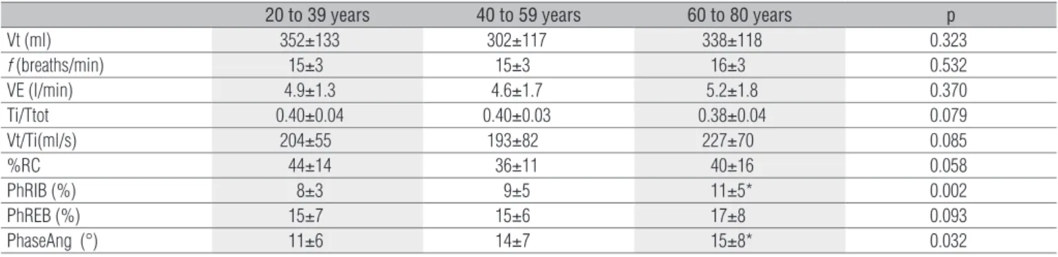
Table 3 presents comparisons of breathing pattern and thoracoabdominal motion between the age groups. No significant differences were observed in any of the variables relating to the breathing pattern. In relation to
thoracoabdominal motion, PhRIB and PhaseAng were sig-nificantly greater in individuals over the age of 60 than in adults between 20 and 39 years.
In addition, comparisons between the age groups were made for the men and women separately. No signiicant difer-ences between the age groups were found among the women. Among the men, there was only a signiicant diference in the
Participants n=104
Age (years) 46.24±19.57
Weight (kg) 67.12±14.45
Height (m) 1.65±0.09
BMI (kg/m2) 24.66±5.36
FEV1 (% predicted) 96.38±10.45
FVC (% predicted) 97.02±10.34
FEV1/FVC 99.67±7.32
FEF25-75% (% predicted) 90.7±25.96
Table 1. Anthropometric, spirometric, and demographic data of the
sample.
Data are presented as mean ± standard deviation. BMI: body mass index; FVC: forced vital capacity; FEV1: forced expiratory volume in the first second; FEV1/FVC: ratio of FEV1 to FVC; and FEF25-75%:forced expiratory flow at 25 and 75% of FVC.
20 to 39 years 40 to 59 years 60 to 80 years
Women Men Women Men Women Men
Vt (ml) 325±127 441±114 * 309±111 325±115 283±83 383±124 *
f (breaths/min) 15±2 13±4 14±2 16±3 15±2 15±3
VE (l/min) 4.69±1.34 5.61±1.13 * 4.43±1.51 4.65±2.08 4.26±1.29 5.98±1.76 *
Ti/Ttot 0.39±0.03 0.42±0.04 * 0.39±0.03 0.41±0.02 0.38±0.04 0.39±0.04
Vt/Ti(ml/s) 199±56 221±46 193±80 192±90 187±49 259±68 *
%RC 46±15 39±10 40±9 32±7 45±18 37±14
PhRIB (%) 7±3 8±3 7±3 11±6 10±4 12±6
PhREB (%) 15±6 12±6 13±6 16±6 16±9 18±8
PhaseAng (°) 11±5 10±6 12±4 17±9 13±7 17±9
Table 2. Respiratory pattern and thoracoabdominal motion data in age and sex subgroups.
Data are presented as mean ± standard deviation. Vt: tidal volume; f: respiratory frequency; VE: minute ventilation; Ti/Tot: inspiratory duty cycle; Vt/Ti: mean inspiratory flow; %RC: rib cage motion; PhRIB: inspiratory phase relation; PhREB: expiratory phase relation; PhaseAng: phase angle. * significant difference (p<0.05) for comparisons between women and men in each age subgroup.
20 to 39 years 40 to 59 years 60 to 80 years p
Vt (ml) 352±133 302±117 338±118 0.323
f (breaths/min) 15±3 15±3 16±3 0.532
VE (l/min) 4.9±1.3 4.6±1.7 5.2±1.8 0.370
Ti/Ttot 0.40±0.04 0.40±0.03 0.38±0.04 0.079
Vt/Ti(ml/s) 204±55 193±82 227±70 0.085
%RC 44±14 36±11 40±16 0.058
PhRIB (%) 8±3 9±5 11±5* 0.002
PhREB (%) 15±7 15±6 17±8 0.093
PhaseAng (°) 11±6 14±7 15±8* 0.032
Table 3. Comparison of respiratory pattern and thoracoabdominal motion variables between the three age subgroups.
Data are presented as mean ± standard-deviation. Vt: tidal volume; f: respiratory frequency; VE: minute ventilation; Ti/Tot: inspiratory duty cycle; Vt/Ti: mean inspiratory flow; %RC: rib cage motion; PhRIB: inspiratory phase relation; PhREB: expiratory phase relation; PhaseAng: phase angle. * significant difference (p<0.05) for comparisons between adult (20 to 39 years) and elderly (60 to 80 years) participants.
variable Ti/Ttot. Men aged 20 to 39 presented signiicantly lower values than did the men over 60 years.
Discussion
he main result from this study was that there were signii-cant diferences in some of the breathing pattern variables be-tween the sexes and in the thoracoabdominal motion variables between the participants in the three age groups evaluated. Comparison between the sexes showed that women presented signiicantly lower values than men for the variables Vt, VE, and Ti/Tot in the age group between 20 and 39 years and for the variables Vt, VE, and Vt/Ti in the elderly group, but without signiicant diferences in the variables related to thoracoab-dominal motion. Comparison between the age groups showed that the participants over the age of 60 presented signiicantly greater PhRIB and PhaseAng, in relation to the participants aged 20 to 39, but without signiicant diferences in the breath-ing pattern variables.
among healthy adults were conducted among populations in other countries and/or using limited numbers of variables or individuals3,7,10. he present study adds important
informa-tion, considering that the values observed among individuals in diferent age groups can be used both in the evaluation process and physical therapy treatment of patients with acute or chronic respiratory dysfunctions. he participants were re-cruited as a convenience sample from residents of the city of Belo Horizonte, Minas Gerais, Brazil, which may be considered to be a limitation of this study. Nonetheless, the number of par-ticipants analyzed is similar to the numbers in other studies re-lated to respiratory function parameters that have put forward reference values36,37.
Regarding the measurement instrument used, it should be emphasized that respiratory inductive plethysmography is an appropriate method for evaluating breathing pattern and thoracoabdominal motion, as proposed in the present study. It is also worth noting that there is a new method for obtaining information that gives greater detail about the operational vol-umes of the thoracic wall. he main innovation of this method is that it provides greater accuracy of analysis during exercise, given that it can produce a three-dimensional analysis that takes into consideration three compartments of the thoracic wall (pulmonary rib cage, abdominal rib cage, and abdomen), thus difering from the respiratory inductive plethysmography that analyzes two compartments. his point is particularly im-portant in the presence of dynamic hyperinlation38,39. It is
un-likely that there was any hyperinlation among the participants in the present study, given that the evaluation was performed on healthy individuals at rest.
Regarding comparisons between the sexes, signiicant dif-ferences in the breathing pattern variables were observed. In relation to the men, the women in the age group 20 to 39 presented signiicantly lower Vt, VE, and Ti/Ttot. To the best of our knowledge, only one other study made a comparison of breathing patterns between men and women, in which indi-viduals aged between 20 and 45 years were evaluated3. Our data
corroborate what was observed previously in relation to Vt, which was signiicantly lower among females. his result can be attributed to the diference in physical constitution between men and women. However, this diference was insuicient to signiicantly inluence VE, which did not present any diference between women and men in these two studies. Regarding the time components of the breathing pattern, Feltrim3 found a
lower inspiratory time in the female group. his result may help to explain the signiicantly lower Ti/Ttot among the women in the present study. In the elderly group, the signiicant difer-ences observed in Vt and VE were similar to those observed among adults aged 20 to 39, and the diference in Vt/Ti seemed to result from the change in Vt, given that no change in Ti/Ttot
was observed. In relation to participants aged 40 to 59, no sig-niicant diference was observed.
In relation to the comparison between the sexes, the only diference was in the Ti/Ttot among the men over 60 years, compared with those aged 20 to 39. Despite the diference ob-served, the values were within the normal range7. Verschakelen
and Demedts10 evaluated the inluence of sex on individuals in
this age group, but only in relation to the variables of thora-coabdominal motion.
Comparison of the variables relating to thoracoabdominal motion between the sexes in the present study did not show any signiicant diference between men and women. his result corroborates the indings in the literature. he inluence of sex on thoracoabdominal motion was previously evaluated in two studies3,10, none of which found any signiicant diference in the
percentage displacement of the chest and abdominal compart-ments between men and women in the supine position at rest. hus, it seems to be well established that displacement of the abdominal compartment is proportionally greater in both men and women in the supine position.
Regarding thoracoabdominal asynchrony, no signiicant diferences were found between men and women. To the best of our knowledge, the inluence of sex on thoracoabdominal asynchrony has not been evaluated in any studies. he values observed in the present study are close to those described in the literature for healthy individuals at rest26.
Comparison between the three age groups did not show any signiicant diferences in the breathing pattern variables. hese results are in agreement with other studies that com-pared breathing pattern variables between healthy adults and elderly individuals. No signiicant diferences were observed in the parameters analyzed at rest in these studies7,11. Recently,
Britto et al.40 evaluated two groups of elderly people, one aged
60 to 69 and one over 69 years, also without signiicant difer-ences in the respiratory pattern.
In relation to thoracoabdominal motion in the present study, the elderly participants presented signiicantly greater PhRIB and PhaseAng than the participants aged 20 to 39. he presence of greater thoracoabdominal asynchrony ob-served among the elderly participants may have been due to structural modiications to the rib cage, weakness of the respiratory muscles, and changes to the respiratory drive41,
given that these factors may increase respiratory overload25.
he inluence of age on thoracoabdominal asynchrony was previously evaluated among 18 elderly individuals7. his study
evaluated the ratio of maximum compartmental amplitude to Vt, which is a parameter measuring thoracoabdominal asynchrony, and did not observe any signiicant diference. It is possible that the limited number of individuals may have inluenced the results.
1. Macklem PT. Clinical assessment on the respiratory muscles. Pneumonol Pol. 1988;56(6):249-53.
2. De Troyer A, Estenne M. Functional anatomy of the respiratory muscles. Clin Chest Med. 1988;9(2):175-93.
3. Feltrim M. Estudo do padrão respiratório e da configuração toracoabdominal em indivíduos normais, nas posições sentada, dorsal e laterais, com o uso da pletismografia por indutância [dissertação]. São Paulo (SP): Universidade Federal de São Paulo; 1994.
4. Kiciman NM, Andreasson B, Bernstein G, Mannino FL, Rich W, Henderson C, et al. Thoracoabdominal motion in newborns during ventilation delivered by endotracheal tube or nasal prongs. Pediatr Pulmonol. 1998;25(3):175-81.
5. Sackner MA, Gonzalez H, Rodriguez M, Belsito A, Sackner DR, Grenvik S. Assessment of asynchronous and paradoxic motion between rib cage and abdomen in normal subjects and in patients with chronic obstructive pulmonary disease. Am Rev Respir Dis. 1984;130(4):588-93.
6. Konno K, Mead J. Measurement of the separate volume changes of rib cage and abdomen during breathing. J Appl Physiol. 1967;22(3):407-22.
7. Tobin MJ, Chadha TS, Jenouri G, Birch SJ, Gazeroglu HB, Sackner MA. Breathing patterns. 1. Normal subjects. Chest. 1983;84(2):202-5.
8. Ragnarsdottir M, Kristinsdottir EK. Breathing movements and breathing patterns among healthy men and women 20-69 years of age. Reference values. Respiration. 2006;73(1):48-54.
9. Maynard V, Bignall S, Kitchen S. Effect of positioning on respiratory synchrony in non-ventilated pre-term infants. Physiother Res Int. 2000;5(2):96-110.
10. Verschakelen JA, Demedts MG. Normal thoracoabdomianl motions. Influence of sex, age, posture, and breath size. Am J Respir Crit Care Med. 1995;151(2 Pt 1):399-405.
11. Britto RR, Vieira DSR, Rodrigues JM, Prado LF, Parreira VF. Comparação do padrão respiratorio em adultos e idosos. Rev Bras Fisioter. 2005;9(3):281-7.
12 Tobin MJ, Perez W, Guenther SM, Lodato RF, Dantzker DR. Does rib cage-abdominal paradox signify respiratory muscle fatigue? J Appl Physiol. 1987;63(2):851-60.
13. Perez T, Becquart LA, Stach B, Wallaert B, Tonnel AB. Inspiratory muscle strength and endurance in steroid-dependent asthma. Am J Respir Crit Care Med. 1996;153(2):610-5.
14. Allen JL, Wolfson MR, McDowell K, Shaffer TH. Thoracoabdominal asynchrony in infants with airflow obstruction. Am Rev Respir Dis. 1990;141(2):337-42.
15. Rusconi F, Gagliardi L, Aston H, Silverman M. Respiratory inductive plethysmography in the evaluation of lower airway obstruction during methacholine challenge in infants. Pediatr Pulmonol. 1995;20(6):396-402.
16. Ashutosh K, Gilbert R, Auchincloss JH Jr, Peppi D. Asynchronous breathing movements in patients with chronic obstructive pulmonary disease. Chest. 1975;67(5):553-7.
17. Delgado HR, Braun SR, Skatrud JB, Reddan WG, Pegelow DF. Chest wall and abdominal motion during exercise in patients with chronic obstructive pulmonary disease. Am Rev Respir Dis. 1982;126(2):200-5.
18. Sharp JT, Goldberg NB, Druz WS, Fishman HC, Danon J. Thoracoabdominal motion in chronic obstructive pulmonary disease. Am Rev Respir Dis. 1977;115(1):47-56.
19. American Thoracic Society; European Respiratory Socitety. ATS/ERS Statement on respiratory muscle testing. Am J Respir Crit Care Med. 2002;166(4):518-624.
20. WHO – World Health Organization. Global database on body mass index: an interactive surveillance tool for monitoring nutrition transition. Geneva: WHO; 2004.
21. Pereira CAC. Espirometria. J Pneumol. 2002;28(Supl 3):S1-S81.
22. Carry PY, Baconnier P, Eberhard A, Cotte P, Benchetrit G. Evaluation of respiratory inductive plethysmography: accuracy for analysis of respiratory waveforms. Chest. 1997;111(4):910-5.
23. Clarenbach CF, Senn O, Brack T, Kohler M, Bloch KE. Monitoring of ventilation during exercise by a portable respiratory inductive plethysmograph. Chest. 2005;128(3):1282-90.
24. Fiamma MN, Samara Z, Baconnier P, Similowski T, Straus C. Respiratory inductive plethysmography to assess respiratory variability and complexity in humans. Respir Physiol Neurobiol. 2007;156(2):234-9.
25. Alves GS, Britto RR, Campos FC, Vilaça AB, Moraes KS, Parreira VF. Breathing pattern and thoracoabdominal motion during exercise in chronic obstructive pulmonary disease. Braz J Med Biol Res. 2008;41(11):945-50.
26. Bloch KE, Li Y, Zhang J, Bingisser R, Kaplan V, Weder W, et al. Effect of surgical lung volume reduction on breathing patterns in severe pulmonary emphysema. Am J Respir Crit Care Med. 1997;156(2 Pt 1):553-60.
27. Mayer OH, Clayton RG Sr, Jawad AF, McDonough JM, Allen JL. Respiratory inductance plethysmography in healthy 3- to 5-year-old children. Chest. 2003;124(5):1812-9.
28. Parreira VF, Tomich GM, Britto RR, Sampaio RF. Assessment of tidal volume and thoracoabdominal motion using volume and flow-oriented incentive spirometers in healthy subjects. Braz J Med Biol Res. 2005;38(7):1105-12.
29. Reber A, Bobbià SA, Hammer J, Frei FJ. Effect of airway opening manoeuvres on thoraco-abdominal asynchrony in anaesthetized children. Eur Respir J. 2001;17(6):1239-43.
30. Reber A, Geiduschek JM, Bobbià SA, Bruppacher HR, Frei FJ. Effect of continuous positive airway pressure on the measurement of thoracoabdominal asynchrony and minute ventilation in children anesthetized with sevoflurane and nitrous oxide. Chest. 2002;122(2):473-8.
31. Tomich GM, França DC, Diório AC, Britto RR, Sampaio RF, Parreira VF. Breathing pattern, thoracoabdominal motion and muscular activity during three breathing exercises. Braz J Med Biol Res. 2007;40(10):1409-17.
32. Tomich G. Exercícios respiratorios após gastroplastia: análise do padrão respiratorio e do movimento toracoabdominal [dissertação]. Belo Horizonte (MG): Universidade Federal de Minas Gerais; 2006.
33. Sackner MA. Diagnostic Techniques in Pulmonary Disease. In: Sackner MA, editor. Lung biology in healthy and disease. New York: Marcel Dekker; 1980. p. 525-37.
34. Sackner MA, Watson H, Belsito AS, Feinerman D, Suarez M, Gonzalez G, et al. Calibration of respiratory inductive plethysmograph during natural breathing. J Appl Physiol. 1989;66(1):410-20.
35. Parreira VF, Coelho EM, Tomich GM, Alvim AMA, Sampaio RF, Britto RR. Avaliação do volume corrente e da configuração toracoabdominal durante o uso de espirômetros de incentivo a volume e a fluxo, em sujeitos saudáveis: influência da posição corporal. Rev Bras Fisioter. 2004;8(1):45-51.
36. Neder JA, Andreoni S, Lerario MC, Nery LE. Reference values for lung function tests. II. Maximal respiratory pressures and voluntary ventilation. Braz J Med Biol Res. 1999;32(6):719-27.
37. Neder JA, Andreoni S, Castelo-Filho A, Nery LE. Reference values for lung function tests. I. Static volumes. Braz J Med Biol Res. 1999;32(6):703-17.
38. Aliverti A, Stevenson N, Dellaca RL, Lo Mauro A, Pedotti A, Calverley PM. Regional chest wall volumes during exercise in chronic obstructive pulmonary disease. Thorax. 2004;59(3):210-6.
39. Vogiatzis I, Georgiadou O, Golemati S, Aliverti A, Kosmas E, Kastanakis E, et al. Patterns of dynamic hyperinflation during exercise and recovery in patients with severe chronic obstructive pulmonary disease. Thorax. 200;60(9):723-9.
40. Britto RR, Zampa CC, de Oliveira TA, Prado LF, Parreira VF. Effects of the aging process on respiratory function. Gerontology. 2009;55(5):505-10.
41. Jassens JP, Pache JC, Nicod LP. Physiological changes in respiratory function associated with ageing. Eur Respir J. 1999;13(1):197-205.
In conclusion, the present study made it possible to de-scribe values for variables relating to breathing pattern and thoracoabdominal motion in men and women of diferent age groups. he data found in this study suggest that the breathing pattern is inluenced by sex whereas the thoracoabdominal motion is inluenced by age.
Acknowledgements
Fundação de Amparo à Pesquisa do Estado de Minas Gerais (FAPEMIG) and Conselho Nacional de Desenvolvimento Cientíico e Tecnológico (CNPq).