Prevalence and Correlates of Vascular Disease at Ultrasound
Examination in Patients on Hemodialysis
Sebastião Baptista Miguel
1,2, Jair Baptista Miguel
1,2, Luis Guillermo Velarde
3, Elisa de Albuquerque Sampaio
2, Jorge
Paulo Strogoff de Matos
2, Jocemir Ronaldo Lugon
2Clinefron Clínica de Doenças Renais1, Santo Antonio de Pádua, RJ, Divisão de Nefrologia, Faculdade de Medicina, Universidade Federal Fluminense2, Niterói, RJ, Departamento de Estatística, Instituto de Matemática e Estatística, Universidade Federal Fluminense3, Niterói, RJ - Brazil
Mailing address: Jocemir Ronaldo Lugon •
Rua Haddock Lobo 369/309 - Tijuca - 20260-131 - Rio de Janeiro, RJ - Brazil E-mail: [email protected]
Manuscript received July 05, 2010; revised manuscript received October 20, 2010; accepted November 30, 2010.
Abstract
Background: Patients on hemodialysis present an increased risk of cardiovascular death. Intimal media thickness (IMT) and presence of arterial calcifications are well-known risk factors for cardiovascular death in hemodialysis patients.
Objective: To assess the prevalence of IMT and arterial calcifications in HD patients and to correlate image findings with clinical and laboratory data.
Methods: Cross-sectional study involving 75 patients on dialysis for > 12 months. Patients underwent B-mode ultrasound scan (US) for determination of IMT of the distal third of the common carotid arteries. Arterial calcifications were assessed by US of carotids, femoral and tibial arteries, and labeled positive if calcification was found in any arterial site.
Results: Patients were 52 ± 13 years old, 57% were males and 16% were diabetics. IMT ≥ 0.9 mm was found in 57% of
cases and arterial calcifications at US in 48%. Aging (decades) and smoking were associated with both increased IMT
(adjusted odds ratio [aOR] = 3.4, p < 0.001; aOR = 4.4, p = 0.045, respectively) and presence of vascular calcifications (aOR = 3.0, p < 0.001; aOR = 6.8, p = 0.011, respectively). High intact parathyroid hormone levels (per each 100 pg/ml) were significantly associated with increased IMT (aOR = 1.7, p = 0.021), but not with vascular calcification. In contrast, Diabetes and time on dialysis (years) were significant determinants for calcifications at US (aOR = 15.0, p = 0.009; aOR = 1.39, p = 0.020), but not for increased IMT.
Conclusion: Increased IMT and calcifications at US are common findings in hemodialysis patients. Aging and smoking are consistent determinants for both image alterations. Parathyroid hormone elevation is associated with increased IMT. Diabetes and time on dialysis substantially increase the risk for arterial calcification. (Arq Bras Cardiol
2011;96(4):260-265)
Keywords: Hemodialysis, vascular diseases, calcinosis, Doppler ultrasonography.
Introduction
End-stage renal disease (ESRD) patients have a high mortality rate, mainly related to cardiovascular disease1. The
development of non-invasive strategies for early detection of vascular disease could provide opportunities for an early patient approach. B-mode ultrasonography (US), which carries the potential for detection of early stages of vascular disease, can be used for the measurement of intimal-media thickness (IMT), considered a marker of atherosclerosis, and for detection of arterial calcifications2, which have
been shown to be associated with a poorer survival rate on dialysis3,4. Currently, other diagnostic methods are available
for the identification of arterial calcification in the clinical setting, including x-rays, echocardiography and electron-beam
computer tomography5-7. However, B-mode ultrasonography
is a low-cost and easy-to-perform technique.
The objectives of the present study were to assess the prevalence of increased IMT and vascular calcifications using B-mode US and to correlate the findings with clinical and laboratory data.
Methods
This is a cross-sectional, single-center study in which patients from a single dialysis center aged between 18 and 75 years, who had been on hemodialysis for at least 12 months, were approached for enrollment. The study was approved by the local Ethics Committee and all patients signed the informed consent form. Evaluation was accomplished between March 2006 and September 2007.
Imaging studies
ultrasonography devices (Medison 8000, and Esaote Caris Plus). IMT measurements were performed in the distal third of the common carotid arteries as previously suggested2. Cases with any value ≥ 0.9 mm, a cutoff previously associated with
elevated incidence of cardiovascular events in a non-dialysis population8,9, were labeled positive. At the ultrasonographic
evaluation of vascular calcification,a highly echogenic vascular wall or intraluminal structure producing bright echoes with shadowing were considered to be calcifications10. The sites
investigated were: the distal third of the common carotid arteries; carotid and proximal third of the internal and external branches; the whole length of common femoral arteries, proximal third of superficial and deep femoral arteries; and distal half of posterior tibial arteries10. If a calcification was
found in any arterial site, the case was labeled positive11.
Demographic, clinical and laboratory data
Demographic and clinical data were derived from chart examination. Hypertension was defined as current
pre-dialysis systolic BP ≥ 140 mmHg and/or diastolic BP ≥ 90
mmHg and/or use of antihypertensive medications. Patients were classified as diabetics if they had diabetes previously to renal replacement therapy. Hemoglobin, creatinine, and equilibrated Kt/V (eKt/V) values represent the mean of the last three determinations before evaluation. To better estimate the impact of mineral metabolism disturbances on findings, cumulative exposure was assessed through calculation of the mean of all values for serum calcium, phosphorus and intact parathyroid hormone (i-PTH) measurements along a 36-month period just before evaluation or since hemodialysis initiation for patients on renal replacement therapy for less than 3 years. Calcium and phosphorus serum levels were measured on a monthly basis and i-PTH every six months. Routine blood analyses were performed in the same laboratory. An ultra-sensitive immunoturbidimetric assay for C-reactive protein (CRP) was specifically performed for the study and the values shown here correspond to a single determination by the time of study enrollment.
Statistical analyses
Continuous variables are presented as mean ± SD or median and range as appropriate. Categorical variables are shown as frequencies. Differences between frequencies were analyzed by the Chi-square test. Associations between variables were assessed by backward conditional logistic regression analysis, in which all potentially relevant variables are included in the first step. Only variables presenting P values lower than 0.10 persisted in the next step. In case of significant collinearity between two variables, only the more relevant persisted in the next step. The decision about which variables should be excluded from the analysis in each step was made by the software. P values lower than 0.05 were considered significant. The software SPSS, version 17.0 was used for all statistics.
Results
Of 128 patients on hemodialysis, 75 were enrolled in this study. The main reasons for exclusions were: dialysis time
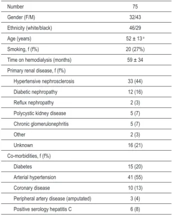
lower than one year (18 patients); age older than 75 years (14 patients); and refusal to participate in the study (12 patients). Patients enrolled in the study were 52 ± 13 years old, 43 (57%) males, 59 ± 34 months on hemodialysis, and 12 (16%) were diabetics. Demographic and clinical characteristics of patients are shown in Table 1. Patients presented a wide range of age (Figure 1) and time on dialysis. Forty patients (53%) were on hemodialysis for less than 5 years, 30 (40%) for 5 to 10 years and 5 (7%) for more than 10 years. Laboratory data are shown in Table 2.
B-mode US was performed in all 75 patients. Increased IMT was present in 43 (57%) and arterial calcifications were detected in 36 (48%), Table 3. Calcification was present in more than three arteries in 23 of these 36 patients and 33 of them also exhibited increased IMT. Ten patients (22%) with increased IMT did not have ultrasonographic calcifications.
In search for determinants of the image findings, two models of backward conditional regression analysis were developed,
with each one using IMT ≥ 0.9 mm and calcifications at
US as the dependent variables. Clinical and laboratory data were used as the independent variables. Age (adjusted odds ratio [aOR] = 3.36 per decade, p < 0.001), smoking (aOR = 4.49, p = 0.046), and high iPTH levels (1.67 per 100 pg/
ml) were significantly associated with IMT ≥ 0.9 mm, Table 4.
Age (aOR = 2.96 per decade, p < 0.001) and smoking (aOR = 6.77, p = 0.011) were also associated with calcifications at US, Table 5. Diabetes (aOR = 1.67, p = 0.021) and time on dialysis (aOR = 1.39, p = 0.020) were associated with a significant risk for calcifications at US (aOR = 1.67, p =
Table 1 - Demographic and clinical characteristics of patients
Number 75
Gender (F/M) 32/43
Ethnicity (white/black) 46/29
Age (years) 52 ± 13 a
Smoking, f (f%) 20 (27%)
Time on hemodialysis (months) 59 ± 34
Primary renal disease, f (f%)
Hypertensive nephrosclerosis 33 (44)
Diabetic nephropathy 12 (16)
Relux nephropathy 2 (3)
Polycystic kidney disease 5 (7)
Chronic glomerulonephritis 5 (7)
Other 2 (3)
Unknown 16 (21)
Co-morbidities, f (f%)
Diabetes 15 (20)
Arterial hypertension 41 (55)
Coronary disease 10 (13)
Peripheral artery disease (amputated) 3 (4)
Positive serology hepatitis C 6 (8)
Table 2 - Laboratory data of patients
Hemoglobin (g/dl) 10.9 ±1.5 a
Creatinine (mg/dl) 10.9 ± 3.4
Calcium (mg/dl) 9.4 ± 1.0
Phosphorus (mg/dl) 4.9 ± 1.1
CaxP (mg2 x dl2) 46.2 ± 11.6
iPTH (pg/ml) 281 (43-1,759)
Equilibrated Kt/V 1.6 ± 0.2
C-reactive protein (mg/l) 6 (0 -150)
a Mean ± standard deviation ormedian (range).
Table 3 - Findings on image studies
IMT ≥ 0.9 mm 43 (57%)
Calciication at US 36 (48%)
IMT - intimal media thickness; US - ultrasonography.
Table 4 - Backward conditional regression analysis for association of clinical and laboratory parameters with increased IMT
Odds ratio 95% conidence
interval p value
Age (decades) 3.36 1.81 - 6.21 <0.001
Smoking (y/n) 4.39 1.03 - 18.77 0.046
iPTH (each 100 pg/ml) 1.67 1.08 - 2.58 0.021
Variables entered on step 1 - gender, time on dialysis, smoking, age, diabetes, i-PTH, C-reactive protein.
Figure 1 -Distribution of the patients according to age.
40 to 49 50 to 59
<40 60 to 69 70 to 75
highest band (> 300 pg/ml) in comparison to the middle band (aOR = 6.26 [1.05 - 37.50], p = 0.045).
Discussion
It is well known that ESRD patients bear an elevated risk for cardiovascular diseases, which represent the leading cause of death in this population1. The arterial disease is generalized
and the presence of carotid structural defects2,8, as well as
peripheral vascular disease2,12 are strongly correlated with
coronary disease and mortality. The objective of this study was to assess vascular disease in ESRD patients using non-invasive imaging studies and to correlate the findings with clinical and laboratory data. The profile of our patients is roughly similar to the overall population on dialysis in Brazil13.
The frequency of IMT ≥ 0.9 mm, a recognized marker of
atherosclerosis, was very high, affecting 43 (57%) of patients. Although a number of studies had already addressed the issue of IMT determination in ESRD patients14,15, we could not find the incidence of IMT ≥ 0.9 mm in a non-selected hemodialysis
sample for an appropriate prevalence comparison. It should be emphasized that the rate of 57% was obtained with a cutoff of 0.9 mm, which is associated with increased incidence of cardiovascular events8, but is, in some way, conservative. In
a recent study, for instance, it was found that the upper limits (97.5 percentile) of IMT in a healthy population aged 50 to 59, and 60 to 75 years were 0.71 and 0.81 mm, respectively16.
Calcifications at US were also common, affecting 36 (48%) of patients, a proportion comparable to the ones reported in previous studies, which varied from 50 to 64%17-19.
Thirty-three out of our 36 patients with calcifications also exhibited increased IMT, whereas 10 patients with increased IMT did not have ultrasonographic calcifications. These findings are 0.021 and aOR = 1.39, p = 0.020, respectively), but not for
increased IMT. Phosphorus, calcium, Ca x P product and iPTH were not found to be risk factors for vascular calcification at US. C-reactive protein levels were not associated with IMT
≥ 0.9 mm or calcifications at US.
To better explore the relationship between i-PTH levels and IMT an additional multivariate regression analysis model was used, in which i-PTH levels were stratified in 3 categorical ranges: < 150 pg/ml, 150 to 300 pg/ml, and > 300 pg/ml. In this analysis, a significant elevated risk was restricted to the
Table 5 - Backward conditional regression analysis for association of clinical and laboratory parameters with vascular calciication
Odds ratio
95% conidence
interval p value
Age (decades) 2.96 1.63 – 5.36 <0.001
Smoking (y/n) 6.77 1.56 - 29.3 0.011
Diabetes (y/n) 1.67 1.08 – 2.58 0.021
Time on dialysis (years) 1.39 1.05 – 1.82 0.020
consistent with the present view that increased IMT can be seen as a preclinical marker of atherosclerosis that precedes plaque formation2,20.
To check for potential determinants of image findings in our study, we resorted to two models of logistic regression analysis, using selected clinical and laboratory data as independent variables against each one of the image findings. Age uniformly influenced image parameters, in a way that each decade of age was associated with approximately a three-fold increase in the chance of a positive finding in the studied image parameters. This finding is consistent with the extensive information available in the literature, highlighting the role of age on the development of vascular disease16,21. Similarly, smoking
influenced both image parameters, with a four-fold increase in the chance of a positive IMT and an almost seven-fold increase in the chance of a calcification. Studies addressing smoking as a risk factor for vascular calcification in ESRD patients are scarce, but the smoking habit is a traditional risk factor for cardiovascular disease, with the risk being strongly dose-related22. The effects of time on dialysis, as well as of
diabetes, however, were only seen towards the presence of calcifications. A recent study showed that patients on hemodialysis have roughly an eight-fold increased risk of having vascular calcification, when matched to subjects in general population23. Diabetes is a well-known risk factor for vascular
calcifications24 and the absence of its influence on increased
IMT allows us to speculate whether the uremic environment can prevail upon glucose metabolism derangements during the early phase of vascular structural abnormalities in ESRD25
patients. However, caution is advised in this regard, as the size of the sample may have contributed to the lack of impact of this factor upon IMT enlargement. High levels of i-PTH levels were associated with increased IMT. In an attempt to better clarify this issue, we resorted to an additional regression model employing i-PTH stratification. Significant association between i-PTH levels and increased IMT was restricted to the comparison between the group with PTH levels > 300 pg/ml and the one with i-PTH values between 150 and 300 pg/ml.
Interestingly, we did not find a correlation between vascular calcifications and cumulative mineral metabolism disturbances in our study. However, this finding cannot be seen as an argument against the reported role of phosphate overload in the development of vascular calcification26,27,
as phosphate serum levels may not be a precise marker of the oral phosphate load28. In addition, it does not refute the
extensive literature placing mineral metabolism disturbances as pivotal risk factors for the high cardiovascular mortality among ESRD patients29,30, as mortality was not addressed in
our study. It should be mentioned that, in agreement to our findings, absence of correlation between Ca x P product and IMT had already been reported15.
Our study carries limitations and the negative data should be interpreted with caution. The cross-sectional character of the design and the low number of patients may have contributed to the lack of significance in some statistics. However, the positive findings reinforce the role of some traditional risk factors such as aging, smoking, and diabetes as determinants of vascular disease in the ESRD population. They also show that some non-traditional factors, such as time on dialysis and elevated i-PTH levels can have an impact upon vascular abnormalities in such patients.
Conclusions
Both increased IMT and calcifications at US are common findings in hemodialysis patients, but increased IMT was more frequent. Smoking was a consistent determinant for both image findings. The major impact was seen with age, so that each decade of age was associated with a three-fold increase in the chance of a positive finding in any of the imaging studies. Diabetes and time on dialysis substantially increase the risk for arterial calcification.
Potential Conflict of Interest
No potential conflict of interest relevant to this article was reported.
Sources of Funding
There were no external funding sources for this study.
Study Association
This article is part of the thesis of master submitted by Sebastião Baptista Miguel, from Universidade Federal Fluminense.
References
1. Foley RN, Parfrey PS, Sarnak M. Clinical epidemiology of cardiovascular disease in chronic renal disease. Am J Kidney Dis. 1998;32(5 Suppl. 3):112-9.
2. Van der Meer IM, Bots ML, Hofman A, del Sol AL, van der Kuip DA, Witterman JC. Predictive value of non-invasive measures of atherosclerosis for incident myocardial infarction. The Rotterdam study. Circulation. 2004;109(9):1089-94.
3. Papagianni A, Dovas S, Bantis C, Belechri AM, Kalovoulos M, Dimitriadis C, et al. Carotid atherosclerosis and endothelial cell adhesion molecules as predictors of long-term outcome in chronic hemodialysis patients. Am J Nephrol. 2008;28(2):265-74.
4. Benedetto FA, Tripepi G, Mallamaci F, Zoccali C. Rate of atherosclerotic plaque formation predicts cardiovascular events in ESRD. J Am Soc Nephrol. 2008;19(4):757-63.
5. Adragão T, Frazão JM. Cardiovascular risk in dialysis patients: an X-ray vision on vascular calcifications. Kidney Int. 2008;74(12):1505-7.
7. Haydar AA, Hujairi NM, Covic AA, Pereira D, Rubens M, Goldsmith DJ. Coronary artery calcification is related to coronary atherosclerosis in chronic renal disease patients: a study comparing EBCT-generated coronary artery calcium scores and coronary angiography. Nephrol Dial Transplant. 2004;19(9):2307-12.
8. Price JF, Tzoulaki I, Lee AJ, Fowkes FG. Ankle brachial index and intima media thickness predict cardiovascular events similarly and increased prediction when combined. J Clin Epidemiol. 2007;60(10):1067-75.
9. Savage T, Clarke AL, Giles M, Tomson CR, Raine AE. Calcified plaque is common in the carotid and femoral arteries of dialysis patients without clinical vascular disease. Nephrol Dial Transplant. 1998; 13(8):2004-12.
10. Guerin AP, London GM, Marchais SJ, Metivier F. Arterial stiffening and vascular calcifications in end-stage renal disease. Nephrol Dial Transplant. 2000;15(7):1014-21.
11. Blacher J, Guerin AP, Pannier B, Marchais SJ, London GM. Arterial calcifications, arterial stiffness, and cardiovascular risk in end-stage renal disease. Hypertension. 2001; 38(4):938-42.
12. Pearte CA, Furberg CD, O’Meara ES, Psaty BM, Kuller L, Power NR, et al. Characteristics and baseline clinical predictors of future fatal versus non-fatal coronary heart disease events in older adults: the Cardiovascular Health Study. Circulation. 2006;113(18):2177-85.
13. Sesso R, Lopes AA, Thomé FS, Bevilacqua JL, Romão Jr JE, Lugon J. Relatório do Censo Brasileiro de Diálise, 2008. J Bras Nefrol 2008; 30(4):233-8
14. Ohkuma T, Minagawa T, Takada N, Ohno M, Oda H, Ohashi H. C-reactive protein, lipoprotein(a), homocysteine, and male sex contribute to carotid atherosclerosis in peritoneal dialysis patients. Am J Kidney Dis. 2003;42(6):355-61.
15. Zoccali C, Benedetto FA, Maas R, Mallamaci F, Tripepi G, Malatino LS, et al. Asymmetric dimethylarginine, C-reactive protein, and carotid intima-media thickness in end-stage renal disease. J Am Soc Nephrol. 2002;13(2):490-6.
16. Lim TK, Lim E, Dwivedi G, Kooner J, Senior R. Normal value of carotid intima-media thickness - a surrogate marker of atherosclerosis: quantitative assessment by B-mode carotid ultrasound. J Am Soc Echocardiogr. 2008;21(2):112-6.
17. London GM. Cardiovascular disease in chronic renal failure: pathophysiologic aspects. Semin Dial. 2003;16(2):85-94.
18. Wang AY, Ho SS, Wang M, Liu EK, Ho S, Lui SF, et al. Cardiac valvular calcification as a marker of atherosclerosis and arterial calcification in end-stage renal disease. Arch Intern Med. 2005;165(3):327-32.
19. Yildiz A, Memisoglu E, Oflaz H, Yazic H, Pusuraglu H, Akkaya V, et al. Atherosclerosis and vascular calcification are independent predictors of left
ventricular hypertrophy in chronic haemodialysis patients. Nephrol Dial Transplant. 2005;20(4):760-7.
20. Zureik M, Ducimetière P, Touboul PJ, Courbon D, Bonithon-Kopp C, Berr C, et al. Common carotid intima-media thickness predicts occurrence of carotid atherosclerotic plaques: longitudinal results from the Aging Vascular Study (EVA) study. Arterioscler Thromb Vasc Biol. 2000;20(6):1622-9.
21. Juonala M, Kähönen M, Laitinen T, Hutri-Kahonen N, Jokinen E, Taittonen L, et al. Effect of age and sex on carotid intima-media thickness, elasticity and brachial endothelial function in healthy adults: the cardiovascular risk in Young Finns Study. Eur Heart J. 2008;29(9):1198-206.
22. Ockene IS, Miller NH. Cigarette smoking, cardiovascular disease, and stroke: a statement for healthcare professionals from the American Heart Association. American Heart Association.Task Force on Risk Reduction. Circulation. 1997;96(9):3243-7.
23. Coll B, Betriu A, Martínez-Alonso M, Amoldo ML, Arcidiacono MV, Borras M, et al. Large artery calcification on dialysis patients is located in the intima and related to atherosclerosis. Clin J Am Soc Nephrol. 2010, Oct 7 [Epub ahead of print].
24. London GM, Marchais SJ, Guérin AP, Metivier F. Arteriosclerosis, vascular calcifications and cardiovascular disease in uremia. Curr Opin Nephrol Hypertens. 2005;14(6):525-31.
25. Jono S, Nishizawa Y, Shioi A, Morili H. Parathyroid hormone-related peptide as a local regulator of vascular calcification: its inhibitory action on in vitro calcification by bovine vascular smooth muscle cells. Arterioscler Thromb Vasc Biol. 1997;17(6):1135-42.
26. Shigematsu T, Kono T, Satoh K, Yokoyama K, Yoshida T, Hosoya T, et al. Phosphate overload accelerates vascular calcium deposition in end-stage renal disease patients. Nephron Dial Transplant. 2003;18(Suppl. 3):S86-9.
27. Neves KR, Graciolli FG, dos Reis LM, Graciolli RG, Neves CL, Magalhães AO, et al. Vascular calcification: contribution of parathyroid hormone in renal failure. Kidney Int. 2007;71(12):1262-70.
28. El-Abbadi MM, Pai AS, Leaf EM, Yang HY, Bartley BA, Quan KK, et al. Phosphate feeding induces arterial medial calcification in uremic mice: role of serum phosphorus, fibroblast growth factor-23, and osteopontin. Kidney Int. 2009;75(12):1297-307.
29. Block GA, Port FK. Re-evaluation of risks associated with hyperphosphatemia and hyperparathyroidism in dialysis patients: Recommendations for a change in management. Am J Kidney Dis. 2000;35(6):1226-37.