77 Magnetic resonance imaging of knee osteonecrosis
Radiol Bras. 2010 Mar/Abr;43(2):77–80 Original Article • Artigo Original
Magnetic resonance imaging of knee osteonecrosis:
a study of 19 cases*
Ressonância magnética da osteonecrose do joelho: estudo de 19 casos
Daniel Leme da Cunha1, Antonio Carlos Pires Carvalho2, Elísio José Salgado Ribeiro3, Romeu Côrtes Domingues4
OBJECTIVE: To describe epidemiological, clinical and magnetic resonance imaging findings of osteonecrosis in the distal femur and proximal tibia. MATERIALS AND METHODS: Evaluation of 19 patients (12 women and 7 men), with no previous history of causative factors, with magnetic resonance imaging findings suggestive of osteonecrosis in the tibial plateau or femoral condyle. RESULTS: Osteochondral abnormalities were observed in 63.1% of the cases; in 73.6% of them, such abnormality was associated with ipsilateral meniscal lesion. Also, a significant association with bone marrow edema (grade III in 16 cases) was observed. CONCLUSION: Magnetic resonance imaging has demonstrated to be a noninvasive method with good sensitivity in the diagnosis of knee osteonecrosis as well as of associated lesions which are most frequently found in women (63% of cases).
Keywords: Osteonecrosis; Magnetic resonance imaging; Subchondral abnormalities.
OBJETIVO: Descrever os achados epidemiológicos, clínicos e de ressonância magnética da osteonecrose das porções distal do fêmur e proximal da tíbia. MATERIAIS E MÉTODOS: Avaliação de 19 pacientes (12 mulheres e 7 homens), sem história prévia de fatores causais, com achados à ressonância magnética suges-tivos de osteonecrose do platô tibial ou côndilo femoral. RESULTADOS: Verificou-se a presença de anorma-lidades osteocondrais em 63,1% dos casos e em 73,6% destes houve associação com lesão meniscal ipsi-lateral. Houve também importante associação com edema na medular óssea em correspondência (grau III em 16 casos). CONCLUSÃO: A ressonância magnética demonstrou ser um método não invasivo com boa sensibilidade no diagnóstico da osteonecrose do joelho, bem como das lesões associadas, sendo mais fre-quente nas mulheres (63% dos casos).
Unitermos: Osteonecrose; Imagem por ressonância magnética; Anormalidades subcondrais. Abstract
Resumo
* Study developed at Faculdade de Medicina da Universidade Federal do Rio de Janeiro (UFRJ) and Clínicas de Diagnóstico Por Imagem (CDPI) and Multi-Imagem, Rio de Janeiro, RJ, Brazil.
1. Master Fellow degree, Program of Post-Graduation, Facul-dade de Medicina da UniversiFacul-dade Federal do Rio de Janeiro (UFRJ), MD, Radiologist at Clínicas de Diagnóstico Por Imagem (CDPI) and Multi-Imagem, Rio de Janeiro, RJ, Brazil.
2. PhD, Associate Professor, Adjunct Coordinator for the Pro-gram of Post-Graduation at Faculdade de Medicina da Universi-dade Federal do Rio de Janeiro (UFRJ), Rio de Janeiro, RJ, Brazil. 3. MD, Radiologist, Clínicas de Diagnóstico Por Imagem (CDPI) and Multi-Imagem, Rio de Janeiro, RJ, Brazil.
4. MD, Radiologist, Director of Clínicas de Diagnóstico Por Imagem (CDPI) and Multi-Imagem, Rio de Janeiro, RJ, Brazil.
Mailing address: Dr. Daniel Leme da Cunha. Rua Miguel de Frias, 77, Bloco 2, ap. 1403, Icaraí. Niterói, RJ, Brazil, 24220-008. E-mail: [email protected]
Received September 27, 2009. Accepted after revision De-cember 3, 2009.
sity, besides the advantage of noninva-siveness and high-definition multiplanar images(3).
Ramnath & Kattapuram(4) have related
the presence of a “linear” subchondral sig-nal abnormality in cases supposedly origi-nated in insufficiency fracture, such factor being absent in cases of osteoarthrosis (which also presents similar abnormalities related to osteonecrosis in the subchondral bone).
The present study was aimed at describ-ing epidemiological, clinical and mainly magnetic resonance imaging findings of osteonecrosis of the knee.
MATERIALS AND METHODS
Nineteen magnetic resonance imaging studies of knees of 12 women and 7 men with no previous history of causal factors, Cunha DL, Carvalho ACP, Ribeiro EJS, Domingues RC. Magnetic resonance imaging of knee osteonecrosis: a study of 19 cases. Radiol Bras. 2010;43(2):77–80.
significant gonalgia not related to local trauma, meniscal surgery or even previous corticoid therapy. Yamamoto & Bullough(1)
indicate that, additionally to the supposed vascular origin associated with increase in the intraosseous pressure, the mechanism of insufficiency fracture would play a role of relevant etiopathogenic factor besides osteoarthrosis, so the term “spontaneous” is questioned, considering that causal fac-tors might by implied.
The diagnosis is based on images that confirm the clinical suspicion. At the early phases of the disease, conventional radiog-raphy presents normal findings or minimal changes(2). In cases where conventional
radiological signs are normal or dubious, magnetic resonance imaging is particularly useful for confirming the diagnostic suspi-cion since it demonstrates suggestive or even typical abnormalities in signal
inten-0100-3984 © Colégio Brasileiro de Radiologia e Diagnóstico por Imagem INTRODUCTION
78
Cunha DL et al.
Radiol Bras. 2010 Mar/Abr;43(2):77–80
with acute symptoms and signs compatible with osteonecrosis at examination were evaluated in the period from March to De-cember/2007. Patients with previous his-tory of trauma, local surgery of corticoid therapy were excluded. All the patients in-cluded in the present study were white, with ages ranging between 45 and 77 years (mean, 61 years) presented gonalgia. The following characteristics were taken into consideration in the clinical analysis: pain pattern (sudden or insidious; focal or dif-fuse) and association with joint edema, blockage and instability.
Magnetic resonance imaging studies were performed in a GE 1.5 T Signa sys-tem (General Electric Medical Syssys-tems, Milwaukee, WI, USA), with acquisition of sagittal, T1-weighted sequence (repetition time [TR] = 405; time of excitation [TE] = 13.9; number of excitations [NEX] = 1; echo train length [ETL] = 2) and with pro-ton density, fat-suppression in the coronal plane (TR = 1775; TE = 14; NEX = 2; ETL = 4), axial plane (TR = 1975; TE = 21; NEX = 2; ETL = 4) and sagittal plane (TR = 2250; TE = 14; NEX = 2; ETL = 4).
Subchondral bone abnormalities were evaluated by two radiologists with experi-ence in musculoskeletal system. Such ab-normalities are defined as focal changes in signal intensity, with hypointense signal on T1-weighted images and variable signal
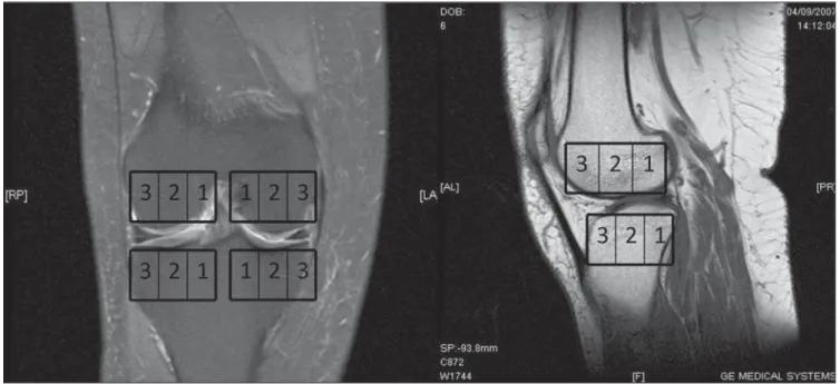
intensity (hyper or hypointense) on T2-weighted images. The lesions location was defined as follows: distal femoral extrem-ity (medial or lateral condyle) or tibial (in-ternal or ex(in-ternal plateau). Subsequently, each of the above described locations was arbitrarily divided into three zones, both in the coronal and sagittal planes to define the lesion epicenter. On the coronal plane (Fig-ure 1), zone 1 would correspond to the in-ternal third; zone 2 to the middle third, and zone 3, to the external third of the femoral condyle/tibial plateau. On the sagittal plane, 1, 2 and 3 would correspond, respec-tively, the posterior, middle and anterior thirds of the evaluated regions. Addition-ally, the presence or not signal intensity lin-ear subchondral abnormality was evalu-ated. The presence of ipsilateral associated bone marrow edema, articular cartilage abnormality and meniscal lesion (whether unstable or not). Bone marrow edema was classified into grades I to III (respectively mild, moderate or significant). Subchon-dral lesions were measured (in millimeters) on the three planes (longitudinal × antero-posterior × cross-sectional planes).
RESULTS
As regards clinical presentation, 14 pa-tients (73.6%) had a sudden pain onset, and in 5 patients the gonalgia onset was
insidi-ous. Eight patients reported joint blockage, two reported increased joint volume (edema), and three reported joint instabil-ity.
All the patients presented lesions with hypointense signal on T1-weighted se-quence and predominantly hyperintense signal (89.4%) on proton density weighted images with fat suppression.
In eight cases, lesions were located in the external tibial plateau, in nine cases, in the medial femoral condyle, and in the in-ternal tibial plateau in only two patients. In-volvement of the lateral femoral condyle was observed in none of the cases. Sagit-tal images demonstrated a higher number of lesions with epicenter in the zone 2 (17 cases; 89.4%), two cases in the zone 3, and none in the zone 1. On coronal images, most of findings were observed in the zone 3 (11 cases; 57.8%), followed by the zone 2 (7 cases) and the zone 1 in a sole case.
The presence of linear subchondral ab-normality (Figure 2) was observed in 15 among the 19 cases (78.9%). Such abnor-mality was absent in the four remainder cases.
Association with grade III bone marrow edema was observed in 16 cases, and grade II in two cases (Figure 3).
Lesions size ranged from 1.5 to 8 mm on the longitudinal axis, from 2 to 30 mm on the anteroposterior axis, and from 6 to
79 Magnetic resonance imaging of knee osteonecrosis
Radiol Bras. 2010 Mar/Abr;43(2):77–80
26 mm on the cross-sectional axis [mean: 4.5 mm (lateral axis); 16.8 mm (ântero-pos-terior axis); and 11.8 mm (cross-sectional axis) – mean estimated volume, 466 mm3]. Chondral abnormalities were found in 63.1% of the patients, while associated ipsilateral meniscal lesion was observed in 14 cases (73.6%), with instability criteria (radial lesion, root ligament lesion) in 6 (31.5%) of these cases.
DISCUSSION
Spontaneous osteonecrosis of the knee is a condition that affects predominantly fe-male individuals, particularly in their sev-enth decade of life(5), with no association
with systemic disorders, alcohol abuse, previous corticoid therapy, meniscal sur-gery or local trauma. In the first known description, Albäck et al.(6) have studied 40
patients (6 men and 34 women) with mean age of 70 years. Eschard et al.(2) have
re-ported a clear female predominance (23 female × 11 male patients). In the present 19-patient sample, 12 were women (63.1%) and 7 (36.9%) were men, with mean age of 61 years.
Lotke & Ecker(7) have defined sudden
acute gonalgia onset in patients aged above 60 years without associated report of trauma as clinical criteria for the diagnosis of osteonecrosis. In the study developed by Eschard et al.(2), acute pain onset was
ob-served in only 25% of the expected inci-dence, differently from the general litera-ture. In the present study, at the time of the magnetic resonance imaging study, the greatest majority of the evaluated patients reported sudden focal pain onset.
In the initial description presented by Albäck et al.(6), that was corroborated by
the studies developed by Lotke & Ecker(7),
the most frequently found site of osteo-chondral abnormalities was the medial femoral condyle. In the series developed by Björkengren et al.(8), this same location was
reported in all of the 16 cases. Similarly, in the present study, there was a subtle pre-dominance of involvement of the medial femoral condyle, followed by the internal tibial plateau.
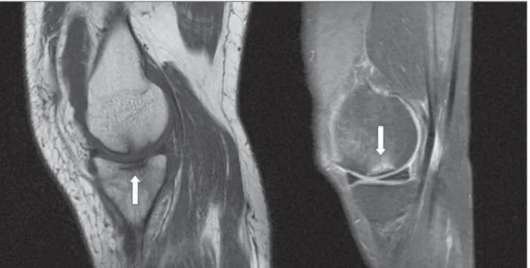
Figure 2. Sagittal (T1-weighted FSE / DP FSE) images showing linear subchondral abnormality (arrows).
80
Cunha DL et al.
Radiol Bras. 2010 Mar/Abr;43(2):77–80
Previous studies had already empha-sized the association of spontaneous os-teonecrosis with meniscal lesion and, pro-vided the diagnosis of the mentioned dis-ease is not made when the patient is sub-mitted to a meniscectomy, the acceleration of the clinical conditions and consequen-tial articular involvement are evident. Ac-cording to Muscolo et al.(9), in five cases with meniscal lesion there was a later on-set of ipsilateral osteochondral abnormali-ties. Corroborating the literature, there was a considerable predominance of cases with homolateral meniscal lesion in association with osteochondral lesions, most fre-quently observed when instability was present.
Because of the low sensitivity and the high negative likelihood ratio in chondral lesions diagnosis by magnetic resonance imaging(10), the absence of findings at this imaging study does not exclude the pres-ence of such lesions. In the present study, more than half of cases (63.1%) presented associated chondral abnormalities. Such rate may be lower as compared with arthroscopy that is considered as the gold standard diagnostic method.
Some studies(4,11) have already related
the presence of insufficiency fracture in previously weakened bones as a trigger of the osteonecrosis process, based on corre-lation with similar findings described for femoral head osteonecrosis. Yamamoto & Bullough(1) have reported histological
find-ings, postulating that one of the primary events that would lead to the development of osteonecrosis would be a subchondral
insufficiency fracture in patients with bones mechanically weakened by non-neo-plastic diseases, specifically osteoporosis, in spite of any confirmation by means of bone mass measurements in the evaluated patients. Ramnath & Kattapuram(4) have proposed that the presence of a linear sub-chondral signal at magnetic resonance im-aging in association with the already de-scribed osteochondral abnormalities would affirm such relation, whereas the absence of this signal would suggest a stronger as-sociation with osteoarthrosis findings, cast-ing a doubt in relation to the validity of the term “spontaneous” that is currently uti-lized. Additionally, in the present study, sudden pain onset was observed preferen-tially in weight-sustaining zones, besides a higher degree of associated bone marrow edema. However, in patients without such characteristics, the pain onset would be in-sidious and osteochondral abnormalities would be more evident, differently from the subchondral edema that is more subtle in such cases. In the greatest majority of pa-tients in the present study the linear sub-chondral signal was observed, as well as a higher rate os association between the pres-ence of this signal and edema as compared with cases without such alteration.
CONCLUSION
Magnetic resonance imaging has dem-onstrated to be a noninvasive method with good sensitivity in the diagnosis of os-teonecrosis of the knee, as well as associ-ated lesions, besides the association of
lin-ear subchondral abnormality with sudden onset of gonalgia and high degree of edema in the adjacent bone marrow. Such condi-tion is most frequently found in women (63% of cases).
REFERENCES
1. Yamamoto T, Bullough PG. Spontaneous osteone-crosis of the knee: the result of subchondral in-sufficiency fracture. J Bone Joint Surg Am. 2000; 82:858–66.
2. Eschard JP, Brochot P, Etienne JC. Osteonecro-sis of the femoral condyles: a retrospective study of 34 cases. Eur J Orthop Surg Traumatol. 1997;7: 267–70.
3. Muglia VF, Simão MN, Elias Júnior J, et al. Er-ros comuns de interpretação de ressonância mag-nética de joelho: como reconhecê-los e evitá-los. Radiol Bras. 2001;34:161–6.
4. Ramnath RR, Kattapuram SV. MR appearance of SONK-like subchondral abnormalities in the adult knee: SONK redefined. Skeletal Radiol. 2004;33:575–81.
5. Lotke PA, Ecker ML, Alavi A. Painful knees in older patients: radionuclide diagnosis of possible osteonecrosis with spontaneous resolution. J Bone Joint Surg Am. 1997;59:617–21. 6. Ahlbäck S, Bauer GCH, Bohne WH.
Spontane-ous osteonecrosis of the knee. Arthritis Rheum. 1968;11:705–33.
7. Lotke PA, Ecker ML. Current concepts review. Osteonecrosis of the knee. J Bone Joint Surg Am. 1988;70:470–3.
8. Björkengren AG, AlRowaih A, Lindstrand A, et al. Spontaneous osteonecrosis of the knee: value of MR imaging in determining prognosis. AJR Am J Roentgenol. 1990;154:331–6.
9. Muscolo DL, Costa-Paz M, Ayerza M, et al. Me-dial meniscal tears and spontaneous osteonecro-sis of the knee. Arthroscopy. 2006;22:457–60. 10. Karam FC, Silva JLB, Fridman MW, et al. A
res-sonância magnética para o diagnóstico das lesões condrais, meniscais e dos ligamentos cruzados do joelho. Radiol Bras. 2007;40:179–82. 11. Kattapuram TM, Kattapuram SV. Spontaneous