7 7
Instituto Dante Pazzanese de Cardiologia, Hospital do Coração and Associação do Sanatório Sírio
Mailing address: José Ribamar Costa Júnior – Rua Botucatu, 221/12 – 04023-060 – São Paulo, SP, Brazil – E-mail: [email protected]
English version by Stela Maris C. e Gandour
Objective - To assess the in-hospital evolution of pa-tients undergoing percutaneous stent placement in the ca-rotid arteries.
Methods - From August 1996 to April 2001, we studied 86 patients with carotid arterial obliterative lesions > 70% who were treated with percutaneous stent placement in the carotid arteries. We assessed the rate of success of the implantation and of the procedure, the types of stents used, mortality rate, and neurological complica-tions.
Results - Successful implantation was obtained in 98.9% of the cases, and the procedure was successful in 91.8%. The Wallstent was the most frequently used stent (73 patients - 77%). Cerebral strokes occurred as follows: 3 (3.2%) transient ischemic attacks, 1 (1.1%) minor stroke, and 3 (3.1%) major strokes. One (1.1%) patient died during hospitalization.
Conclusion - The high rate of success of stent implan-tation (98.9%) in addition to the low rate of cerebral stroke/death (4.2%) showed the efficiency and safety of percutaneous stent placement in carotid arteries.
Key words: placement, stent, carotid arteries
Arq Bras Cardiol, volume 80 (nº 1), 77-82, 2003
José Ribamar Costa Jr., Manuel N. Cano, Dinaldo C. Oliveira, Antonio M. Kambara, Adriana C. Moreira, Rogério Queiroz, Silvia Cano, Amanda G.M.R. Sousa,
J. Eduardo M.R. Sousa
São Paulo, SP - Brazil
Percutaneous Implantation of Endoprostheses in the
Carotid Arteries
Cerebral stroke is currently one of the most prevalent diseases. In the United States of America, 800 thousand new cases of cerebral stroke are estimated to occur every year, of which, one third result from atherosclerotic disease with significant carotid artery stenosis 1,2. Cerebral stroke
causes some degree of disability in daily activities in appro-ximately 2 million patients, causing an increase in the number of economically unproductive citizens, which results in a heavy burden on the state 3,4.
In Brazil, this disease is extremely common. It was com-puted at first among the afflictions of cerebral origin (cranial traumas excluded), and then, among the general causes of death in the population 5.
In the 1950s, Eascott, DeBakey, and Cooley were the pioneers in surgical carotid endarterectomy, which, in its initial phase, had some discouraging results. However, with improvement in the surgical technique and in pre-and postoperative care, the results became more favora-ble, leading surgical endarterectomy to play a relevant role in the treatment of carotid atherosclerotic disease from the 1970s on 2,6. It is estimated that 200 thousand surgical
carotid endarterectomies were performed in the USA in 2001 7.
At the end of the 1970s, Mathias et al 8,9 introduced
ca-rotid balloon catheter angioplasty as a therapeutical alterna-tive to surgery.
In the 1990s, with the endoprostheses, the carotid percu-taneous treatment became more consolidated, and the short, medium-, and long-term results became extremely en-couraging. It is estimated that 3 thousand carotid percuta-neous interventions were performed in the USA in 2001 7,10.
Methods
From August 1996 to April 2001, 86 patients most of whom were males (51.2%), with a mean age of 64.3 years (±22 years), underwent percutaneous implantation of caro-tid endoprosthesis. These patients were referred for treatment because they had previous neurological symp-toms (amaurosis, presyncope, syncope, and others), or sig-nificant atherosclerotic disease in other areas (coronary ar-teries, lower limbs, renal arar-teries, and others), or a signifi-cant carotid stenosis diagnosed on a noninvasive evalua-tion (Doppler ultrasound, computerized tomography, nu-clear magnetic resonance, and others).
The inclusion criteria were as follows: asymptomatic patients with stenotic lesion m 70%, according to quantita-tive coronary angiography (QCA); patients with a previous history of transient ischemic attack in the last year charac-terized by episodes of dizziness, syncope, paresis, and tem-porary paresthesia or amaurosis reported in the side of the angiographic lesion and stenosis m 60%; patients with a history of contralateral ischemic cerebral stroke with steno-tic lesion m 60% and patent cerebral arteries.
The exclusion criteria were as follows: patients with marked tortuosity at the origin of the supraaortic branches or aorto-iliac obstruction, not allowing the passage of ca-theters; patients with stenotic lesions, but with mobile thrombi inside; existence of diffuse stenotic lesions in the intracranial portion of the internal carotid artery or in the cerebral arteries; existence of cerebral aneurysms or intra-cranial arteriovenous malformations; recent history of di-gestive, pulmonary, or tumor hemorrhages, which contrain-dicate therapy with antiplatelet agents.
The following patients were considered as high-risk patients for the procedure: those older than 70 years; those with concomitant coronary artery disease; those with signi-ficant neurological antecedents (history of convulsion, stroke); those with contralateral carotid occlusion; those undergoing previous surgical carotid endarterectomy and evolving to restenosis; those with cervical neoplasia ope-rated upon or undergoing radiation therapy, or both (cha-racterizing the so-called “hostile neck”).
Successful implantation was defined as a residual lesion < 30% on quantitative digital angiography and ab-sence of important dissections or thrombi, while success of the procedure was characterized by successful implantation in the absence of transient ischemic attack, and of minor and major ischemic stroke according to the definition of the Na-tional Institutes of Health.
Transient ischemic attack was defined as any neurolo-gical deficit reverted in the first 24 hours after the procedure with no residual neurological damage.
Minor ischemic cerebral stroke was defined as a new neurological event resulting in a mild reduction in neurolo-gical functions (speech, motor or sensorial function, or both) with complete reversion in the first 7 days after the procedure or reaching up to 4 points in the cerebral stroke scale of the National Institutes of Health. A new
neurologi-cal deficit persisting longer than 7 days or reaching 4 or more points in the cerebral stroke scale of the National Ins-titutes of Health was classified as major ischemic cerebral stroke.
The patients were premedicated (24 hours) with ticlo-pidine (500 mg) and acetylsalicylic acid (200 mg), and, after endoprosthesis implantation, they continued to take this association of antiplatelet agents for 30 days, when ticlopi-dine was suspended and acetylsalicylic acid was maintai-ned indefinitely.
Prior to hospital discharge, all patients were assessed by at least 2 neurologists from the Hospital do Coração and Instituto Dante Pazzanese.
The hemodynamics laboratory was equipped with a chest X-ray device with a 3,000-milliampere generator, 50 to 150 kilovolt peak, high-definition image intensifier (1,024-line monitor), and a digital converter for electronic image processing. The images were documented in the digital system with a laser compact disc (CD) or in the analog sys-tem in a cinephotographic 35-mm film.
The patient’s blood pressure (invasive), oxygen saturation (pulse oximeter), and heart rate (electrocardio-gram) were continuously monitored.
After antisepsis, local anesthesia was performed with subcutaneous injection of 20 mL of a 2% lidocaine solution in the right and left inguinal regions. Then, using Sel-dinger’s technique, the right femoral artery and then the left femoral artery were punctured and maintained with 8 and 6 French (F) introducers with valves, respectively. A tempo-rary electrode of a cardiac endocavitary pacemaker was placed in the right ventricle, to which a temporary VVI-mode pacemaker generator with a heart rate of 50 bpm was con-nected.
The primitive carotid artery was catheterized via the punctured common femoral artery and an 8/9F guide ca-theter or a 7F long introducer with valves was placed. With injection of a low-osmolarity iodinated contrast agent, a ca-rotid angiography in 2 views (right and left anterior oblique) was obtained. The following parameters were measured with quantitative angiography (SMS): the reference diame-ter of the vessel, the extension of the lesion, and the mini-mum luminal diameter prior to the procedure.
Anticoagulation was performed with the intravenous injection of 10,000 units of unfractioned heparin immedia-tely before the use of the 0.014” guidewire.
After the 0.014” guidewire was passed through the lesion, the balloon catheter (4x20 mm) was positioned for predilation, which was performed with the aid of a mano-meter and lasted 10 seconds at the most. Then a new carotid angiography was performed to confirm the absence of com-plications in the vessel.
7 9 Control carotid angiography and a shoot of the
cere-bral circulation were performed. In the absence of thrombi or dissections and confirmation of residual lesion < 30%, the procedure was finished.
The patients were referred to the semi-intensive care unit with cardiac monitoring and maintained on intravenous hydration with saline solution at a velocity of 50 to 70 mL/h for 8 to 12 hours. The patients were allowed to eat 4 hours after their arrival at the unit. The introducers and the cardiac pacemaker electrode were removed 4 to 6 hours after the end of the procedure. Hemostasia was performed with local ma-nual compression for approximately 20 minutes, which was complemented with a compressive dressing and immobiliza-tion of the lower limb for approximately 8 hours. In case of an uneventful evolution, the patients were transferred to a room after 24 hours, being discharged from the hospital on the following day.
Results
Ninety-four carotid lesions in 86 patients were treated. Implantation was successful in 98.9% of the cases, and the procedure was successful in 91.8% of the cases.
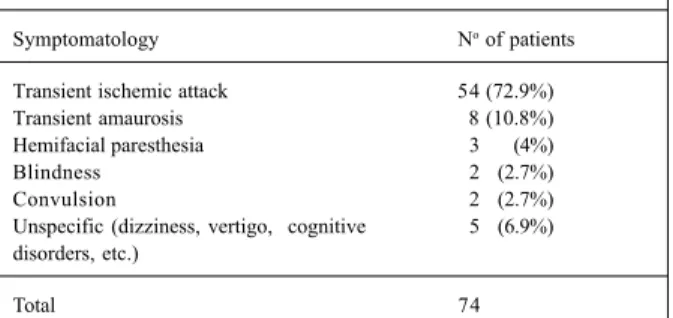
Prior to the procedure, the most common symptom was transient ischemic attack present in 72.9% of the patients, followed by transitory amaurosis in 10.8% (tab. I). Most of the 94 endoprostheses implanted were of the metallic self-expanding type, the Wallstent, used 73 (77.6%) times. In order of frequency, the other endoprostheses used were as follows: the Smart (self-expanding), the Palmaz (expandable balloon), the Symphony (self-expanding), the Memotherm (self-expanding), and the Herculink (expanda-ble balloon) (fig. 1).
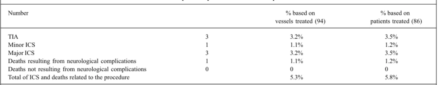
The 7 cases of in-hospital neurological complications were as follows: 3 (3.2%) transient ischemic attacks, 1 (1.1%) minor ischemic cerebral stroke, and 3 (3.2%) major ischemic cerebral strokes. These complications occurred when the patients were still in the hemodynamic laboratory and may have been related to microembolism. One patient died at the hospital (14th day).
The combined rate of minor and major ischemic cere-bral strokes was 4.2% (tab. II).
When analyzing the presence of neurological compli-cations according to the presence of symptoms prior to the procedure, we observed that the asymptomatic patients
nei-ther had any type of cerebral stroke nor died, while the sym-ptomatic patients had a mortality rate of 1.1% and an inci-dence of major ischemic cerebral stroke of 3.2% (fig. 2).
Pictures 1, 2, and 3 depict the procedure of percuta-neous implantation of carotid endoprosthesis in a patient in our series.
Only 1 patient had a complication at the site of access (right and left inguinal regions); it was a hematoma of 6 cm in diameter in the right inguinal region, which resolved sponta-neously.
Three patients evolved with total atrioventricular block, the cardiac rhythm being dictated by the temporary pacemaker with recovery of sinus rhythm in the first 24 hours in all cases.
Discussion
It is believed that an asymptomatic patient with a carotid obstruction m 80% has a 6% annual risk of having an ische-mic cerebral stroke, while a symptomatic patient has a 10% risk in the first year and a 40% risk by the end of 5 years 12.
Surgical carotid endarterectomy should have a rate of cerebral stroke/death < 6% in symptomatic patients and 3% in asymptomatic patients 13.
The rate of ischemic cerebral stroke after surgical caro-tid endarterectomy has ranged from 1.5% to 9%, depending on the case series analyzed 14,15. In the European carotid
sur-gery trial (ECST), the rate of ischemic cerebral stroke/death with this type of surgery was 7.5% 15. The North-American
Symptomatic Carotid Endarterectomy Trial (NASCET),
Table I – Clinical presentation of symptomatic patients
Symptomatology No of patients
Transient ischemic attack 54 (72.9%) Transient amaurosis 8 (10.8%) Hemifacial paresthesia 3 (4%)
Blindness 2 (2.7%)
Convulsion 2 (2.7%)
Unspecific (dizziness, vertigo, cognitive 5 (6.9%) disorders, etc.)
Total 74
Fig. 1 – Types of stents used.
Fig. 2 – Complications related to the procedure in symptomatic versus asymptomatic patients.
3 (3,2%)
1 (1.1%)
which randomized symptomatic patients with 50 to 69% ca-rotid stenosis for clinical treatment or surgical caca-rotid endar-terectomy, revealed a rate of ischemic cerebral stroke/death of 5.8% in the surgical group 16. On the other hand, the
Asymptomatic Carotid Atherosclerosis Study Group (ACAS) reported a 2.3% rate of ischemic cerebral stroke/ death related to surgery 17.
The Carotid and Vertebral Artery Transluminal Angio-plasty Study (CAVATAS) randomized 504 patients, 253 for surgical carotid endarterectomy and 251 for percutaneous carotid intervention. The rate of cerebral stroke/death at 30 days was 6.4% for surgical carotid endarterectomy and 5.9% for percutaneous carotid intervention. After a 3-year clinical follow-up, the authors obtained equivalence of treatments 18.
Carotid angioplasty with implantation of endoprosthe-ses is a promising alternative technique to the classic sur-gical carotid endarterectomy. The study by Whole et al 19,
who analyzed 2,569 patients undergoing carotid endopros-thesis implantation, reported a 30-day mortality of 1.2%, a combined rate of cerebral stroke/death of 4.3%, and an inci-dence of restenosis at 180 days of 4.8%.
The use of cerebral protection in percutaneous carotid
intervention aiming at reducing the microembolic phenome-na has been proposed by some groups. Currently, several systems provide cerebral protection.
In 1990, Theron et al 20 were the pioneers in the use of
cerebral protection with the introduction of a system of distal carotid occlusion with a balloon mounted on a tri-coa-xial catheter, therefore, reducing the distal embolization of microfragments of the atherosclerotic plaque. Although effective in reducing embolic phenomena, this method cau-ses a temporary interruption in cerebral blood flow, which is not always well tolerated by patients (especially in the pre-sence of contralateral carotid occlusion or anomalies in the circle of Willis). The good results initially reported by Theron et al could not be universally reproduced, and seve-ral cases of cerebseve-ral ischemia resulting from the use of this system were reported 19,21. In addition, operational technical
difficulties made it less and less used.
In 1996, Kachel 22 who used a 9F balloon reported a
re-verse blood flow system through the external carotid artery based on occlusion of the common carotid artery close to its bifurcation. Despite being simple and of easy applicabi-lity, later studies showed that this system was neither safe
Table II – In-hospital complications related to the procedure
Number % based on % based on
vessels treated (94) patients treated (86)
TIA 3 3.2% 3.5%
Minor ICS 1 1.1% 1.2%
Major ICS 3 3.2% 3.5%
Deaths resulting from neurological complications 1 1.1% 1.2%
Deaths not resulting from neurological complications 0 0 0
Total of ICS and deaths related to the procedure 5.3% 5.8%
TIA- transient ischemic attack; ICS- ischemic cerebral stroke.
Picture 2 – Endoprosthesis (stent) implantation of the self-expanding type (Wallstent) extending from the left common carotid artery to the left internal carotid artery. The origin of the left external carotid artery was not obstructed, maintaining a good flow. According to the on-line digital angiography, the degree of residual lesion was < 10%.
Left External Carotid
Left Internal Carotid
Immediate result of stent implantation in the common and internal left carotid
Picture 1 – On quantitative angiography, an 80% lesion can be seen obstructing the left internal carotid artery ostium (ICA).
Left External Carotid
Left Common Carotid
Left Internal Carotid
8 1 nor efficient in preventing distal microembolization,
espe-cially in the presence of cerebral collateral circulation, which currently makes it rarely used 23.
In 1999, carotid filters appeared and provided the following advantages: effectiveness in preventing micro-embolization (fragments of up to 100 micrometers), no inter-ruption of the cerebral blood flow during its use, and easy manipulation 24. Among the deficiencies of the device, we
can cite the following: the filters are rigid, making their pas-sage into more significant stenoses difficult; the difficulty in adjusting the size of the filters to the carotid diameter in each patient; and, mainly, their still extremely elevated costs to allow their inclusion in our current practice 25.
It is worth noting that at the time these data were gat-hered, these devices were not available for use at the hospi-tals where these patients were treated, and that, up to March 2001, no model had been approved for use in clinical practice by the agencies that regulate medical practice in the USA (Federal Drug Association – FDA) and in Brazil (Mi-nistry of Health).
The most appropriate patients for carotid percuta-neous interventions are as follows: those with isolated
ca-Picture 3 – After 18 months, the patient underwent a new carotid angiographic stu-dy, which showed maintenance of the immediate result with residual lesion < 10%. After the end of the process of endothelialization of the prosthesis, patency of the left external carotid artery was observed. A 50% stenosis in the left external carotid artery ostium was noted on quantitative angiography, but this finding did not result in cli-nical manifestations.
rotid disease and severe flow obstruction close to the angle of mandible, inaccessible to surgery; those with radical dis-section of the neck followed by radiation therapy (the cases of cervical neoplasias, for example); those with restenosis after carotid endarterectomy; and those with fibromuscular dysplasia 12,21.
In our group of patients, the 98.9% rate of success re-vealed the efficiency of the percutaneous treatment, and, if the patients are analyzed according to the presence or absence of symptoms, the combined rate of (major/minor) ischemic cere-bral stroke was 5.4% for symptomatic patients and 0% for asymptomatic patients. In the group of asymptomatic patients, the estimated rate of complication was considerably low and inferior to that defined for carotid endarterectomy according to the American Heart Association 12.
These data become more relevant when we consider that of the 86 patients analyzed in this study, 43 (50%) were in the subgroup classified as high risk for interventions, be-cause they were old, had contralateral carotid occlusion (5.8%), had already had a previous cerebral stroke (5.8%), had already been revascularized (4.6%), or because they had the so-called “hostile neck”, ie, they had had cervical neoplasia or had undergone previous radiation therapy in the cervical region, or both (3.5%).
Currently, the Stent versus Carotid Endarterectomy (CREST) study is being carried out by the National Institu-tes of Health under the leadership of Dr. Hobson. In this study, 2,500 patients will be randomized. The results of the CREST study will define whether one technique is superior to the other.
Carotid stent implantation already plays a significant role in the treatment of carotid atherosclerotic disease. It is un-deniable that the 98.8% rate of success and the 4.2% rate of cerebral stroke/death in our study show the efficiency and safety of this type of procedure. We also know that the im-provement in the materials used (introducers, catheters, gui-dewires, endoprostheses, etc.) and the appearance of new devices have significantly contributed to improve the results of percutaneous implantation of carotid endoprosthesis.
In some studies, the devices used for cerebral protec-tion have had the ability to reduce the indices of neurologi-cal complications 26. This has increased interest in the
scien-tific community, generating more clinical trials that will shor-tly define the actual role played by these devices in carotid endoprosthesis implantation.
References
1. Dorros G. Carotid arterial obliterative disease; should endovascular revascula-rization (stent supported angioplasty) today supplant carotid endarterectomy. J Intervent Cardiol 1996; 9: 193-6.
2. Debakey MH. Carotid endarterectomy revisited. J Endovasc Surg 1996; 3: 4. 3. Englewood CO. Patient outcomes research teams study groups. Stroke Clinical
Updates. Nat Stroke Assoc 1994; 5: 9-12.
4. Americam Heart Association. Heart and Stroke Facts Statistical Supplement. New York: American Heart Association, 1994: 12.
5. Andrade LAF, Tilbery CP, Pimentel PCA, et al. Acidente Vascular Cerebral In: Knobel E. Condutas no Paciente Grave. São Paulo: Atheneu, 1999: 630-47. 6. Diethich EB. Cerebrovascular disease therapy; the past, the present, and the
future. J Endovasc Surg 1996; 3: 7-9.
7. Joye DJ. Carotid Up to Date. Fellow Courses Transcatheter Cardiovascular Treatment, 2001.
9. Mathias K, Mittermayer C, Ensinger H, et al. Perkutane katheterdilatation von karotisstenosen. Rofo 1980; 133: 258-61.
10. Joye DJ. Carodit Up to Date. Fellow Courses Transcatheter Cardiovascular Treat-ment, 2000.
11. Brott T, Adams HP, Olinger CP, et al. Measurement of acute cerebral infarction: a clinical examination Scale. Stroke 1989; 20: 864-70.
12. Moore WS, Barnett HJM, Beebe HG, et al. Guidelines for carotid endarterectomy: a multidisciplinary consensus statement from the ad hoc committee. American Heart Association. Stroke 1995; 26: 188-200.
13. Zarins CK. Carotid endarterectomy:the gold standart. J Endovasc Surg 1996; 3: 10-15.
14. Lusby RJ, Wylie EJ. Complications of carotid endarterectomy. Surg Clin Nort Am 1983; 63: 1293-301.
15. Farrell B, Fraser A, Sandercock P, et al. European carotid surgery trial (ECST). Lancet 1998; 351: 1379-86.
16. Bernett H. North American Symptomatic Carotid Endarterectomy Trial Colabo-rators (NASCET). Beneficial effect of carotid endarterectomy in symptomatic patients with higth-grade carotid stenosis. N Engl J Med 1991; 325: 453-5. 17. White CJ. Asymptomatic Carotid Atherosclerosis Study Group (ACAS).
Endar-terectomy for asymptomatic carotid artery stenosis. JAMA 1995; 273: 1421-8.
18. Brow MM. For the Carotid and vertebral artery transluminal angioplasty study investigators (CAVATAS). Results of the carotid and vertebral artery angio-plasty study. Br J Surg 1999; 86; 710-11.
19. Whole M, Roubin G, et al. Global experience in cervical carotid artery stent pla-cement. Cathet Cardiovasc Interven 2000; 50: 160-7.
20. Théron JG, Payelle GG, Coskun O, et al. Carotid artery stenosis: Treatment with pro-tected balloon angioplasty and stent placement. Radiology 1996; 201: 627-36. 21. Henry M, Amor M, Masson I, et al. Angioplasty and stenting of the extracranial
carotid arteries. J Endovasc Surg 1998; 6: 293-304.
22. Kachel R. Results of balloon angioplasty in carotid arteries. J Endovasc Surg 1996; 3: 22-30.
23. Théron JG. Cerebral protection during carotid angioplasty. J Endovasc Surg 1996; 3: 484-5.
24. Iyer SS. Treatament of extracranial carotid disease: Stents reign supreme. Transca-theter Cardiovascular Treatment. Washington DC, USA, 2000
25. Diethrich EB. Indications for carotid stenting; a preview of the potential derived from early clinical experience. J Endovasc Surg 2000; 3: 132-9.