ABSTRACT
Removal of calcium hydroxide from Weine Type
II systems using photon-induced photoacoustic
streaming, passive ultrasonic, and needle
irrigation: a microcomputed tomography study
Adam LLOYD1, Geraldine NAVARRETE1, Melissa Andreia MARCHESAN1, David CLEMENT2
1- University of Tennessee Health Science Center, College of Dentistry, Department of Endodontics, Memphis, TN, USA. 2- The University of Oklahoma, College of Dentistry, Oklahoma City, OK, USA.
Corresponding address: Adam Lloyd - University of Tennessee Health Science Center - College of Dentistry - Department of Endodontics - Union Ave. -
Memphis TN 38163 - United States - Phone: (901) 448-1731 - Fax: (901) 448-1799 - email: [email protected]
6XEPLWWHG$SULO0RGL¿FDWLRQ-XO\$FFHSWHG$XJXVW
O
bjective: This study compared the effectiveness of Er:YAG laser-activated irrigation (PIPS), passive ultrasonic irrigation (PUI) with EndoUltra and standard needle irrigation (SNI) in the removal of calcium hydroxide [Ca(OH)2] from the mesial roots of Weine Type II mandibular molars. Material and Methods: Thirty mandibular molars were screened by μCT for the presence of mesial roots with complex intra-canal anatomy and a commonwith Ca(OH)2 paste. Specimens were divided into three groups (n=10) according to the technique used for Ca(OH)2 removal: PIPS, at 15 Hz and 20 mJ using a 9 mm long, 600 μm diameter tip; PUI using a 15/.02 tip; and SNI (30 Ga. side-vented needle). Equal volumes of 8.25% NaOCl and 17% EDTA were used in all groups. μCT was used to measure the initial amount of Ca(OH)2 present and to assess the residual volume of Ca(OH)2 following each irrigation protocol. Data were analyzed using Tukey HSD and Kruskal-Wallis tests (D=5%). Results: The mean volume of Ca(OH)2
in the coronal third than in the middle and apical third (p<0.001). Ca(OH)2 was similarly removed from the coronal and middle thirds with the three methods used (p>0.05). PIPS
2 removal in the apical third
than PUI (median 100%, IQR: 85-100) and SNI (median 47%; IQR: 16-72) (p<0.001). Conclusions: PIPS laser-activation was more effective for the removal of Ca(OH)2 from
EndoUltra and SNI.
Ke y w or ds: Endodontics. Lasers. Ultrasonics. Intracanal dressing. Irrigants.
I N TROD UCTI ON
Intracanal medicaments have been used to further reduce the bacterial load of the root canal system that chemomechanical debridement may not remove20. Among these, calcium hydroxide
[Ca(OH)2] has been the most widely accepted intracanal medicament because of its high pH (12.5)
and endodotoxin concentrations in teeth with apical periodontitis1,25. Concerns have been raised about
the potential interactions of the remaining Ca(OH)2
and endodontic sealers. Margelos, et al.19 (1997)
demonstrated incomplete setting and increased brittleness of zinc oxide-eugenol based sealers. Barbizam, et al.3 (2008) reported diminished
adhesion of Epiphany sealer to root canal walls. Kim and Kim11 (2002) reported that the potential
for Ca(OH)2
Different techniques have been proposed for the removal of Ca(OH)2 from the root canal system,
including a combination of irrigating solutions and devices6,18,23. The most frequently described
technique includes a combination of irrigation
23. In
this instance, only the Ca(OH)2 in the main canal
system extensions or irregularities relying entirely on irrigation for the removal of Ca(OH)2 from these regions. While some investigators have shown a combination of ethylenediaminetetraacetic acid (EDTA) and sodium hypochlorite (NaOCl) irrigants completely remove Ca(OH)2 from single systems6,
others report that complete removal of medicaments from such teeth is unlikely7. Furthermore, they
suggest depth of needle penetration plays a
7.
The effectiveness of irrigation depends on the chemical action of the irrigant and the ability to bring the irrigant in contact with the complex structures within the root canal system22,26. Energizing irrigants
with activated devices has been used to enhance fluid interchange when compared with needle irrigation. Passive ultrasonics have demonstrated enhanced irrigation through the creation of eddy currents and microstreaming along the instrument shaft and improved Ca(OH)2 removal2,18. A recent
study evaluated the effect of the GentleWave system (Sonendo Inc, Laguna Hills, California, USA) in the removal of Ca(OH)2 from mandibular molars with two separate mesial canals18. The investigators
found that this treatment was able to render mesial canals free of Ca(OH)2 when compared with passive ultrasonic irrigation (PUI) and standard needle irrigation (SNI). Although this system shows encouraging results, there are drawbacks to its
a customized disposable handpiece to individual teeth for stabilization creating a closed system for delivery of the multisonic stream of irrigants. Promising results for the removal of Ca(OH)2 have also been shown using an Erbium:Yttrium-Aluminum-Garnet (Er:YAG) laser coupled with a short radial-stripped tip in a technique known as photon-induced photoacoustic streaming (PIPS; Fidelis; Fotona, Ljubljana, Slovenia). Arslan, et al.2 (2015) demonstrated that PIPS irrigation
provided complete removal of Ca(OH)2
grooves in single-rooted teeth when compared with ultrasonic, sonic, and SNI techniques, which did not.
Multiple investigations have examined Ca(OH)2 removal from non-complex single-rooted teeth that may present misleading information not representative of the complex anatomy encountered by endodontists2,6,23. Recent microcomputed
tomographic (μCT) studies on the anatomical complexities of the mesial roots of mandibular
molars have shown a prevalence of isthmuses of approximately 80% between 3-6 mm from the apex9,26. These anatomical structures can retain
debris from instrumentation8,21; microbes5; and
Ca(OH)217,18.
The use of μCT imaging to volumetrically assess the removal of Ca(OH)2 allows for a three
dimensional quantitative evaluation based on gray values with higher sensitivity than sectioning techniques28. The purpose of this study was to use
activated irrigation, a PUI with EndoUltra and SNI in the removal of Ca(OH)2 from the mesial roots of Weine Type II mandibular molars with complex anatomy.
M ATERI AL AN D M ETH OD S
Tooth selection followed protocol approval from an Institutional Review Board with no patient health
(14-03540-XM). Mandibular molars were screened for the presence of isthmuses and Weine Type II
one apical foramen) using a high-resolution μCT scanning system (ACTIS BIR 150/130, Varian Medical Systems, Palo Alto, California, USA). The images were acquired at 75 kV and 100 μA through 360° of rotation around the vertical axis resulting in a cross-sectional pixel size of approximately 30 μm. Each backscatter projection had a 16-bit addressable 1,024×1,024 area and was used to create a volume-rendered representation (VG Studio Max 2.3; Volume Graphics GmbH, Heidelberg, Germany). Thirty teeth were selected and the distal root of each sample was sectioned with a diamond
resin. Samples were individually mounted in a
the μCT stage.
All endodontic procedures were performed under
Pico, Carl Zeiss Meditec Inc., Jena, Germany) by
clinical experience. Canals were instrumented in
Gates-Glidden drills (Dentsply Maillefer, Ballaigues, Switzerland), followed by 25/.12 and 25/.10 K3
Vortex (Dentsply Tulsa Dental Specialties, Tulsa, Oklahoma, USA) instruments. The apical preparation was standardized to a 25/.06 at 0.5 mm from the
laser-activated irrigation at a wavelength of 2,940 nm in 30 s exposure intervals at 15 Hz and 20 mJ. A 9 mm long, 600 μm diameter quartz tip, with the polyamide tip stripped back 3 mm was used. All samples were dried with sterile paper points and a premixed calcium hydroxide/barium sulfate paste (Ultracal XS, Ultradent Products Inc, South Jordan, Utah, USA) was injected into the mesial canals using
of the binding point in a slow-speed handpiece until Ca(OH)2 was visualized at the apical foramen27. A
chamber and the access cavity was sealed (Cavit, 3M ESPE, Seefeld, Germany). A proximal radiograph
2 length
and density. The apices of all specimens were sealed with sticky wax. The specimens were stored at 37°C in 100% humidity for seven days and a second μCT scan was performed to quantify the percentage volume of Ca(OH)2 occupying the root canal system.
The specimens were accessed again and a
Switzerland) along with 3 mL of NaOCl was inserted into each canal for the working length to loosen the Ca(OH)214,23. The specimens were then randomly
distributed into three experimental groups (n=10):
Gr ou p 1
Laser-activated irrigation using an Er:YAG laser and a PIPS tip was performed according to the manufacturer’s instructions. The tip was placed into the access cavity only and activated with each of the following irrigating solutions as they were introduced into the chamber with a 28 Ga side-vented irrigation needle:
Step 1: Three 30 s cycles of 6 mL/interval of 8.25% NaOCl interrupted by a 30 s wait between each cycle
Step 2: 30 s cycle of 6 mL of water Step 3: 30 s cycle of 6 mL of 17% EDTA Step 4: 30 s cycle of 6 mL of water
Gr ou p 2
The PUI with EndoUltra protocol was the same as for PIPS following the steps previously described. However, PUI was delivered using nickel-titanium activator tips (15/.02) in a cordless ultrasonic handpiece (EndoUltra, Vista, Racine, Wisconsin, USA) and activated as far apically as achievable without binding in 2 mm amplitude motions.
Gr ou p 3
SNI was conducted with a 30 Ga side-vented needle delivering 18 mL of 8.25% NaOCl over a period of 90 s at a distance of 2 mm close to the working length with 2 mm amplitude movements, followed by 6 mL of water, 6 mL of 17% EDTA
water.
The total volume of irrigation was the same in all experimental groups.
irrigation. Three-dimensional (3D) volumes were automatically generated from thresholding and region growing based on grey values, separating Ca(OH)2 from dentin. The unchanged outer root
precision alignment of both overlaid 3D datasets to subvoxel accuracy (VG Studio Max 2.3, Volume Graphics GmbH, Heidelberg, Germany). Total Ca(OH)2 volume from each specimen was derived
of the radiopaque canal walls. Each specimen was
divided into coronal, middle, and apical thirds. Regions of interest for each third were created based on slice position and volumes recorded.
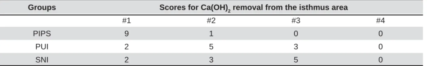
The images were qualitatively evaluated in a double-blind manner by two independent precalibrated evaluators to classify the removal of Ca(OH)2 from the isthmus area. A four-level score system was employed:
1= Clean isthmus, no Ca(OH)2. 2= Clusters of Ca(OH)2 volume.
3= Clusters of Ca(OH)2 filling >50% of the volume.
2.
The Shapiro-Wilk test was used to assess data normality. The mean volume of Ca(OH)2per third before removal had a normal distribution and data were compared with one way-ANOVA and Tukey HSD tests. Kruskal-Wallis one way-ANOVA test was used to compare the mean percentage volume of Ca(OH)2 remaining after the different irrigation
between the interaction root canal thirds and irrigation techniques was demonstrated by Tukey’s
determine the interobserver reproducibility for the removal of Ca(OH)2 from the isthmus area. Statistical analysis was performed using SigmaPlot 13.0 (Systat, San Jose, California, USA).
RESULTS
The mean Ca(OH)2 volume before removal was 9.21±2.19, 9.69±2.34, and 10.83±2.50 mm3 for
SNI, PIPS, and PUI respectively. The mean volume of Ca(OH)2
in the coronal third than in the middle and apical third, and the middle was higher than the apical third (p<0.001).
roots of Weine Type II mandibular molars was
techniques (p<0.0001) (Figure 1). PUI (median 8.33%) and SNI (median 4.78%) showed the highest amounts of remaining Ca(OH)2 when
compared with PIPS (median 0.00%), which showed the lowest (p<0.01). None of the specimens in the PUI group were completely free of Ca(OH)2,
with the apical third showing the highest percentage of remaining Ca(OH)2 (median 100%). SNI
Coronal Middle Apical
PIPS 0% (0%, 0%)aA 0% (0%, 0%)aA 0% (0%, 0%)aA
PUI 0% (0%, 0%)aA 0.50% (0%, 5.82%)aA 100% (85%, 100%)bB
SNI 0.073% (0%, 0.416%)aA 1.7% (0.8%, 4.5%)aA 47% (16%, 72%)aB
Lowercase superscript letters indicate homogeneous subsets between canal thirds Uppercase superscript letters indicate homogeneous subsets between methods
Table 1- Median (interquartile range) percentage of remaining Ca(OH)2 after irrigation with different techniques for canal thirds
Figure 1-7KUHHGLPHQVLRQDOUHFRQVWUXFWLRQVRIPHVLDOURRWVRIPDQGLEXODUPRODUVZLWK:HLQH7\SH,,FDQDOFRQ¿JXUDWLRQV illustrating remaining Ca(OH)2 in the different canal thirds (coronal – red; middle – yellow; apical – green) before (A1–C1) and after irrigation protocols (A2) PIPS, (B2) PUI, (C2) SNI.
showed a median of 47% of remaining Ca(OH)2 in the apical third and no differences between the thirds (p>0.05). PIPS showed a median of 0% of remaining Ca(OH)2 and no differences between the evaluated thirds (p>0.05). The interaction between the irrigation methods and the root canal thirds showed that all irrigation methods used were statistically similar in removing Ca(OH)2 from the coronal and middle thirds (Table 1). However, PIPS
Ca(OH)2 in the apical third when compared with both PUI and SNI (p<0.001).
for the removal of Ca(OH)2 from the isthmus
(p<0.05) when compared with SNI and PUI, which were statistically similar (p>0.05), whereas higher scores, which imply more Ca(OH)2 remaining in the isthmus area, were found for SNI and PUI (Table 2).
D I SCUSSI ON
In the present study, all specimens were irrigated with PIPS prior to the placement of Ca(OH)2. PIPS has demonstrated the ability to remove debris
interchange through expanding and collapsing cavitational bubbles along the root canal system12,16. s A pilot study showed that PIPS irrigation allowed
for better penetration of the intracanal medicament into isthmuses and interconnections between main canals for the type of teeth selected for this study.
This is the first qualitative investigation to examine remaining Ca(OH)2 in the isthmus area,
volumes of Ca(OH)2 in the different thirds. The highest amount of remaining Ca(OH)2 occurred with SNI and PUI. The rationale for such occurrence is the surface tension barrier created in the apical part
these complex non-separated areas18.
Irrigation with the recently marketed PUI device showed an overall 8.33% of Ca(OH)2 remaining in the root canal system, with no removal of medicament from the apical third. The isthmus area showed clusters of Ca(OH)2
the volume. We believe this is due to the inability of the EndoUltra tip to reach beyond the buccal-lingual curvature in spite of the 25/.06 apical enlargement.
The tips of the EndoUltra activator are a smooth 15/.02 wire, claimed by the manufacturer to be constructed from nickel-titanium. Manufacturer’s instructions recommend tip placement at 2 mm from the canal terminus. In our study, the tip was placed in the canal as far apically as achievable without binding to move freely and improve the
root canal system13
of the effectiveness of EndoUltra as an adjunct to irrigation in the literature. EndoUltra did not
acoustic streaming and hydrodynamic shear stress propagation to eliminate Ca(OH)2 from the apical third. This is in contrast to other PUI with EndoUltra irrigation devices that have shown more effective removal of Ca(OH)2 when compared with SNI18,24.
For the SNI group, the tip of the needle was placed at a distance 2 mm close to the working length without binding. A 30 Ga needle has an external diameter of 0.31 mm, which is equivalent to the dimensions of a canal prepared to a 25/.06 at 1.0 mm less than the working length. Fluid
interchange 0.75 mm from the tip of the needle4 and
explains the resulting 47% of Ca(OH)2 remaining in the apical third.
Unlike SNI and PUI, the PIPS tip remains in the access cavity providing, expanding, and collapsing cavitational bubbles that progress as shear forces along the canal walls12. Our results
showed no remaining Ca(OH)2 throughout the root canal system in mesial roots of mandibular molars with Weine Type II canal anatomy and isthmuses. Even the most apical area of the root canal, which
10,
was free of Ca(OH)2
velocity from the middle to apical third may have contributed to these results12. Additionally, samples
tested were Weine Type II canal systems that allow
between the mesio-buccal and mesio-lingual
velocity shear stresses could have resulted from the inherent canal anatomy, potentiating Ca(OH)2 removal from the apical third.
Under the conditions of the present study, the use of PIPS laser-activated irrigation demonstrated consistent removal of Ca(OH)2 from the entire canal Groups Scores for Ca(OH)2 removal from the isthmus area
#1 #2 #3 #4
PIPS 9 1 0 0
PUI 2 5 3 0
SNI 2 3 5 0
system of mandibular molars with Weine Type II
isthmus areas found in the apical half. The new PUI and SNI did not consistently remove Ca(OH)2 from
removing Ca(OH)2 from mandibular molars with
in future studies.
CON CLUSI ON S
suggest that PIPS laser-activation was more effective for the removal of Ca(OH)2 from mesial roots of mandibular molars with Weine Type II canal
ACKN OW LED GEM EN TS
We are grateful for the assistance and expertise provided by Dr. Daniela Handl, Volume Graphics GmbH, in the evaluation of the 3D datasets. This study was supported by a resident research grant from AAE Foundation – American Association of Endodontists Foundation and is submitted in partial
Master of Dental Science.
REFEREN CES
1- Adl A, Motamedifar M, Shams MS, Mirzaie A. Clinical investigation of the effect of calcium hydroxide intracanal dressing on bacterial lipopolysaccharide reduction from infected root canals. Aust Endod J. 2015;41:12-6.
2- Arslan H, Akcay M, Capar ID, Saygili G, Gok T, Ertas H. An in vit ro comparison of irrigation using photon-initiated photoacoustic
streaming, ultrasonic, sonic and needle techniques in removing calcium hydroxide. Int Endod J. 2015;48:246-51.
3- Barbizam JV, Trope M, Teixeira EC, Tanomaru-Filho M, Teixeira FB. Effect of calcium hydroxide intracanal dressing on the bond strength of a resin-based endodontic sealer. Braz Dent J. 2008;19:224-7.
4- Boutsioukis C, Gogos C, Verhaagen B, Versluis M, Kastrinakis E, Van der Sluis LWM. The effect of apical preparation size on irrigant
Fluid Dynamics model. Int Endod J. 2010;43:874-81.
5- Byström A, Claesson R, Sundqvist G. The antibacterial effect of camphorated paramonochlorophenol, camphorated phenol and calcium hydroxide in the treatment of infected root canals. Endod Dent Traumatol. 1985;1:170-5.
6- Calt S, Serper A. Dentinal tubule penetration of root canal sealers after root canal dressing with calcium hydroxide. J Endod. 1999;25:431-3.
7- Chou K, George R, Walsh LJ. Effectiveness of different intracanal irrigation techniques in removing intracanal paste medicaments. Aust Endod J. 2014;40:21-5.
8- Endal U, Shen Y, Knut A, Gao Y, Haapasalo M. A high-resolution computed tomographic study of changes in root canal isthmus
9- Gu LS, Wei X, Ling JQ, Huang XY. A microcomputed tomographic
molars in a Chinese population. J Endod. 2009;35:353-6.
10- Kenee DM, Allemang JD, Johnson JD, Hellstein J, Nichol BK. A
removal techniques. J Endod. 2006;32:563-5.
medication on apical seal. Int Endod J. 2002;35:623-8.
photon-induced photoacoustic streaming (PIPS) using Particle Image Velocimetry (PIV). Clin Oral Investig. 2015;20:381-6. 13- Krell KV, Johnson RJ, Madison S. Irrigation patterns during
1988;14:65-8.
14- Lambrianidis T, Kosti E, Boutsioukis C, Mazinis M. Removal
from the root canal. Int Endod J. 2006;39:55-61.
of calcium hydroxide dressing from the root canal. J Endod. 1999;25:85-8.
16- Lloyd A, Uhles JP, Clement DJ, Garcia-Godoy F. Elimination of intracanal tissue and debris through a novel laser-activated system assessed using high-resolution micro-computed tomography: a pilot study. J Endod. 2014;40:584-7.
17- Ma J, Shen Y, Yang Y, Gao Y, Wan P, Gan Y, et al. I n vit r o study
of calcium hydroxide removal from mandibular molar root canals. J Endod. 2015;41:553-8.
18- Ma JZ, Shen Y, Al-Ashaw AJ, Khaleel HY, Yang Y, Wang ZJ, et al. Micro-computed tomography evaluation of the removal of calcium hydroxide medicament from C-shaped root canals of mandibular second molars. Int Endod J. 2015;48:333-41.
19- Margelos J, Eliades G, Verdelis C, Palaghias G. Interaction of calcium hydroxide with zinc oxide-eugenol type sealers: a potential clinical problem. J Endod. 1997;23:43-8.
20- Nair PN, Henry S, Cano V, Vera J. Microbial status of apical
apical periodontitis after “one-visit” endodontic treatment. Oral Surg Oral Med Oral Pathol Oral Radiol Endod. 2005;99:231-52.
created by conventional rotary versus self-adjusting file instrumentation in mesial root canal systems of mandibular molars. Int Endod J. 2012;45:413-8.
22- Rosenfeld EF, James GA, Burch BS. Vital pulp tissue response to sodium hypochlorite. J Endod. 1978;4:140-6.
23- Salgado RJ, Moura-Netto C, Yamazaki AK, Cardoso LN, Moura AA, Prokopowitsch I. Comparison of different irrigants on calcium hydroxide medication removal: microscopic cleanliness evaluation. Oral Surg Oral Med Oral Pathol Oral Radiol Endod. 2009;107:580-4.
24- Silva LJ, Pessoa OF, Teixeira MB, Gouveia CH, Braga RR. Micro-CT evaluation of calcium hydroxide removal through passive ultrasonic irrigation associated with or without an additional instrument. Int Endod J. 2015;48:768-73.
25- Sjögren U, Figdor D, Spångberg L, Sundqvist G. The antimicrobial effect of calcium hydroxide as a short-term intracanal dressing. Int Endod J. 1991;24:119-25.
26- Teixeira FB, Sano CL, Gomes BP, Zaia AA, Ferraz CC, Souza-Filho FJ. A preliminary in vit r o study of the incidence and position
Int Endod J. 2003;36:276-80.
27- Torres CP, Apicella MJ, Yancich PP, Parker MH. Intracanal placement of calcium hydroxide: a comparison of techniques, revisited. J Endod. 2004;30:225-7.
28- Wiseman A, Cox TC, Paranjpe A, Flake NM, Cohenca N,
of calcium hydroxide from mesial canals of mandibular molars: a microtomographic study. J Endod. 2011;37:235-8.