ABSTRACT
http://dx.doi.org/10.1590/1678-775720150535
Risk of developing palat ally displaced canines in
pat ient s w it h ear ly det ect able dent al anom alies:
a r et r ospect ive cohor t st udy
Daniela Gamba GARIB1, Melissa LANCIA2, Renata Mayumi KATO2, Thais Marchini OLIVEIRA1, Lucimara Teixeira
das NEVES3
1- Universidade de São Paulo, Faculdade de Odontologia de Bauru, Departamento de Odontopediatria, Ortodontia e Saúde Coletiva; Hospital de Reabilitação de Anomalias Craniofaciais, Bauru, SP, Brasil.
2- Universidade de São Paulo, Hospital de Reabilitação de Anomalias Craniofaciais, Bauru, SP, Brasil.
3- Universidade de São Paulo, Faculdade de Odontologia de Bauru, Departamento de Ciências Biológicas; Hospital de Reabilitação de Anomalias Craniofaciais, Bauru, SP, Brasil.
Corresponding address: Daniela Gamba Garib - Departamento de Odontopediatria, Ortodontia e Saúde Coletiva - Faculdade de Odontologia de Bauru -
Universidade de São Paulo - Alameda Octávio Pinheiro Brisolla, 9-75 - Bauru - SP - Brazil - 17012-901 - Phone/Fax: 55 14 2344480 - e-mail: [email protected]
6XEPLWWHG1RYHPEHU0RGL¿FDWLRQ$SULO$FFHSWHG$SULO
T
he early recognit ion of risk fact ors for t he occurrence of palat ally displaced canines ( PDC) can incr ease t he possibilit y of im pact ion pr event ion. Obj ect ive: To est im at e t he r isk of 3'&RFFXUUHQFHLQFKLOGUHQZLWKGHQWDODQRPDOLHVLGHQWL¿HGHDUO\GXULQJPL[HGGHQWLWLRQ Mat er ial and Met hods: The sam ple com pr ised 730 longit udinal or t hodont ic r ecor ds fr om childr en ( 448 fem ales and 282 m ales) w it h an init ial m ean age of 8.3 year s ( SD= 1.36) . The dent al anom aly gr oup ( DA) included 263 r ecor ds of pat ient s w it h at least one dent al DQRPDO\LGHQWL¿HGLQWKHLQLWLDORUPLGGOHPL[HGGHQWLWLRQ7KHQRQGHQWDODQRPDO\JURXS ( NDA) was com posed of 467 r ecor ds of pat ient s w it h no dent al anom alies. The occur r ence of PDC in bot h gr oups was diagnosed using panoram ic and per iapical radiographs t aken in t he lat e m ixed dent it ion or ear ly per m anent dent it ion. The pr evalence of PDC in pat ient s w it h and w it hout ear ly diagnosed dent al anom alies was com par ed using t he chi- squar e t est ( p< 0 . 0 1 ) , r elat iv e r isk assessm ent s ( RR) , and posit iv e and negat iv e pr edict iv e values ( PPV and NPV) . Result s: PDC fr equency was 16.35% and 6.2% in DA and NDA JURXSVUHVSHFWLYHO\$VWDWLVWLFDOO\VLJQL¿FDQWGLIIHUHQFHZDVREVHUYHGEHWZHHQJURXSV ( p< 0.01) , w it h gr eat er r isk of PDC developm ent in t he DA gr oup ( RR= 2.63) . The PPV and NPV was 16% and 93% , r espect ively. Sm all m axillar y lat eral incisor s, deciduous m olar infraocclusion, and m andibular second prem olar dist oangulat ion were associat ed wit h PDC. Conclusion: Childr en w it h dent al anom alies diagnosed dur ing ear ly m ixed dent it ion have an appr oxim at ely t w o and a half fold incr eased r isk of developing PDC dur ing lat e m ixed dent it ion com par ed w it h childr en w it hout dent al anom alies.Ke y w or ds: Toot h abnor m alit ies. Canine t oot h. Et iology. Or t hodont ics.
I N TROD UCTI ON
Ap a r t f r o m t h e t h i r d m o l a r s, t h e ca n i n e s represent t he perm anent t eet h t hat m ost com m only show er upt ive disor der s14. The pr evalence of cases in w hich m axillar y ect opic canines palat ally deviat e is 1.7%14, com m only affect ing t hr ee fem ales for each m ale2 4 , 2 5 , 2 7. Less f r equ en t ly, t h e m ax illar y canines ar e buccally im pact ed and t his ir r egular it y seem s t o be a clinical m anifest at ion of ant er ior cr ow ding20. The rat io bet w een buccal and palat al
im pact ion of perm anent m axillary canines report ed in lit erat ur e is 1: 620.
bet w een gender s and et hnical backgr ounds, and incr eased fr equencies of ot her concom it ant dent al anom alies24, 26. The sear ch for associat ed dent al an o m al i es w as co n si d er ed t h e m o st r el ev an t m et hod t o invest igat e t he genet ic det er m inant s of PDC2,24. Peck, Peck and Kat aj a25 ( 1996) found t hat pat ient s w it h PDC have incr eased pr evalence of per m anent t oot h agenesis, excluding t hir d m olar s ( 17% ) , and show t he m andibular second pr em olar as t he m ost fr equent ly absent t oot h. Addit ionally, t hese aut hor s found t hat appr ox im at ely 20% of pat ient s w it h PDC have sm all lat eral incisor s not necessar ily at t he sam e ar ch side of t he ect opic canine.
The st udy by Sacer dot i and Baccet t i27 ( 2004) d oes n ot of f er su p p or t t o t h e h y p ot h esis t h at local condit ions m ay be a cause for PDC7,8, since t h ey d i d n o t d et ect a sso ci a t i o n b et w een t h e occur r ence of bilat eral PDC and t he occur r ence of bilat eral agenesis or m icr odont ia of lat eral incisor s. Addit ionally, unilat eral PDC in cases w it h unilat eral agenesis of m axillar y incisor s rar ely occur s at t he sam e arch side27. Sigler, Baccet t i and McNam ara Jr30 ( 2011) show ed t hat individuals w it h PDC exhibit ed
VLJQL¿FDQWO\KLJKHUSUHYDOHQFHRIVPDOOPD[LOODU\
lat eral incisor s ( six- fold higher ) , dist oangulat ion of m andibular second pr em olar s ( t hr ee- fold higher ) , and infraocclusion of deciduous m olar s ( t w o- fold h ig h er ) com p ar ed w it h a con t r ol g r ou p. Ot h er
VWXGLHV YHUL¿HG DQ LQFUHDVHG SUHYDOHQFH RI 3'&
in pat ient s scr eened for ot her dent al anom alies such as second pr em olar agenesis, sm all lat eral incisor s, infraocclusion of deciduous m olar s, and enam el hypoplasia2,19,29. Fam ily hist or y has alr eady
EHHQLGHQWL¿HGDVDULVNIDFWRUIRU3'&DQGRWKHU
her it able dent al anom alies, as w ell as t he gender bias m ent ioned24,26,27.
Ect opic er upt ion of m axillar y canines has t w o m aj or clinical concer ns: t he consequent im pact ion of t he canine and t he possibilit y of incisor ext er nal root resorpt ion9,15- 18.The t reat m ent prot ocol for PDC during perm anent dent it ion is oft en canine t ract ion, w hich m ay pr esent som e collat eral effect s such as r oot r esor pt ion of neighbor ing t eet h, cr est bone loss at t he m esial aspect of t he canine, and t oot h discolorat ion9,13.Ext ract ion m ay also be indicat ed for canines w it h init ial unfavorable posit ion, or in case of t oot h ankyloses16.
Conver sely, w hen t her e is an ear ly or t hodont ic diagnosis of PDC, sim pler clinical appr oaches such as deciduous canine ext ract ion and rapid m axillar y expansion can lead t o spont aneous canine erupt ion in a high per cent age of childr en3,5,23,30. These ear ly appr oaches can pr event canine im pact ion, incisor r oot r esor pt ion, and collat eral effect s r elat ed t o t oot h t ract ion. Ther efor e, t he r ecognit ion of r isk fact or s for t he occur r ence of PDC can incr ease t he possibilit y of ear ly diagnosis and int er vent ion. The
obj ect ive of t his st udy was t o evaluat e longit udinal r ecor ds of pat ien t s w it h som e ear ly - diagn osed dent al anom alies t o est im at e r isks of developing PDC dur ing t he lat e m ixed dent it ion.
M ATERI AL AN D M ETH OD S
Th i s r e t r o sp e ct i v e l o n g i t u d i n a l st u d y w a s approved by t he Research Et hical Com m it t ee of t he Hospit al for Rehabilit at ion of Craniofacial Anom alies, Univer sit y of São Paulo ( HRAC- USP) ( 379/ 2010) . Th e pat ien t r ecor ds w er e an ony m ized an d
de-LGHQWL¿HGSULRUWRDQDO\VLV7KHLQLWLDOVDPSOHZDV FRPSRVHGRIWKHRUWKRGRQWLF¿OHVRIFKLOGUHQ
t r eat ed fr om 1980 t o 2005 at t he Societ y for t he Social Pr om ot ion of Cleft Lip and Palat e Pat ient s ( PROFI S) . I nclusion cr it er ia w er e: pr esence of an
LQLWLDOSDQRUDPLFUDGLRJUDSKWDNHQGXULQJWKH¿UVW
t ransit ional per iod or int er- t ransit ional per iod of m ixed dent it ion, accor ding t o t he Van der Linden31
FODVVL¿FDWLRQDQGSUHVHQFHRIDWOHDVWRQH
m or e panoram ic radiograph t aken eit her dur ing t he second t ransit ional per iod of m ixed dent it ion or dur ing t he ear ly per m anent dent it ion. Exclusion crit eria were: poor qualit y records ( dark or dist ort ed p an or am ic r ad iog r ap h s; ab sen ce of p er iap ical radiographs in cases show ing ect opic canines) and pr esence of syndr om es or craniofacial anom alies. Eight y individuals w er e excluded based on t hese exclusion cr it er ia.
7KH¿QDOVDPSOHZDVFRPSRVHGRIRUWKRGRQWLF
r ecor ds fr om childr en w it h an init ial m ean age of 8 . 3 y ear s ( SD= 1 . 3 6 ) , f r om bot h gen der s ( 4 4 8 fem ales and 282 m ales) . A r ough est im at e of t he et hnic backgr ound of t he sam ple based on facial phot ograph was: Whit e ( 84% ) , Black ( 12% ) , and Asian ( 4% ) . The exper im ent al and cont r ol gr oups included 263 and 467 r ecor ds, r espect ively, and w er e com p osed b ased on t h e an aly ses of t h e init ial panoram ic radiographs and dent al cast s t o invest igat e t he pr esence of t he follow ing dent al anom alies: 1. Agenesis of any per m anent t eet h, except for t hir d m olar s; 2. Micr odont ia of m axillar y lat er al in cisor s; 3 . I n f r aocclu sion of decidu ou s m olar s; 4. Dist oangulat ion of m andibular second pr em olar s; 5. Toot h t ransposit ions.
init ial dent al cast s and panoram ic radiograph series. A deciduous m olar was considered in infraocclusion w h en m or e t h an 1 m m of v er t ical discr epan cy was m easur ed fr om t he m esial m ar ginal r idge of
WKHFORVHVWSHUPDQHQW¿UVWPRODU29. Maxillar y and
PDQGLEXODU¿UVWDQGVHFRQGGHFLGXRXVPRODUVZHUH
consider ed in t he analy sis of infraocclusion. The diagnosis of dist oangulat ion of m andibular second prem olars followed t he crit eria described by Shalish, et al.28 ( 2002) .
The sam ple was divided int o t w o gr oups. The dent al anom aly gr oup ( DA) was com posed of 263
SDWLHQWVZLWKDWOHDVWRQHGHQWDODQRPDO\LGHQWL¿HG
in t he init ial or m iddle m ixed dent it ion. Recor ds from children wit hout t hese dent al anom alies in t he ear ly/ m iddle m ixed dent it ion ( n= 467) com posed t he non- dent al anom aly gr oup ( NDA) . Age and gender dist r ibut ion in bot h gr oups is pr esent ed in Table 1.
Panoram ic radiographs from lat e m ixed dent it ion and/ or ear ly per m anent dent it ion w er e evaluat ed t o assess r isk s f or t h e dev elopm en t of PDC in
ERWK JURXSV &RQVLGHULQJ WKH ¿QGLQJV RI (ULFVRQ
and Kur ol15 ( 1986) show ingt hat t he at t em pt t o r ad i o g r ap h i cal l y d et er m i n e t h e er u p t i o n p at h of m ax illar y can in es is gen er ally of lit t le valu e in childr en younger t han 10 year s old, w e only exam ined panoram ic radiographs in r ecor ds fr om childr en aged 10 year s or older. The PDC diagnosis follow ed t he radiographic param et er s suggest ed by Lindauer, et al.22 DQG ZDV FRQ¿UPHG t hrough t he int erpret at ion of periapical radiographs according t o t he Clark’s t echnique12. Rapid m axillary
ex p an sion ( RME) p er f or m ed d u r in g t h e m ix ed dent it ion was r egist er ed in bot h gr oups because
50(PD\KDYHDSRVLWLYHLQÀXHQFHRQ3'&FDVHV6. Th e f r e q u e n c y o f PD C d e v e l o p m e n t w a s calcu lat ed in DA an d NDA g r ou p s. I n t er g r ou p com par isons w er e per for m ed using t he Chi- squar e
WHVW ZLWK D VLJQL¿FDQFH OHYHO RI ,Q RUGHU WR
m easu r e t h e st r en gt h of associat ion s bet w een occur r ences of ear ly- diagnosed dent al anom alies a n d PD C, t h e r e l a t i v e r i sk ( RR) a t t h e 9 5 %
FRQ¿GHQFHLQWHUYDODQGWKHSRVLWLYHDQGQHJDWLYH
pr edict ive values ( PPV and NPV) w er e calculat ed. Addit ionally, t he fr equency of PDC developm ent was separat ely calculat ed for each dent al anom aly and com pared wit h t he cont rol group using t he Chi-squar e t est ( p< 0.01) and r elat ive r isk assessm ent .
RESULTS
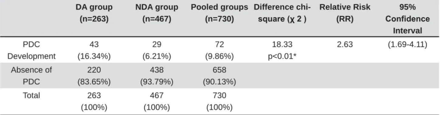
Sevent y- t w o individuals w er e affect ed by PDC ( 9.86% ) w it h a m ale: fem ale rat io of 1: 3 in t he com bined DA and NDA gr oups ( n= 730) . I n t his subgr oup of individuals w it h PDC, 29.1% ( n= 21) sh o w e d b i l a t e r a l e x p r e ssi o n , 3 1 . 9 % ( n = 2 3 ) u n ilat er al r igh t ex pr ession , an d 3 8 . 9 % ( n = 2 8 ) unilat eral left expr ession.
Th e DA g r ou p p r esen t ed PDC f r eq u en cy of 1 6 . 3 % com par ed w it h 6 . 2 % of t h e NDA gr ou p
7DEOH7KLVGLIIHUHQFHZDVVWDWLVWLFDOO\VLJQL¿FDQW
and indicat ed a t w o and a half fold incr ease in r isk of PDC in pat ien t s w it h ear ly- diagn osed den t al anom aly ( Table 2) . Posit ive pr edict ive value ( PPV) corresponded t o 16% and negat ive predict ive value
0HDQDJHDW¿UVWHYDOXDWLRQ (SD)
Mean age at second evaluation (SD)
Male Female
DA group (n=263) 8y2m (1.46) 10y10m (0.88) 95 168
NDA group (n=467) 8y6m (1.26) 10y4m (0.42) 187 280
Total (n=730) 8y4m (1.36) 10y8m (0.79) 282 448
Table 1- Age and gender distribution in dental anomaly (DA) and non-dental anomaly (NDA) groups
DA group (n=263)
NDA group (n=467)
Pooled groups (n=730)
Difference chi-VTXDUHȤ
Relative Risk (RR)
95% &RQ¿GHQFH
Interval
PDC Development
43 (16.34%)
29 (6.21%)
72 (9.86%)
18.33 p<0.01*
2.63 (1.69-4.11)
Absence of PDC
220 (83.65%)
438 (93.79%)
658 (90.13%)
Total 263 (100%)
467 (100%)
730 (100%)
6WDWLVWLFDOO\VLJQL¿FDQWGLIIHUHQFHDWS
( NPV) was 93% .
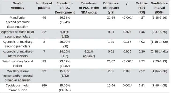
St a t i st i ca l l y si g n i f i ca n t a sso ci a t i o n s w e r e obser ved bet w een incr eased fr equencies of PDC developm ent and som e of t he dent al anom alies w er e separat ely evaluat ed ( Table 3) . The r elat ive
ULVNRI3'&LQFKLOGUHQZLWKWKHVHVSHFL¿FGHQWDO
anom alies var ied fr om 2.4 t o 4.3 ( Table 3) . Toot h t ransposit ion was absent in t he sam ple.
The fr equency of RME per for m ed dur ing t he m ixed dent it ion was sim ilar in bot h DA and cont r ol gr ou ps ( Table 4 ) . No ot h er t y pe of t r an sv er sal ex pansion was r egist er ed except RME. Ex t raoral t r act ion w as per f or m ed in 3 8 . 8 % of DA gr ou p and 40.8% of NDA gr oup. Ser ial ex t ract ion was
SHUIRUPHGLQRQO\RQHFDVHRI'$JURXSDQG¿YH
cases of NDA gr oup.
D I SCUSSI ON
This st udy evaluat ed longit udinal r ecor ds fr om pat ient s w it h ear ly- diagnosed dent al anom alies t o est im at e r isks for developing PDC dur ing t he lat e
m ixed dent it ion.
Pr e v i o u s c r o s s - s e c t i o n a l s t u d i e s s h o w e d an associat ion b et w een PDC an d ot h er d en t al anom alies including sm all m axillary lat eral incisors, t oot h agenesis, deciduous m olar infraocclusion, and ot her slight t oot h ect opia2,19,25.These st udies evaluat ed t he concom it ant occur r ence of canine ect opic er upt ion and ot her dent al anom alies, and point ed t o som e r isk indicat or s for PDC.
7KH SUHVHQW VWXG\ LV WKH ¿UVW WR HYDOXDWH D
lar ge sam ple w it h longit udinal r ecor ds for dent al an o m al i es t h at co u l d b e u sed as m ar k er s t o est im at e PDC r isk s1 1. Ou r r esu lt s sh ow ed t h at childr en w it h ear ly r ecognizable dent al anom alies have an incr eased r isk of 2.5 fold t o develop PDC lat er in life com par ed w it h childr en w it hout t hese anom alies ( Table 2) . According t o posit ive predict ive v alu e ( PPV) , t h e f r eq u en cy of p osit iv e r esu lt s ( pr esence of an ear ly- diagnosed dent al anom aly) t hat w er e t r ue posit ive ( pat ient s w ho developed PDC) was 16% . Considering t he negat ive predict ive value ( NPV) , 93% of pat ient s w it h negat ive r esult s Dental
Anomaly
Number of patients
Prevalence of PDC Development
Prevalence of PDC in the
NDA group
Difference chi-square
Ȥ
p Relative Risk (RR)
&RQ¿GHQFH interval
(95%)
Mandibular second premolar
distoangulation
49 26.53%
(13/49)
21.85 <0.001* 4.27 (2.38-7.66)
Agenesis of mandibular second premolars
22 9.09%
(2/22)
0.01 0.925 1.46 (0.37-5.75)
Agenesis of maxillary second premolars
8 25.00%
(2/8)
1.99 0.158 4.03 (1.15-14.06)
Agenesis of maxillary lateral incisors
7 14.29%
(1/7)
6.21% (29/467)
0.01 0.929 2.30 (0.36-14.61)
Small maxillary lateral incisor
82 23.17%
(19/82)
23.07 <0.001* 3.73 (2.20-6.33)
Maxillary lateral incisor and/or second
premolar agenesis
32 15.63%
(5/32)
2.83 0.093 2.52 (1.04-6.06)
Deciduous molar infraocclusion
159 15.09%
(24/159)
10.96 0.001* 2.43 (1.46-4.05)
6WDWLVWLFDOO\VLJQL¿FDQWGLIIHUHQFHDWS
Table 3- Prevalences of palatally displaced canines (PDC) development associated with each separate dental anomaly compared with the non-dental anomaly group
RME Non RME Total Difference chi-VTXDUHȤ
p
DA group 144 (58.5%) 102 (41.5%) 246 (100%) 3.02 0.082
NDA group 219 (52.3%) 208 (48.7%) 427 (100%)
( absence of dent al anom aly) w er e t r ue negat ive and did not develop PDC. The r esult s of t his st udy cor r oborat es t hat PDC belongs t o a spect r um of in t er r elat ed d en t al an om alies2 6. Th e lit er at u r e show s t he occur r ence of ot her dent al anom alies concom it ant w it h PDC2,19,24. Addit ionally, a higher pr evalence of dent al anom alies is obser v ed not
RQO\LQSDWLHQWVZLWK3'&EXWDOVRLQWKHLU¿UVWDQG
second- degr ee r elat ives26.
Sm all m axillar y lat eral incisor s and m andibular second prem olar dist oangulat ion were t he m ain risk fact or s for PDC am ong t he ear ly- diagnosed dent al an om alies ( Table 3 ) . Th ese r esu lt s cor r obor at e previous cross- sect ional st udies t hat dem onst rat ed
VLJQL¿FDQW DVVRFLDWLRQ EHWZHHQ VPDOO PD[LOODU\
lat eral incisor and PDC2,25,27. Dist oangulat ion of t he m andibular second pr em olar was ear ly descr ibed as a m ild expr ession of t he sam e genet ic or igin id en t if ied f or an t im er e ag en esis2 8.Recen t ly, a cr oss- sect ional st udy dem onst rat ed a st at ist ically
VLJQL¿FDQW GLIIHUHQFH EHWZHHQ WKH SUHYDOHQFH RI
PDC ( 2 8 % ) in pat ien t s w it h dist oan gu lat ion of t he m andibular second pr em olar s and in a cont r ol gr oup ( 4.2% )4.
D e ci d u o u s m o l a r i n f r a o ccl u si o n w a s a l so
FRQ¿UPHG WR EH D ULVN IDFWRU IRU 3'& 7DEOH
t his associat ion was pr eviously r epor t ed in a cr oss-sect ion al st u dy2 9. Th e pr ev alen ce of decidu ou s m olar infraocclusion, report ed from cross- sect ional st udies in a w hit e populat ion, var ies fr om 1.3% t o 8.9%1,10,21. The pr evalence of deciduous m olar infraocclusion in our com bined sam ple ( 21. 8% ) was m uch higher t han t he fr equency r epor t ed in pr evious st udies ( Table 3) and could be explained by our longit udinal per iod of obser vat ion.
1R VLJQL¿FDQW GLIIHUHQFHV ZHUH REVHUYHG
b et w een PDC d ev elop m en t in in d iv id u als w it h agenesis of m ax illar y lat eral incisor s or second pr em olar s, and t he NDA gr oup ( Table 3) . Peck , Peck and Kat aj a25 ( 1996) also showed t hat agenesis
RI PD[LOODU\ ODWHUDO LQFLVRUV ZDV QRW VLJQL¿FDQWO\
associat ed w it h PDC. On t he ot her hand, pr evious cr o ss- sect i o n st u d i es h av e sh o w n si g n i f i ca n t associat ions bet w een PDC and second pr em olar agenesis2,19,25.Alt hough second pr em olar agenesis
KDVEHHQSUHYLRXVO\LGHQWL¿HGDVDULVNLQGLFDWRU
for PDC in cr oss- sect ional st udies, t he associat ion
ZDVQRWVLJQL¿FDQWLQRXUFRKRUWHYDOXDWLRQWKLV
differ ence could be explained by r efer ence values, w hich considered general populat ion frequencies in t he for m er.
A lim it at ion of t his st udy is t he possibilit y of false- posit ive diagnosis for PDC using panoram ic radiographs22. However, t he false- posit ive rat e is low ( 4.22% ) and seem s not t o com pr om ise t he st udy r esult s22. The 80 indiv iduals t hat w er e excluded fr om t he sam ple w er e not analyzed eit her because t he absence or bad qualit y of radiographs. Ot her
lim it at ion of t his st udy could be t he bias of sam ple select ion becau se t h e st u dy w as r et r ospect iv e. How ever, t he sam ple select ion follow ed t he cr it er ia of a t im e int er val w hen t he pat ient s st ar t ed t he o r t h o d o n t i c t r ea t m en t ( f r o m 1 9 8 0 t o 2 0 0 5 ) . Anot her concern regarding t he m et hodology is t hat t he ear ly or t hodont ic t r eat m ent w it h RME m ight
KDYHKDGDQLQÀXHQFHRQWKHVSRQWDQHRXVHUXSWLRQ
of ect opic canines. How ever, bot h DA and cont r ol gr oups show ed sim ilar RME fr equencies ( Table 4)
DQG WKH SUHYDOHQFH RI 3'& ZDV VWLOO VLJQL¿FDQWO\
higher in DA gr oup.
Our r esult s show t hat sm all m ax illar y lat eral in cisor s, dist oan gu lat ion of m an dibu lar secon d pr em olar, and deciduous m olar infraocclusion ar e early risk m arkers for PDC. Pediat ric and ort hodont ic populat ion w it h such dent al anom alies diagnosed during t he early m ixed dent it ion should be carefully m onit or ed dur ing t he cr it ical age per iod for ear ly d iag n osis an d in t er v en t ion of m ax illar y can in e ect opic erupt ion. The recognit ion of risk m arkers for t he occurrence of PDC can increase t he possibilit y of early diagnosis and int ervent ion. Fut ure longit udinal st udies could cont r ibut e t o ident ify ot her pot ent ial r isk indicat or s for PDC including fam ily hist or y, fem ale gender, hypodiver gent pat t er n, and enam el hypoplasia2,27.
CON CLUSI ON
Childr en w it h som e dent al anom alies diagnosed d u r i n g t h e e a r l y m i x e d d e n t i t i o n h a v e a n appr oxim at ely t w o and a half fold incr ease in r isk of developing PDC dur ing t he lat e m ixed dent it ion co m p ar ed w i t h ch i l d r en w i t h o u t t h ese d en t al anom alies. Microdont ia of m axillary lat eral incisors, m andibular second pr em olar s dist oangulat ion, and deciduous m olar infraocclusion const it ut e early risk m ar ker s for PDC developm ent . When t he m axillar y canines ar e not palpable, a panoram ic radiograph is highly recom m ended in 10- year- old children wit h clinically or radiographically diagnosed DA in or der t o invest igat e PDC.
REFEREN CES
1- Andlaw RJ. Subm er ged deciduous m olar s: a pr evalence sur vey in Som er set . J I nt Assoc Dent Child. 1977; 8: 42- 5.
2- Baccet t i T. A cont r olled st udy of associat ed dent al anom alies. Angle Or t hod. 1998; 68: 267- 74.
3- Baccet t i T, Leonar di M, Ar m i P. A random ized clinical st udy of t w o int er cept ive appr oaches t o palat ally displaced canines. Eur J Or t hod. 2008; 30: 381- 5.
4- Baccet t i T, Leonar di M, Giunt ini V. Dist ally displaced pr em olar s: a dent al anom aly associat ed w it h palat ally displaced canines. Am J Or t hod Dent ofacial Or t hop. 2010; 138: 318- 22.
6- Baccet t i T, Sigler LM, McNam ara JA Jr. An RCT on t r eat m ent of palat ally displaced canines w it h RME and/ or a t ranspalat al ar ch. Eur J Or t hod. 2011; 33: 601- 7.
7- Becker A, Sharabi S, Chaushu S. Maxillar y t oot h size var iat ion in dent it ions w it h palat al canine displacem ent . Eur J Or t hod. 2002; 24: 313- 18.
8- Becker A, Zilber m an Y, Tsur B. Root lengt h of lat eral incisor s adj acent t o palat ally- displaced m axillar y cuspids. Angle Or t hod. 1984; 54: 218- 25.
9- Bishara SE. I m pact ed m axillary canines: a review. Am J Ort hod Dent ofacial Or t hop. 1992; 101: 159- 71.
10- Br ear ley LJ, McKibben DH Jr. Ank y losis of pr im ar y m olar t eet h . I . Pr evalen ce an d ch ar act er ist ics. ASDC J Den t Ch ild. 1973; 40: 54- 63.
11- Bur t BA. Concept s of r isk in dent al public healt h. Com m unit y Dent Oral Epidem iol. 2005; 3: 240- 7.
12- Clar ck CA. A m et hod of ascer t aining t he r elat ive posit ion of
XQHUXSWHGWHHWKE\PHDQVRI¿OPUDGLRJUDSKV3URF56RF0HG
1910; 3: 87- 90.
13- Cr escini A, Nier i M, Rot undo R, Baccet t i T, Cor t ellini P, Prat o GP. Com bined sur gical and or t hodont ic appr oach t o r epr oduce t he physiologic er upt ion pat t er n in im pact ed canines: r epor t of 25 pat ient s. I nt J Periodont ics Rest orat ive Dent . 2007; 27: 529- 37. 14- Dachi SF, How ell FV. A sur vey of 3,874 r out ine full- m ont h radiographs. I I . A st udy of im pact ed t eet h. Oral Sur g Oral Med Oral Pat hol. 1961; 14: 1165- 9.
15- Ericson S, Kurol J. Radiographic assessm ent of m axillary canine erupt ion in children w it h clinical signs of erupt ion dist urbance. Eur J Or t hod. 1986; 8: 133- 40.
1 6 - Er icson S, Kur ol J. Ear ly t r eat m ent of palat ally er upt ing m ax illar y canines by ex t ract ion of t he pr im ar y canines. Eur J Or t hod. 1988; 10: 283- 95.
17- Er icson S, Kur ol J. I ncisor r oot r esor pt ions due t o ect opic m ax illar y can in es im ag ed b y com p u t er ized t om og r ap h y : a com parat ive st udy in ext ract ed t eet h. Angle Ort hod. 2000; 70: 276-83.
18- Ericson S, Kurol PJ. Resorpt ion of incisors aft er ect opic erupt ion of m axillar y canines: a CT st udy. Angle Or t hod. 2000; 70: 415- 23.
19- Gar ib DG, Peck S, Gom es SC. I ncr eased occur r ence of dent al an om alies associat ed w it h secon d- pr em olar agen esis. An gle Or t hod. 2009; 79: 436- 41.
2 0 - Jacob y H. Th e et iolog y of m ax illar y can in e im p act ion s. Am er ican J Or t hod. 1983; 84: 125- 32.
21- Kurol J. I nfraocclusion of prim ary m olars: an epidem iologic and fam ilial st udy. Com m unit y Dent Oral Epidem iol. 1981; 9: 94- 102. 22- Lindauer SJ, Rubenst ein LK, Hang WM, Andersen WC, I saacson
5-&DQLQHLPSDFWLRQLGHQWL¿HGHDUO\ZLWKSDQRUDPLFUDGLRJUDSKV
J Am Dent Assoc. 1992; 123: 91- 2,95- 7.
23- Naoum ova J, Kur ol J, Kj ellber g H. Ext ract ion of t he deciduous can in e as an in t er cept iv e t r eat m en t in ch ildr en w it h palat al displaced canines - par t I : shall w e ext ract t he deciduous canine or not ? Eur J Or t hod. 2015; 37: 209- 18.
24- Peck S, Peck L, Kat aj a M. The palat ally displaced canine as a dent al anom aly of genet ic or igin. Angle Or t hod. 1994; 64: 249- 56. 25- Peck S, Peck L, Kat aj a M. Pr evalence of t oot h agenesis and peg- shaped m ax illar y lat eral incisor associat ed w it h palat ally displaced canine ( PDC) anom aly. Am J Ort hod Dent ofacial Ort hop. 1996; 110: 441- 3.
26- Pir inen S, Ar t e S, Apaj alaht i S. Palat al displacem ent of canine is genet ic and r elat ed t o congenit al absence of t eet h. J Dent Res. 1996; 75: 1742- 6.
27- Sacerdot i R, Baccet t i T. Dent oskelet al feat ures associat ed wit h unilat eral or bilat eral palat al displacem ent of m axillar y canines. Angle Or t hod. 2004; 74: 725- 32.
28- Shalish M, Peck S, Wasser st ein A, Peck L. Malposit ion of uner upt ed m andibular second pr em olar associat ed w it h agenesis of it s ant im er e. Am J Or t hod Dent ofacial Or t hop. 2002; 121: 53- 6. 29- Shalish M, Peck S, Wasserst ein A, Peck L. I ncreased occurrence of dent al anom alies associat ed w it h infraocclusion of deciduous m olar s. Angle Or t hod. 2010; 80: 440- 5.
30- Sigler LM, Baccet t i T, McNam ara JA Jr. Effect of rapid m axillary ex p an sion an d t r an sp alat al ar ch t r eat m en t associat ed w it h deciduous canine ext ract ion on t he er upt ion of palat ally displaced canines: a 2- cent er pr ospect ive st udy. Am J Or t hod Dent ofacial Or t hop. 2011; 139: e235- 44.