Rev Odontol UNESP. 2014 Sep.-Oct.; 43(5): 333-337 © 2014 - ISSN 1807-2577 Doi: http://dx.doi.org/10.1590/rou.2014.053
Radiographic evaluation of root canal cleaning, main and laterals,
using dif erent methods of i nal irrigation
Avaliação radiográi ca da limpeza de canais radiculares, principal e laterais,
utilizando diferentes métodos de irrigação i nal
Gisselle Moraima CHÁVEZ-ANDRADE
a, Juliane Maria GUERREIRO-TANOMARU
a,
Lucas Martinati MIANO
a, Renato de Toledo LEONARDO
a, Mario TANOMARU-FILHO
a*
aFaculdade de Odontologia, UNESP – Univ Estadual Paulista, Araraquara, SP, Brasil
Resumo
Objetivo: O objetivo deste estudo foi avaliar a eficácia da irrigação ultrassônica passiva (IUP), utilizando os fluxos intermitente ou contínuo, e da irrigação manual convencional (IMC) na limpeza de canais radiculares, principal e laterais simulados. Material e método: Os canais radiculares de 24 dentes artificiais foram preparados e os canais laterais foram confeccionados nos terços médio e apical. Os espécimes foram divididos em três grupos: G1- IMC, G2- IUP 1 (fluxo intermitente) e G3- IUP 2 (fluxo contínuo). Os canais radiculares foram preenchidos com uma solução de contraste e as raízes foram radiografadas antes e após a irrigação. As imagens digitais foram importadas para o programa Image Tool 3.0 e as áreas do canal totalmente preenchidas com o contraste, e após a irrigação, com remanescente do contraste, foram mensuradas para obtenção de dados percentuais. A análise estatística entre os grupos foi realizada por meio dos testes ANOVA e Tuckey. Resultado: No terço apical, os grupos G2 e G3 (IUP) mostraram melhor limpeza do que o grupo G1 (IMC) (p<0.05). Não houve diferenças estatisticamente significantes entre os grupos G2 e G3 (p>0.05). Conclusão: Irrigação ultrassônica passiva com fluxo intermitente promoveu melhor limpeza dos canais laterais simulados do que a irrigação manual convencional no terço apical do canal radicular. Não houve diferenças entre os grupos no canal principal e no terço médio. Descritores: Endodontia; irrigantes do canal radicular; ultrassom.
Abstract
Objective: To evaluate the efficacy of passive ultrasonic irrigation (PUI) using intermittent or continuous flushing
and conventional manual irrigation (CMI) on the cleaning of main and simulated lateral root canals. Material and
method: The root canals of 24 artificial teeth were prepared and simulated lateral canals were made in the medium
and apical thirds of the root. The specimens were divided into three groups: G1- CMI, G2- PUI 1 (intermittent flushing) and G3- PUI 2 (continuous flushing). Root canals were filled with contrast solution and the roots were radiographed pre- and post-irrigation. The digital images were transferred to Image Tool 3.0 software and the areas of root canal completely filled with contrast, and after irrigation with contrast remnant, were measured to obtain percentage data. Statistical analysis between groups was performed by ANOVA and Tukey tests. Result: In the apical third, G2 and G3 (PUI) groups showed higher percentage of cleaning than G1 (CMI) (p<0.05). Conclusion: Passive ultrasonic irrigation using intermittent flushing promoted a higher cleaning of simulated lateral canals than conventional manual irrigation in the apical third. There were no differences between groups in the main root canal and the middle third.
Descriptors: Endodontics; root canal irrigants; ultrasonics.
INTRODUCTION
h e persistence of microorganisms and their products within the root canal system is the main etiologic factor of periradicular pathology1-3. h e endodontic irrigation aims to promote cleaning
and disinfection of the root canal system2,4,5. h e ei cacy of root
canal irrigation depends on the method employed4, enabling
cleaning of the main and lateral canals as well as the isthmus area4,6-8.
Passive Ultrasonic Irrigation (PUI), used as a i nal irrigation protocol, has demonstrated ei cacy on the removal of debris and microorganisms when compared to conventional manual irrigation (CMI) using syringe and needle4,8-12.
Dif erent methodologies have been used for evaluation of root canal irrigation ei cacy. Abou-Rass, Piccinino13 employed
from extracted molars. De Gregorio et al.6 evaluated the efects
of PUI within main and simulated lateral root canals in single-rooted cleared human teeth. Rödig et al.14 assessed PUI eicacy
on removing dentin debris from simulated irregularities in root canals, and showed greater debris removal for PUI comparing with CMI.
he aim of this study was to evaluate the eicacy of passive ultrasonic irrigation by using two methods of irrigation solution lushing (continuous or intermittent) on the cleaning of main and simulated lateral root canals in the medium and apical thirds. he null hypothesis is that the action of cleaning by both methods of passive irrigation shows similarity in relation to conventional manual irrigation.
MATERIAL AND METHOD
Twenty-four single-rooted artiicial resin teeth (Fábrica de Sorrisos, Dental Rossetto Ltda., Arujá, SP, Brazil) were used in this study. Teeth had the crowns removed by using a cutting machine (Isomet 1000, Buelher Ltda., Lake Bluf, Il, USA). he roots were standardized in 14 mm of length.
he working length (WL) was established 1 mm short from the apex and the root canal instrumentation was realized by using a rotary system (MTwo, VDW, Munich, Germany). he basic sequence of the system was used (10/.04, 15/.05, 20/.06, 25/.06), followed by size 25/.07 instrument and apical preparation with size 30/.05, 35/.04 and 40/.04 instrument. All instruments were used to the working length. Ater each ile change, the root canals were irrigated with 2 mL of distilled water using syringe (Ultradent Products, USA) and a 30G needle (NaviTip Tips, Ultradent Products, USA).
Ater root canal preparation, four lateral canals were made in each root, in the labial and lingual surfaces, at 2 mm and 7 mm short from the apex, according to the methodology reported by Almeida et al.15. A cylinder-shaped bur with 0.20 mm diameter
(Brocas Undercut, Union Tool Co., Pluritec, SP, Brazil) was used. he root canals were illed using a radiographic contrast solution (Meglumine/sodium diatrizoate – 76%, Pielograf, BerliMed SA, Madrid, Spain) thickened with propylene glycol and bismuth oxide at the following proportion: 1 g of bismuth oxide, 1 mL of contrast solution and 1 mL of propylene glycol, according with modiied methodology of Guerreiro-Tanomaru et al.16. Digital periapical radiographs were performed
(Kodak RVG 6100 Digital Radiography System, France) with teeth placed in a standardization device. A radiograph was used to conirm the complete illing of main and lateral canals by the
contrast solution. he specimens were put in a glass lask illed with a silicon-based impression material (Zetaplus, Zhermack, Italy), immersed 1 mm short of the cervical surface, and stored until the experiment. he specimens were divided according to the irrigation protocols (Table 1).
1. Irrigation Protocols
It was used 5 mL of 1% NaOCl for each specimen and the total period of irrigation was 2 minutes for each root canal. In G1, conventional manual irrigation (CMI) was used with syringe and 30G needle (NaviTip Tips, Ultradent Products, USA) at 1 mm short from the working length, followed by the simultaneous aspiration.
In G2, the passive ultrasonic irrigation (PUI 1) was carried out through using the irrigating solution activated by an ultrasonic tip IRRI S (smooth wire) size 25/.00 (VDW, Endo Ultrasonic Files, Endodontic Synergy, Germany), at 1 mm short from the working length. PUI was performed using a piezoelectric device with 30 kHz frequency (CVDent 1000, CVD Vale, São José dos Campos, SP, Brazil) according to Al-Jadaa et al.17 and
van der Sluis et al.18. he irrigation period was divided as follow:
2 mL of solution in the irst 30 seconds using CMI followed by 20 seconds using PUI; 1 mL of the solution for 20 seconds using CMI followed by 20 seconds using PUI; 2 mL of the irrigating solution using CMI for 30 seconds, according to Bhuva et al.1.
he root canals were illed with the irrigating solution by using a 31G needle (NaviTip – Double Sideport Irrigator Tip, Ultradent Products, USA).
In G3, PUI 2 was used similarly to G2, except for a simultaneous irrigation performed during the ultrasonic activation (continuous lushing). he continuous lushing was carried out with an irrigating syringe and 31G needle (NaviTip – Double Sideport Irrigator Tip, Ultradent Products, USA) at 2 mm short from the working length and simultaneous aspiration, following the method used by Bhuva et al.1 and modiied by
Cameron19.
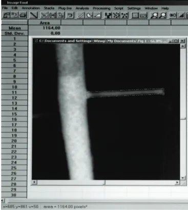
Ater the irrigation protocols, new digital radiographs were transferred to Image Tool 3.0 sotware. he areas of root canal illed with contrast and ater the irrigation (contrast remnant) were delimited as shown in Figure 1. he evaluation was performed by an examiner trained to properly use the sotware and blind to experimental groups. he images were imported into the Image Tool 3.0 sotware and analyzed using the tools of the sotware. his procedure allows an automatic delineation ater calibration of parameters. he measurements were obtained in mm2 for both
the main and lateral canals, and the percentage of cleaning was
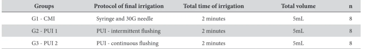
Table 1. Protocols of inal irrigation used for the experimental groups
Groups Protocol of inal irrigation Total time of irrigation Total volume n
G1 - CMI Syringe and 30G needle 2 minutes 5mL 8
G2 - PUI 1 PUI - intermittent lushing 2 minutes 5mL 8
G3 - PUI 2 PUI - continuous lushing 2 minutes 5mL 8
calculated for each group. Data were submitted to the normality test and comparison among groups by using ANOVA and Tukey tests ( p < 0.05).
RESULT
he results demonstrated that there was no statistically signiicant diference among groups regarding to the eicacy of contrast solution removal from the main root canal (p>0.05). For the simulated lateral canals, there was no diference among groups in the medium third (p>0.05). In the apical third, PUI using intermittent lushing showed the highest cleaning when compared to CMI group (p<0.05), however there was no diference among G2 and G3 (p>0.05) (Table 2).
DISCUSSION
he radiographic analysis has been used for the assessment of the irrigation eicacy in root canals from extracted teeth13,20. In this
present study, the digital radiographic system and the evaluation using image sotware analysis were used. he use of image
sotware enables the assessment of the evolution or regression of periapical lesions21, the cleaning and penetration capacity of
irrigating solutions into root canal system6,19. Bronnec et al.22
evaluated the penetration of the irrigant in curved root canals of lower molars during the root canal preparation, by using a radiographic contrast solution (sodium diatrizoate), 3% NaOCl, syringe and 27G needle. he specimens were radiographed in a standardization device and analyzed by using Image J sotware.
Simulated lateral canals can be made in either resin teeth or blocks23-27 or natural teeth15,28 followed by radiographic analysis29.
In the present study, simulated lateral canals were created in artiicial resin teeth, as already described by Tanomaru-Filho et al.26. A 0.20 mm diameter bur was used as conducted
by Al-Jadaa et al.11 in resin blocks and Almeida et al.15 in natural
teeth. he radiographic contrast solution was thickened with propylene glycol and bismuth oxide to provide consistency and radiopacity16.
Either in vitro or ex vivo studies evaluated the penetration capacity of contrast solutions or dyes in simulated lateral canals using human teeth or resin blocks6,22,30. Kahn et al.30 using a video
camera, evaluated the presence or absence of a dye within root canals ater irrigation methods. De Gregorio et al.6 observed the
dye penetration in lateral canals in cleared specimens. Saber Sel, Hashem31, using single-rooted human mandibular premolars,
compared PUI, CMI, and other irrigation systems, regarding to smear layer removal in the cervical, medium, and apical thirds of root canals, using SEM and score systems. he results did not show diference between PUI and CMI, which was in agreement with other studies32 demonstrating that PUI was not able to
completely remove the smear layer from the apical third of root canal.
PUI (intermittent lushing) was also assessed regarding to the efect of dissolution of bovine pulp tissue of simulated lateral canals (0.20 mm diameter) in the apical third of root canal in epoxy resin models11. he results showed a higher efect of PUI
on the pulp dissolution when compared to other irrigation systems (including the non-activation of the irrigant), suggesting a greater apical third cleaning, corroborating the results of the present study.
Few studies compared the two methods of PUI application: intermittent and continuous lushing. Van der Sluis et al.18
determined the inluence of irrigation period on dentinal debris removal in simulated irregularities in the apical third during PUI by using the two methods. he authors concluded that PUI associated with the intermittent lushing for 1 minute was more
Table 2. Comparison among groups for the main and lateral root canals (mean and standard deviation of the cleaning percentage)
Groups Main Canal Lateral Canals (medium third) Lateral Canals (apical third)
G1- CMI 94.54 (± 1.92)A 53.87 (± 13.28)A 51.95 (± 24.23)B
G2- PUI 1 97.52 (± 1.67)A 73.47 (± 18.32)A 77.56 (± 18.68)A
G3- PUI 2 96.08 (± 2.95)A 69.50 (± 24.91)A 73.29 (± 13.15)A,B
Diferent letters indicate statistically signiicant diferences (P < 0.05) in the same column; PUI, passive ultrasonic irrigation; CMI, conventional manual irrigation; G, group.
Figure 1. Image representative of measurements in simulated lateral
efective on dentinal debris removal than PUI associated with continuous lushing for 3 minutes. Using a similar methodology, van der Sluis et al.33 compared the two methods of PUI application
showing that there was no diference between PUI (continuous lushing) with 50 mL of NaOCl and PUI (intermittent lushing) with 12 mL of the irrigant, in the dentinal debris removal from apical third. In the present study, both PUI methods promoted root canal cleaning.
he efective apical irrigation is one of the most important procedures during root canal treatment34. Notwithstanding, the
use of intermittent lushing was the most efective method on cleaning of apical lateral canal. his result suggests the use of this
method because the apical root area presents the greatest amount of accessory canals and apical ramiications, resulting in greater diiculty for cleaning and disinfection.
CONCLUSION
According to the methodology employed, passive ultrasonic irrigation (PUI) and conventional manual irrigation (CMI) promotes a similar cleaning of main and lateral root canals in the medium third. However, PUI with intermittent lushing was the most efective method for cleaning simulated lateral canals in the apical third when compared to CMI.
REFERENCES
1. Bhuva B, PatelS, WilsonR, NiaziS, BeightonD, Mannocci F. The effectiveness of passive ultrasonic irrigation on intraradicular Enterococcus
faecalis biofilms in extracted single-rooted human teeth.Int Endod J. 2010March; 43(3): 241-50. http://dx.doi.org/10.1111/j.1365-2591.2009.01672.x. PMid:20158536
2. MohammadiZ, GiardinoL, MombeinipourA. Antibacterial substantivity of a new antibiotic-based endodontic irrigation solution.Aust Endod J. 2012
April; 38(1): 26-30. http://dx.doi.org/10.1111/j.1747-4477.2010.00263.x. PMid:22432823
3. SeetAN, ZilmPS, GullyNJ, CathroPR. Qualitative comparison of sonic or laser energisation of 4% sodium hypochlorite on an Enterococcus faecalis
biofilm grown in vitro.Aust Endod J. 2012December; 38(3): 100-6. http://dx.doi.org/10.1111/j.1747-4477.2012.00366.x. PMid:23211068
4. MozoS, LlenaC, FornerL. Review of ultrasonic irrigation in endodontics: increasing action of irrigating solutions.Med Oral Patol Oral Cir Bucal. 2012 May; 17(3): e512-6. http://dx.doi.org/10.4317/medoral.17621. PMid:22143738
5. UlusoyOI, GörgülG. Effects of different irrigation solutions on root dentine microhardness, smear layer removal and erosion.Aust Endod J. 2013August;
39(2): 66-72. http://dx.doi.org/10.1111/j.1747-4477.2010.00291.x. PMid:23890262
6. de GregorioC, EstevezR, CisnerosR, ParanjpeA, CohencaN. Efficacy of different irrigation and activation systems on the penetration of sodium
hypochlorite into simulated lateral canals and up to working length: an in vitro study.J Endod. 2010July; 36(7): 1216-21. http://dx.doi.org/10.1016/j.
joen.2010.02.019. PMid:20630302
7. Ordinola-ZapataR, BramanteCM, GarciaRB, de AndradeFB, BernardineliN, de MoraesIG, et al. The antimicrobial effect of new and conventional
endodontic irrigants on intra-orally infected dentin.Acta Odontol Scand. 2013May-July; 71(3-4): 424-31. http://dx.doi.org/10.3109/00016357.2012.6905
31. PMid:22607322
8. Passarinho-NetoJG, MarchesanMA, FerreiraRB, SilvaRG, Silva-SousaYT, Sousa-NetoMD. In vitro evaluation of endodontic debris removal as
obtained by rotary instrumentation coupled with ultrasonic irrigation.Aust Endod J. 2006December; 32(3): 123-8.
http://dx.doi.org/10.1111/j.1747-4477.2006.00035.x. PMid:17201755
9. LeeSJ, WuMK, WesselinkPR. The effectiveness of syringe irrigation and ultrasonics to remove debris from simulated irregularities within prepared root
canal walls.Int Endod J. 2004October; 37(10): 672-8. http://dx.doi.org/10.1111/j.1365-2591.2004.00848.x. PMid:15347291
10. van der SluisLW, VersluisM, WuMK, WesselinkPR. Passive ultrasonic irrigation of the root canal: a review of the literature.Int Endod J. 2007June; 40(6): 415-26. http://dx.doi.org/10.1111/j.1365-2591.2007.01243.x. PMid:17442017
11. Al-JadaaA, PaquéF, AttinT, ZehnderM. Necrotic pulp tissue dissolution by passive ultrasonic irrigation in simulated accessory canals: impact of canal location and angulation.Int Endod J. 2009January; 42(1): 59-65. http://dx.doi.org/10.1111/j.1365-2591.2008.01497.x. PMid:19125981
12. JiangLM, VerhaagenB, VersluisM, LangedijkJ, WesselinkP, van der SluisLW. The influence of the ultrasonic intensity on the cleaning efficacy of passive ultrasonic irrigation.J Endod. 2011May; 37(5): 688-92. http://dx.doi.org/10.1016/j.joen.2011.02.004. PMid:21496672
13. Abou-RassM, PiccininoMV. The effectiveness of four clinical irrigation methods on the removal of root canal debris.Oral Surg Oral Med Oral Pathol.
1982September; 54(3): 323-8. http://dx.doi.org/10.1016/0030-4220(82)90103-7. PMid:6957828
14. RödigT, SedghiM, KonietschkeF, LangeK, ZiebolzD, HülsmannM. Efficacy of syringe irrigation, RinsEndo and passive ultrasonic irrigation in
removing debris from irregularities in root canals with different apical sizes.Int Endod J. 2010July; 43(7): 581-9.
http://dx.doi.org/10.1111/j.1365-2591.2010.01721.x. PMid:20636517
15. AlmeidaJF, GomesBP, FerrazCC, Souza-FilhoFJ, ZaiaAA. Filling of artificial lateral canals and microleakage and flow of five endodontic sealers.Int Endod J. 2007September; 40(9): 692-9. http://dx.doi.org/10.1111/j.1365-2591.2007.01268.x. PMid:17608677
16. Guerreiro-TanomaruJM, LoiolaLE, MorgentalRD, LeonardoRT, Tanomaru-FilhoM. Efficacy of four irrigation needles in cleaning the apical third of
root canals.Braz Dent J. 2013; 24(1): 21-4. http://dx.doi.org/10.1590/0103-6440201302153. PMid:23657408
17. Al-JadaaA, PaquéF, AttinT, ZehnderM. Acoustic hypochlorite activation in simulated curved canals.J Endod. 2009October; 35(10): 1408-11. http://
dx.doi.org/10.1016/j.joen.2009.07.007. PMid:19801241
19. CameronJA. The effect of ultrasonic endodontics on the temperature of the root canal wall.J Endod. 1988November; 14(11): 554-9. http://dx.doi.
org/10.1016/S0099-2399(88)80090-6. PMid:3249194
20. RamZ. Effectiveness of root canal irrigation.Oral Surg Oral Med Oral Pathol. 1977August; 44(2): 306-12.
http://dx.doi.org/10.1016/0030-4220(77)90285-7. PMid:268582
21. CarvalhoFB, GonçalvesM, Guerreiro-TanomaruJM, Tanomaru-FilhoM. Evaluation of periapical changes following endodontic therapy: digital subtraction
technique compared with computerized morphometric analysis. Dentomaxillofac Radiol. 2009 October; 38(7): 438-44. http://dx.doi.org/10.1259/
dmfr/53304677. PMid:19767513
22. BronnecF, BouillaguetS, MachtouP. Ex vivo assessment of irrigant penetration and renewal during the cleaning and shaping of root canals: a digital
subtraction radiographic study.Int Endod J. 2010April; 43(4): 275-82. http://dx.doi.org/10.1111/j.1365-2591.2009.01677.x. PMid:20487446
23. SilverGK, LoveRM, PurtonDG. Comparison of two vertical condensation obturation techniques: Touch ’n Heat modified and System B.Int Endod J. 1999
August; 32(4): 287-95. http://dx.doi.org/10.1046/j.1365-2591.1999.00215.x. PMid:10551120
24. Gurgel-FilhoED, FeitosaJP, GomesBP, FerrazCC, Souza-FilhoFJ, TeixeiraFB. Assessment of different gutta-percha brands during the filling of
simulated lateral canals.Int Endod J. 2006February; 39(2): 113-8. http://dx.doi.org/10.1111/j.1365-2591.2006.01054.x. PMid:16454791
25. KarabucakB, KimA, ChenV, IqbalMK. The comparison of gutta-percha and Resilon penetration into lateral canals with different thermoplastic delivery
systems.J Endod. 2008July; 34(7): 847-9. http://dx.doi.org/10.1016/j.joen.2008.03.024. PMid:18570993
26. Tanomaru-FilhoM, Sant’anna-JuniorA, BossoR, Guerreiro-TanomaruJM. Effectiveness of gutta-percha and Resilon in filling lateral root canals using the
Obtura II system.Braz Oral Res. 2011May-June; 25(3): 205-9. http://dx.doi.org/10.1590/S1806-83242011000300003. PMid:21670852
27. Tanomaru-FilhoM, BossoR, Sant’AnnaAJr, BerbertFLCV, Guerreiro-TanomaruJM. Effectiveness of gutta-percha and Resilon in filling lateral root
canals using thermomechanical technique.Rev Odontol UNESP. 2013; 42(1): 37-41. http://dx.doi.org/10.1590/S1807-25772013000100007.
28. Tanomaru-FilhoM, Sant’AnnaAJr, BerbertFL, BossoR, Guerreiro-TanomaruJM. Ability of gutta-percha and Resilon to fill simulated lateral canals by using the Obtura II system.J Endod. 2012May; 38(5): 676-9. http://dx.doi.org/10.1016/j.joen.2012.01.007. PMid:22515901
29. GoldbergF, ArtazaLP, De SilvioA. Effectiveness of different obturation techniques in the filling of simulated lateral canals.J Endod. 2001May; 27(5):
362-4. http://dx.doi.org/10.1097/00004770-200105000-00015. PMid:11485258
30. KahnFH, RosenbergPA, GliksbergJ. An in vitro evaluation of the irrigating characteristics of ultrasonic and subsonic handpieces and irrigating needles and probes.J Endod. 1995May; 21(5): 277-80. http://dx.doi.org/10.1016/S0099-2399(06)80998-2. PMid:7673832
31. SaberS-D, HashemAA. Efficacy of different final irrigation activation techniques on smear layer removal.J Endod. 2011September; 37(9): 1272-5. http://
dx.doi.org/10.1016/j.joen.2011.06.007. PMid:21846546
32. CiucchiB, KhettabiM, HolzJ. The effectiveness of different endodontic irrigation procedures on the removal of the smear layer: a scanning electron
microscopic study.Int Endod J. 1989January; 22(1): 21-8. http://dx.doi.org/10.1111/j.1365-2591.1989.tb00501.x. PMid:2513277
33. van der SluisLW, GambariniG, WuMK, WesselinkPR. The influence of volume, type of irrigant and flushing method on removing artificially placed
dentine debris from the apical root canal during passive ultrasonic irrigation.Int Endod J. 2006June; 39(6): 472-6.
http://dx.doi.org/10.1111/j.1365-2591.2006.01108.x. PMid:16674742
34. ParkE, ShenY, HaapasaloM. Irrigation of the apical root canal.Endod Topics.2013; 27(1): 54-73. http://dx.doi.org/10.1111/etp.12028
CONFLICTS OF INTERESTS
he authors declare no conlicts of interest.
*CORRESPONDING AUTHOR
Mário Tanomaru Filho, Departamento de Odontologia Restauradora, Faculdade de Odontologia, UNESP - Univ Estadual Paulista, Rua Humaitá, 1680, CP 331, Centro, 14801-903 Araraquara - SP, Brazil, e-mail: [email protected]