ISSN 0102-695X http://dx.doi.org/10.1590/S0102-695X2012005000064 Received 1 Nov 2011 Accepted 8 Jan 2012 Available online 24 May 2012
strains of
Hypnea musciformis
from i
eld-collected and
in vitro
cultured samples
Daniela R. P. Fernandes,
1,2Vanessa S. Caetano,
2,3Márcio M. B.
Tenório,
4Fernanda Reinert,
2,3Yocie Yoneshigue-Valentin
*,1,21Laboratório de Botânica Marinha, Instituto de Biologia, Centro de Ciências
da Saúde, Universidade Federal do Rio de Janeiro, Brazil,
2Programa de Pós-graduação em Biotecnologia Vegetal, Decania do Centro de
Ciências da Saúde, Universidade Federal do Rio de Janeiro, Brazil,
3Laboratório de Fisiologia Vegetal, Instituto de Biologia, Centro de Ciências
da Saúde, Universidade Federal do Rio de Janeiro, Brazil,
4Laboratório de Fitoplâncton, Instituto de Biologia, Universidade Federal do
Rio de Janeiro, Centro de Ciências da Saúde, Brazil.
Abstract:Hypnea musciformis (Wulfen) JV Lamour. is a species of great economic interest as it produces κ-carrageenan and has shown biological activities against HIV and HSV viruses. This species displays different colour strains in its natural habitat, which may have implications for the biotechnological potential of the species. The aim of this study was to characterize the photosynthetic apparatus and pigment profile of three colour strains of H. musciformis (green, brown and red) in their natural
habitat and in culture. Chlorophyll a fluorescence of photosystem II was measured with a pulse-amplitude modulated fluorometer and pigments were quantified by spectrofluorimetry (chlorophyll a) and spectrophotometry (phycobiliproteins). In the natural habitat, we detected significant differences between the colour strains for the following photochemical parameters: the green strain had a higher effective quantum yield (ΦPSII) than the red strain and a higher maximum relative electron transport rate (rETRmax) than the brown and red strains. Saturation irradiances were 1000 μE.m-2.s-1 (green) and 500 μE.m-2.s-1 (brown and red). Concerning in
vitro culture, the green strain presented the lowest ΦPSII, rETRmax, and α rETR, while the brown strain presented the highest values for these same parameters. The chlorophyll a content of the cultured green strain was the lowest. The phycoerythrin contents of the three colour strains were unchanged by either natural of in vitro conditions: lower in green, intermediate in brown and higher in the red strain, ensuring the chromatic identity of the strains. Our results suggest that the green strain has a better performance when exposed to high irradiance, but a lower efficiency under low irradiance compared to the brown and red strains.
Keywords:
photosystem II chlorophyll a
phycobiliproteins colour strains Rhodophyta
Introduction
The photosynthetic capacity of an organism can
be characterized by chlorophyll a l uorescence analysis
(Bolhàr-Nordenkampf & Öquist, 1993; Baker, 2008). This
is a sensitive method, enabling instant and non-invasive
measurements of living organisms in both the i eld and
laboratory. It provides information about the ability of an organism to endure environmental stress by indicating, for example, damage to the photosynthetic apparatus. In photosynthesis, light energy is absorbed by a light-harvesting antenna complex and transferred to the reaction center of photosystem II (PSII) and then to photosystem
I (PSI), both located in the thylakoid membrane - constituting the photochemical event. Excess energy not absorbed by the photochemical event is dissipated as heat
and l uorescence, the non-photochemical events. Non-photochemical quenching reduces the effective absorption
of energy by the photosystem, thus avoiding damage to the photochemical apparatus (Bolhàr-Nordenkampf &
Öquist, 1993; Baker, 2008). Both photochemical and
non-photochemical events of light dissipation, determined
by PSII l uorescence analysis in association with determination of the pigment proi le, are valuable tools
for the characterization of the photosynthetic apparatus of
several organisms (Häder et al., 1997; Bautista & Necchi
2007; Yokoya et al., 2007).
PSII luorescence, oxygen evolution and pigment proile analysis were used to characterize and evaluate the photosynthetic apparatus of two colour strains of cultured
Hypnea musciformis (Wulfen) JV Lamour. (Yokoya et
al., 2007). The strains were obtained from a single thallus with brown and green branches, the latter probably being a mutant (Yokoya et al., 2007). Both strains showed similar potential quantum yield of photosystem II (Fv/Fm) and effective quantum yield (F/Fm' = ΦPSII) (Yokoya et al., 2007). However, the brown strain showed a higher maximum photosynthetic rate and photosynthetic eficiency than the green strain. The pigment proile also differed between the colour strains; for instance, no phycoerythrin was detected in the green strain. Phycoerythrin gives a red
colour to algae and can completely mask the green colour of chlorophyll a. Phycoerythrin plays an important role in the light-harvesting complexes (Neveux et al., 2006) and is one of the main nitrogen reserves in algae (Martínez & Rico, 2002).
There are natural populations of H. musciformis
in which the thalli have a single, apparently ixed colour
at a given beach, such as the green epilitic colour strains
and brown epiphytic colour strains in the State of Rio de Janeiro, Brazil (Reis & Yoneshigue-Valentin, 1998). At another beach, only the red epilitic strain was found. The occurrence of such colour strains is a relection of
the relative concentration of chlorophyll a and accessory
pigments, sometimes of a deiciency of the latter (Yokoya et al., 2007). The occurrence of colour strains is common
among Rhodophyta in genera such as Gracilaria Greville
(Costa & Plastino, 2011; Ursi & Plastino, 2001); Eucheuma J. Agardh and Kappaphycus Doty (Gerung & Ohno, 1997). Colour strains can be a result of either phenotypic acclimation to various light intensities (photoacclimation) or different genotypic adaptations (Kirk, 1994). Specimens
with different pigment compositions may present different
biochemical and physiological responses to abiotic
factors (Kursar et al., 1983; Yokoya et al., 2007), different
compositions of polysaccharides (Guimarães, 2000) and
also different growth rates and pattern of photosynthesis (Guimarães, 2000; Ursi & Plastino, 2001; Yokoya et al.,
2007).
Photosynthetic characterization of the different naturally occurring colour strains of H. musciformis is of major importance for environmentally sustainable exploitation of such resources. H. musciformis has great
economic value for the production of κ-carrageenan, an
industrially important sulfated polysaccharide (Oliveira,
1998; Reis et al., 2006), and has shown antiviral activity against HSV (herpes simplex virus) and HIV (human immunodeiciency virus) (Neushul, 1990). H. musciformis
also contains lectins, which have anti-inlammatory
and hemagglutinating activity, besides being useful in
cancer diagnosis (Nagano et al., 2002; Nagano et al.,
2005). Extracts of H. musciformis also proved to have
anthelmintic, inlammatory, myo-relaxing and anti-fungal action (Salimabi, 1980; Davies et al., 1984; Melo
et al., 1997).
This study characterizes the photosynthetic apparatus and the pigment proiles of three colour strains
of H. musciformis-green, brown, and red in both the ield and in in vitro culture.
Materials and Methods
Algal material
Three colour strains of Hypnea musciformis
(Wulfen) JV Lamour. were collected at the sublittoral fringe in Rio de Janeiro (RJ), Brazil. Green and brown strains were collected at Praia Rasa, Armação de Búzios (22°44'02"S-41°57'29"W), and the red strain at Prainha, Arraial do Cabo (22°57'30"S-42º01'30"W). Some samples were kept alive for luorescence analysis in the ield and in culture, while others samples were kept in a freezer for pigment analysis. To implement the culture, the apical segment of each colour strain was propagated by fragmentation of a single tetrasporophyte thallus, which yielded the clones. Three unialgal cultures were obtained
from different colour strain clones using the protocol
of Fernandes et al. (2011). The clones were maintained in controlled culture conditions with an irradiance of 35±5 μE.m-2.s-1 (measured with a LICOR LI-190 SA
quantameter) at 22±1 °C in 12.5% Von Stosch stagnated culture medium (Edwards, 1970) with a salinity of 32 and a 12 h photoperiod. Ten apices of each colour strain (7 mm long) were placed in different lasks containing 500
mL of culture medium.
Analysis of the photosynthetic apparatus
The photosynthetic apparatus of the three colour
strains of H. musciformis from the ield and the in vitro
culture was evaluated by measuring PSII luorescence (FMS-2, Hansatech Instruments Ltd., KingsLynn, UK) with an ampliied and modulated amber (540 nm) light pulse. Maximal quantum yield (Fv/Fm = (Fm-Fo)/Fm) were obtained after 30 min of dark adaptation (Mouget & Tremblim, 2002). Initial luorescence was obtained using modulated light <0.05 μE.m-2.s-1 (Mouget &
Tremblim, 2002), and maximum luorescence by using a saturating light pulse (0.7 s; 15,300 μE.m-2.s-1). Then,
the same samples were measured in the light to evaluate the effective quantum yield (ΦPSII = (Fm'-Fs)/Fm') and non-photochemical quenching (NPQ = (Fm-Fm')/Fm') (Mouget & Tremblim, 2002). Rapid light curves (RLC) were obtained from the relative electron transport rate (rETR = ΦPSII.PAR.0.5) (Durako & Kunzelman, 2002;
were made on apical segments exposed to actinic light with a photosynthetically active radiation (PAR) range of 18-1033 μE.m-2.s-1.
All parameters reported were obtained
approximately one hour after the thalli had been collected
in the ield, while measurements of in vitro cultured
thalli were made 30 min after the lights were turned on. All measurements were made in a crystallizing dish with
specimens immersed in 100 mL of the original medium:
seawater for ield specimens or Von Stosch culture medium
for in vitro specimens. The light-emitting extremity of the
ibre optic was immersed in the medium at a distance of 5 mm from the thallus apex. Four replicates were measured
for each different strain/treatment.
Pigment extraction and proile
Chlorophyll a (Chl a) and phycobiliproteins
(PHB) were analysed in the ield samples and in vitro
cultured samples. Wet frozen thalli were used (n=4); 20-80 mg of thalli were used for chlorophyll extraction and 150-270 mg for PHB extraction. Extraction procedures were performed in the dark. For chlorophyll extraction, thalli were macerated wrapped in a GF/F glass microiber ilter (Whatman) in a test tube with 6 mL of acetone 90% (Merck). The tubes was kept at 4 °C for 18 h for total
extraction of Chl a, centrifuged at 1800 rpm for 5 min and
the supernatant analysed by spectroluorimetry (Varian Cary Eclipse spectroluorimeter) according to Neveux & Lantoine (1993), with the following modiications: 1) acquisition of a series of ifteen luorescence emission spectra ranging in excitation wavelengths from 390-432 nm every 3 nm. From each emission spectrum, we recovered a value every 2 nm between 659 to 715 nm, thus totaling 435 measured luorescence data points. The luorescence excitation and emission slit widths were
set at 5 and 10 nm, respectively, and the photomultiplier
voltage to 800 V; 2) elimination of negative solutions was performed by using the least-squares approximation technique.
For PHB extraction, thalli were ground in a mortar with 6 mL of phosphate buffer (0.1 M, pH 6.5) and poured into a test tube. The PHBs extracted were phycoerythrin
(PE), phycocyanin (PC), and alophycocyanin (APC). Prior
to analysis, the extracts were maintained at 4 °C for 16 h
and centrifuged at 1800 x g for 5 min. The concentrations
of PE, PC and APC were obtained by measuring the absorbance of the supernatants with a spectrophotometer (80 Cirrus ST, Femto) and using the equations of Kursar
et al. (1983).
Data analysis
Following normality (Kolmogorov-Smirnov’s) and homoscedasticity (Levene's) tests, data were analysed
using factorial analysis of variance (one-way and two-way ANOVA) and, if necessary, Tukey`s test (p<0.05%) using
Statistica software v.7 (StatSoft, Inc., 2004).
Results and Discussion
Photosynthetic parameters
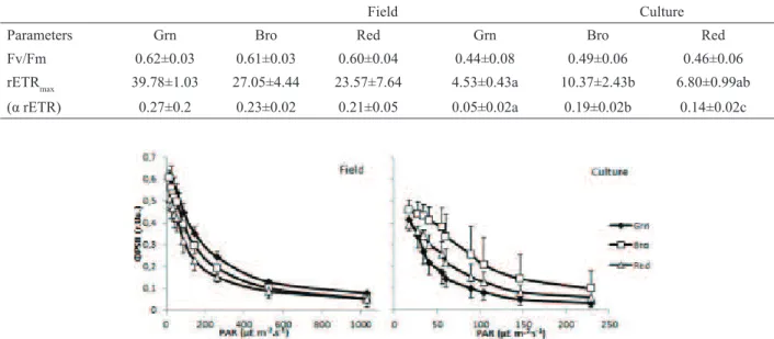
The potential quantum yield (Fv/Fm) of the three colour strains did not differ between ield samples (F=0.45,
p=0.645) or in vitro cultured specimens (F=0.57, p=0.577)
(Table 1). Previous studies reported similar Fv/Fm values
under natural conditions (around 0.6 r.u.) for Rhodophyta
species (Mouget & Tremblim, 2002; Figueroa et al., 2009; Chaloub et al., 2010). Higher Fv/Fm values were repoted
for in vitro cultured Hypnea musciformis (Wulfen) JV
Lamour. strains (Yokoya et al., 2007) than those reported
here. In the natural habitat, the green colour strain had
the highest effective quantum yield (ΦPSII), while the red strain had the lowest (F=79.12, p<0.010). Under the low light conditions of the in vitro culture, the green colour
strain had the lowest Φ PSII among the colour strains (F=68.02, p<0.010) (Figure 1). The rapid light curves are
shown in Figure 1. In the natural habitat, the green strain showed the highest rETRmax (F=10.95, p=0.002) (Figure 2) and the highest light saturation point (around 1,000
μE.m-2.s-1). However, the photosynthetic eficiencies (α
rETR) were similar among the three strains (Table 1). The brown and red strains showed similar light saturation points, approximately 50% lower than that of the green strain (around 500 μE.m-2.s-1). In culture, the brown strain
had the highest rETRmax and the green strain the lowest (F=18.40, p<0.001). The same pattern was observed for
α rETR. All colour strains saturated at around 56 μE.m -2.s-1. The green strain was the most eficient under natural
high light conditions among the three colour strains
and showed the lowest photosynthetic yield under low light culture condition. Non-photochemical quenching (NPQ) drains excitation energy and prevents damage to the photochemical apparatus (Mouget & Tremblim, 2002; Baker, 2008). The NPQ was equivalent for all colour strains under both growth conditions, although, as expected, considerably lower in culture (Figure 3). Under low light, only a small portion of the energy entering the photosynthetic process (3%) is dissipated through heat emission and luorescence (Bolhár-Nordenkampf & Oquist, 1993). Large amounts of light energy cannot
be fully utilized by the photochemical process and the
excess energy therefore has to be dissipated (Falkowski & Raven, 2007). The photosynthetic characterization of
the three strains suggests that the green strain copes more
eficiently under high light conditions but less eficiently under low light conditions than the brown and red strains. Additionally, the ΦPSII and rETRmax parameters appear
photochemical performance of the colour strains because
it was able to differentiate between them both in situ and
in vitro.
Pigment proiles
The content of Chl a did not differ signiicantly
between the three colour strains in the natural habitat (F=0.67, p=0.545). Under in vitro culture conditions, the concentration of Chl a differed signiicantly only
between the green strain and the others two colour strains (F=112.89, p<0.001), with a reduction of 32.6%
in the content (Figure 4). The photochemical process is inluenced primarily by irradiance and the Chl a
concentration (Dring, 1982). The observed decrease in
the Chl a concentration in the green strain in culture may
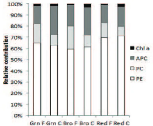
be related to the lower Φ PSII shown by this strain in vitro. Regarding the phycobiliproteins, PE was the major pigment in the three colour strains and, being the dominant
pigment in Rhodophyta, (Kirk, 1994; Van den Hoek et al., 1995). The colour of each strain is directly related to the observed PE concentration gradient (Figure 4). However, only the green strain differed from the other two strains (Field: F=28.55, p<0.001; In vitro F=16.31, p=0.004).
The phycoerythrin content was lower in the in vitro
cultured strains compared to the natural habitat: a 13.81% decrease for the green, 53.53% for the brown and 61.15%
for the red strain (Figure 4). Importantly, the green colour strain of H. musciformis, unlike other green strains found
in Rhodophyta (Yokoya et al., 2007; Costa & Plastino,
Table 1. Maximal quantum yield (Fv/Fm-r.u), relative electron transport rate (rETRmax-µE m-2s-1), photosynthetic eficiency (α
rETR) of green (Grn), brown (Bro) and red (Red) colour strains of Hypnea musciformis in the ield (F) and under culture (C) conditions. Values shown are the means and (±) standard deviations. The letters indicate signiicant differences according to two-way ANOVA (p<0.05).
Field Culture
Parameters Grn Bro Red Grn Bro Red
Fv/Fm 0.62±0.03 0.61±0.03 0.60±0.04 0.44±0.08 0.49±0.06 0.46±0.06
rETRmax 39.78±1.03 27.05±4.44 23.57±7.64 4.53±0.43a 10.37±2.43b 6.80±0.99ab
(α rETR) 0.27±0.2 0.23±0.02 0.21±0.05 0.05±0.02a 0.19±0.02b 0.14±0.02c
Figure 1. Effective quantum yield (Φ PSII) as a function of the irradiance for green (Grn), brown (Bro) and red (Red) colour strains of Hypnea musciformis in the ield and under culture conditions. The symbols are the means and the bars indicate the standard
deviations.
Figure 2. Rapid light curves of the relative electron transport rate (rETR) as a function of the irradiance for green (Grn), brown (Bro) and red (Red) colour strains of Hypnea musciformis in the ield and under culture conditions. The symbols are the means and
2011) showed the presence of PE. Regarding the content of PC, there was no evidence of signiicant differences between the strains for ield samples (F=5.07, p=0.051)
or in the in vitro culture (F=2.08, p=0.206). There was a
reduction of 76.66% in the concentration of PC for the in vitro cultured strains in relation to the ield ones (Figure 4). Alophycocyanin concentrations in the green strain differed from that of the red strain in the natural habitat
(F=6.91, p=0.278). Under culture conditions, the green strain had a concentration of APC signiicantly different from that of the other two strains (F=112.89, p<0.001).
Compared to the natural habitat, the concentration of APC
was reduced 48.39% in the three colour strains in culture. The relative contributions of each pigment for the three strains are shown in Figure 5.
Figure 4. Concentrations (µg.g-1 fresh weight) of chlorophyll
a and the phycobilliproteins (phycoerythrin (PE), phycocyanin (PC) and alophycocyanin (APC) for green (Grn), brown (Bro) and red (Red) colour strains of Hypnea musciformis in the ield
(F) and under culture (C) conditions.
During approximately one year of in vitro
culture, the colour of the three strains was maintained, although less intense in the brown and the red strains
compared to the natural habitat. The maintenance of
the proportionality of phycobiliproteins, especially phycoerythrin, ensures the chromatic identity of the colour
strains. This was found previously in other Rhodophyta colour strains (Costa & Plastino, 2001, 2011; Yokoya et al., 2007). This suggests that different colours are not
the result of chromatic adaptation to naturally occurring
environmental variations. To better characterize these strains, morphological and molecular analysis, as well as the photosynthetic responses to variations in the quality and/or quantity of light are needed.
Figure 5. Relative contributions of chlorophyll a and the phycobilliproteins-phycoerythrin (PE), phycocyanin (PC) and alophycocyanin (APC) for green (Grn), brown (Bro) and red (Red) colour strains of Hypnea musciformis in the ield (F) and
under culture (C) conditions.
Acknowledgments
The authors are grateful to the Coordination
of Superior Level Staff Improvement (CAPES) and
the Brazilian National Council for Scientiic and Technological Development (CNPq) for PhD grants to the irst and second authors [Post-Graduate Programme in Plant Biotechnology, Federal University of Rio de Janeiro
Figure 3. Non-photochemical quenching (NPQ) as a function of the irradiance for green (Grn), brown (Bro) and red
(UFRJ)] and for inancial support of the ifth author (CNPq-Process number 309929/2009-1).
References
Baker N 2008. Chlorophyll Fluorescence: A probe of photosynthesis in vivo. Annu Rev Plant Biol 59: 89-113. Bautista AIN, Necchi O 2007. Photoacclimation in three species
of freshwater red algae. Braz J Plant Physiol 19: 23-34. Bolhàr-Nordenkampf HR, Öquist G 1993. Chlorophyll
luorescence as a tool in photosynthesis research. In: Hall DO, Scurlock JMO, Bolhàr-Nordenkampf HR, Leegood RC, Long SP (org.). Photosynthesis and production in a changing environment: a ield and laboratory manual. London: Chapman & Hall, p. 193-206.
Chaloub RM, Reinert F, Nassar CAG, Fleury BG, Mantuano DG, Larkum AWD 2010. Photosynthetic properties of three Brazilian seaweeds. Rev Bras Bot 33: 371-374. Costa VL, Plastino EM 2001. Histórico de vida de espécimens
selvagens e variantes cromáticas de Gracilaria birdiae
(Gracilariales, Rhodophyta). Rev Bras Bot 24: 491-500. Costa VL, Plastino EM 2011. Color inheritance and pigment
characterization of red (wild- type), greenish-brown, and green strains of Gracilaria birdiae (Gracilariales, Rhodophyta). J Appl Phycol 23: 599-605.
Davies LP, Jamieson DD, Baird-Lambert JA, Kazlauskas R 1984. Halogenated pyrrolopyrimidine analogs of adenosine from marine organisms, pharmacological activities and potent inhibition of adenosine kinase.
Biochem Pharmacol 33: 347-356.
Dring MJ 1982. Photosynthesis in the sea. In: Dring MJ. The Biology of Marine Plants. Great Britain: Edward Arnold, p. 44-64.
Durako MJ, Kunzelman JI 2002. Photosynthetic characteristics of Thalassia testudinum measured in situ by pulse-amplitude modulated (PAM) fluorometry: methodological and scale-based considerations. Aquat Bot 73: 173-185.
Edwards P 1970. Illustrated guide to the seaweeds and sea grasses in the vicinity of Porto Aransas, Texas. Contrib Mar Sci 15: 1-228.
Falkowski PG, Raven JA 2007. Aquatic photosynthesis. Princeton: Princeton University Press.
Fernandes DRP, Yokoya NS, Yoneshigue-Valentin Y 2011. Protocol for seaweed decontamination to isolate unialgal cultures. Rev Bras Farmacogn 21: 313-316.
Figueroa FL, Martínez B, Israel A, Neori A, Malta EJ, Ang Jr P, Inken S, Marquardt R, Rachamim T, Arazi U, Frenk S, Korbee N 2009. Acclimation of red sea macroalgae to solar radiation: photosynthesis and thallus absorbance.
Aquat Biol 7: 159-172.
Gerung GS, Ohno M 1997. Growth rates of Eucheuma denticulatum (Burman) Collins et Harvey and Kappaphycus striatum (Schmitz) Doty under different conditions in warm waters of Southern Japan. J Appl
Phycol 9: 413-415.
Guimarães M 2000. Aspectos isiológicos de Gracilaria domingensis (Gracilariales, Rhodophyta): subsídios para a compreensão da manutenção do polimorismo pigmentar. São Paulo, 89 p. Tese de Doutorado, Universidade de São Paulo.
Häder DP, Lebert M, Flores-Moya A, Jiménez Z, Mercado J, Salles S, Aguilera J, Figueroa FL 1977. Effects of solar radiation on the photosynthetic activity of the red alga
Corallina elongate Ellis et Soland. J photoch photobio B 37: 196-202.
Kirk JTO 1994. Light & photosynthesis in aquatic ecosystems. Cambridge: Cambridge University Press.
Kursar TA, Van Der Meer J, Alberte RS 1983. Light-harvesting system of the red alga Gracilaria tikvahiae. I. Biochemical analyses of pigment mutations. Plant Physiol 73: 353-360.
Martínez B, Rico JM 2002. Seasonal variation of P content and major N pools in Palmaria palmata (Rhodophyta). J Phycol 38: 1082-1089.
Melo VMM, Medeiros DA, Rios FJB, Castelar LIM, Carvalho AD 1997. Antifungal properties of proteins (agglutinins) from the red alga Hypnea musciformis (Wulfen) Lamouroux. Bot Mar 40: 281-284.
Mouget J, Tremblim G 2002. Suitability of the luorescence monitoring system (FMS, Hansatech) for meansurement of photosynthetic characteristics in algae. Aquat Bot 74: 219-231.
Nagano CS, Moreno FBMB, Bloch C, Prates MV, Calvete JJ, Saker-Sampaio S, Farias WRL, Tavares TD, Nascimento KS, Grangeiro TB, Cavada BS, Sampaio AH 2002. Puriication and characterization of lectins from the red marine alga Hypnea musciformis. Prot Pept Letters 9: 159-165.
Nagano CS, Debray H, Nascimento KS, Pinto VPT, Cavada BS, Saker-Sampaio S, Farias WRL, Sampaio AH, Calvete JJ 2005. HCA and HML isolated from the red marine algae Hypnea cervicornis and Hypnea musciformis deine a
novel lectin family. Prot Sci 14: 2167-2176.
Neushul M 1990. Antiviral carbohydrates from marine red algae. Hydrobiologia 204/205: 99-104.
Neveux J, Lantoine F 1993. Spectroluorometric assay of chlorophylls and phaeopigments using the least squares approximation technique. Deep-Sea Res Oceanogr Res Pap 40: 1747-1765.
Neveux J, Tenório, MMB, Dupouy C, Villareal TA 2006. Spectral diversity of phycoerythrins and diazotroph abundance in tropical waters. Limnol Oceanogr 51: 1689-1698. Oliveira EC 1998. The seaweed resources of Brazil. In: Crithcley
AT, Ohno M (org.). Seaweeds resources of the world. Yokosuka: Japan International Cooperation Agency, p. 366-371.
Reis RP, Caldeira AQ, Miranda APS, Barros-Barreto MB 2006. Potencial para maricultura da carragenóita Hypnea musciformis (Wulfen) J.V. Lamour. (Gigartinales - Rhodophyta) na Ilha da Marambaia, Baía de Sepetiba, RJ, Brasil. Acta Bot Bras 20: 763-769.
Reis RP, Yoneshigue-Valentin Y 1998. Variação espaço-temporal de populações de Hypnea musciformis (Rhodophyta, Gigartinales) na Baía de Sepetiba e Armação dos Búzios,
Rio de Janeiro, Brasil. Acta Bot Bras 12: 465-483. Salimabi BD 1980. Antispasmodic and anti-inlammatory
activity of carrageenan from Hypnea musciformis
Wulfen. Indian J Pharmacol 1: 259-261.
Ursi S, Plastino EM 2001. Crescimento in vitro de linhagens de coloração vermelha e verde clara de Gracilaria birdiae
(Gracilariales, Rhodophyta) em dois meios de cultura: análise de diferentes estádios reprodutivos. Rev Bras Bot 24: 587-594.
Van den Hoek C, Mann DG, Jahns HM 1995. Algae: An introduction to phycology. Cambridge: University
Press.
Yokoya YS, Necchi Jr O, Martins AP, Gonzalez SF, Plastino EM 2007. Growth responses and photosynthetic characteristics of wild and phycoerythrin-deicient strains of Hypnea musciformis (Rhodophyta). J Appl Phycol 19: 197-205.
*Correspondence
Yocie Yoneshigue-Valentin