762
RESUMO: “Efeito vasorelaxante do extrato diclorometano de Hyptis fruticosa Salzm. ex Benth., Lamiaceae, em artéria mesentérica de ratos”. O efeito vasorelaxante do extrato diclorometano de Hyptis fruticosa Salzm. ex Benth., Lamiaceae (HFDE), em anéis isolados de artéria mesentérica de ratos foi avaliado nesse estudo. Em anéis intactos, pré-contraídos com fenilefrina (10 µM), HFDE (0,1-3000 µg/mL) induziu vasorelaxamento de maneira dependente de concentração (Emax = 119±14%; n = 6), o qual não foi afetado após remoção do endotélio (Emax = 116±6%; n = 6), após KCl 20 mM (Emax = 135±9%; n = 6) ou em anéis pré-contraídos com KCl 80 mM (Emax = 125±4%; n = 6). Em anéis sem endotélio, HFDE (300 ou 1000 µg/mL) inibiu as contrações induzidas por CaCl2 (inibição máxima = 25±7% e 95±1%, respectivamente). Além disso, HFDE promoveu um vasorelaxamento adicional (15±3%; n = 7) sobre o relaxamento máximo de 10 µM de nifedipina (78±3%, n = 7). Em conclusão, HFDE induz efeito vasorelaxante através de uma via independente de endotélio, possivelmente devido à inibição do inluxo de Ca2+ através de canais de Ca2+ operados
por voltagem.
Unitermos: Hyptis fruticosa, extrato diclorometano, efeito vasorelaxante, artéria mesentérica, ratos.
ABSTRACT: Vasorelaxant effect of Hyptis fruticosa dichloromethane extract (HFDE) on isolated rings of rat mesenteric artery was evaluated in this study. In intact rings, HFDE (0.1-3000 µg/ mL) induced concentration-dependent vasorelaxations (Emax = 119±14%; n = 6) of phenylephrine tonus that were not modiied after endothelium removal (Emax = 116±6%; n = 6), after KCl 20
mM (Emax = 135±9%; n = 6) or in rings pre-contracted with KCl 80 mM (Emax = 125±4%; n = 6). In endothelium denuded rings, HFDE (300 or 1000 µg/mL) inhibited contractions induced by CaCl2 (maximal inhibition = 25±7% and 95±1%; respectively). Furthermore, HFDE promoted an additional vasorelaxation (15±3%; n = 7) after maximal response of 10 µM nifedipine (78±3%; n = 7). In conclusion, HFDE induces vasorelaxant effect through an endothelium-independent pathway, which mostly seems to occur due inhibition of the Ca2+ inlux through voltage-operated
Ca2+ channels.
Keywords:Hyptis fruticosa, dichloromethane extract, vasorelaxant effect, mesenteric artery, rats. Brazilian Journal of Pharmacognosy
20(5): 762-766, Out./Nov. 2010
Article
Received 6 Jul 2009; Accepted 4 Dec 2009; Available online 19 Nov 2010.
Vasorelaxant effect of
Hyptis fruticosa
Salzm. ex Benth., Lamiaceae,
dichloromethane extract on rat mesenteric artery
Ítalo J. A. Moreira,
1Maria P. N. Moreno,
2Maria F. G. Fernandes,
2João B. Fernandes,
2Flávia V. Moreira,
1Ângelo R. Antoniolli,
1Márcio R.V. Santos
*,11Departamento de Fisiologia, Universidade Federal de Sergipe, CCBS, Av Marechal Rondo s/n, Jd. Rosa Elze,
49100-000 São Cristóvão-SE, Brazil
2Departamento de Química, Universidade Federal de São Carlos, Rodovia Washington Luís, km 235, 1
3.565-905 São Carlos-SP, Brazil.
ISSN 0102-695X DOI: 10.1590/S0102-695X2010005000003
*E-mail: [email protected], Tel. +55 79 2105 6842, Fax +55 79 21056474.
INTRODUCTION
The use of medicinal plants for the treatment of human diseases has increased considerably worldwide. Evaluation of the effects of these plants on organs and systems has contributed to the development of the
scientiic basis for their therapeutic application, and also
has enriched considerably the therapeutic arsenal for the treatment of a number of diseases (Elizabetsky, 1986).
Hyptis genus, Lamiaceae, is composed by four
hundred species distributed at all American Continent. In Brazil, this species is mainly distributed at the central region (Harley, 1988).Various species of this genus are
used in the folk medicine because of its antiinlammatory,
antinociceptive, anticonvulsant and antiulcerogenic actions (Barbosa & Ramos, 1992; Akah & Nwambie, 1993; Kuhnt et al., 1995; Bispo et al., 2001).
763
Rev. Bras. Farmacogn. Braz. J. Pharmacogn. 20(5): Out./Nov. 2010
grows up to 1.5 m found on the Brazilian northeastern coast. Phytochemical studies performed in our laboratory have demonstrated that leaves of this plant present tanins, terpenes, steroids and alkaloids, and absence of saponins (unpublished data). Previous pharmacological studies have demonstrated that H. fruticosa presented analgesic (Silva et al., 2006; Cândido, 2006; Menezes et al., 2007), larvicidal (Silva et al., 2008) and hypotensive activities (Santos et al., 2007). Thus, the aim of this work was to evaluate the vasorelaxant effect of Hyptis fruticosa dichloromethane extract (HFDE) and its action mechanism in rats.
MATERIAL AND METHODS
Drugs
The drugs used were: Acetylcholine chloride (Ach), L-phenylephrine chloride (Phe) and cremophor (a derivative of castor oil and ethylene oxide used to emulsify water-insoluble substances) (SIGMA). All compounds were dissolved in distilled water.
Extraction
Hyptis fruticosa Salzm. ex Benth., Lamiaceae, collected near São Cristóvão (S 10o 56` W 37o 11`),
Brazilian State of Sergipe, was identiied by Prof. Dr.
Adauto Souza Ribeiro, Botanist in the Biology Department, Universidade Federal de Sergipe. A voucher specimen was deposited in the Herbarium of the Biology Departament, Universidade Federal de Sergipe (code n° ASE 01137). Aerial parts of H. fruticosa were dried at 40 ºC in an oven with air circulation and pulverized. The powder (500 g) was exhaustively extracted with methanol (1:5 p/v) by 8
dias to room temperature. After iltration, the solvent was
removed under reduced pressure, yielding 78.5 g of the methanol extract. The HFDE was obtained from methanol extract by using the following solvents: dichloromethane, ethyl acetate and methanol yielding: 8.4 g (1.68%), 5.4 g (1.08%) and 29.5 g (5.90%), respectively.
Animals
Male Wistar rats (200-300 g) were used in all experiments. They were housed in conditions of controlled temperature (21±1 ºC) and exposed to a 12 h light-dark cycle with free access to food (Purina-Brazil) and tap water. All procedures described in the present work are in agreement with Animal Research Ethics Committee from Universidade Federal de Sergipe.
Solutions
The composition of the normal Tyrode’s solution
used was: NaCl 158.3, KCl 4.0, CaCl22H2O 2.0, NaHCO3
10.0, C6H12O6 5.6, MgCl2.6H2O 1.05 and NaH2PO4H2O
0.42 mM. K+-depolarizing solutions (KCl 20, 60 and 80
mM) were prepared by replacing 20, 60 or 80 mM KCl in the Tyrode’s solution with equimolar NaCl, respectively and nominally without Ca2+ solution was prepared by
omitting CaCl2.
Tissue preparation
Rats were euthanized by cervical dislocation and exsanguination. The superior mesenteric artery was removed, cleaned from connective tissue and fat, and sectioned in rings (1-2 mm), which were suspended in organ baths containing 10 mL of Tyrode´s solution, gassed with a mixture of 95% O2 and 5% CO2 and maintained at 37 oC. Isometric tension was recorded under a resting
tension of 0.75 g. During the stabilization period the solution was changed every 15 min (Altura & Altura, 1970). The isometric tension was recorded through a force transducer (Gould, Model GM2, USA) coupled to
an ampliier-recorder (Gould, USA). Endothelium was
removed by gently rubbing the intimal surface of the vessels. The presence of functional endothelium was assessed by the ability of acetylcholine (ACh) (10 µM) to induce more than 70% relaxation of pre-contracted vessels with phenylephrine (10 µM). The absence of the relaxation to ACh was taken as evidence that the vessel segments were functionally denuded of endothelium.
HFDE effect on phenylephrine (10 μM) induced tonus in isolated rat superior mesenteric artery rings with or without endothelium
After the stabilization period, two successive contractions of similar magnitude were induced with 10 µM Phe in rings with or without endothelium. During the tonic phase of the third contraction, different concentrations of HFDE (0.1; 0.3; 1; 3; 10; 30; 100; 300; 1000 and 3000 µg/mL) were added cumulatively to the organ bath. The relaxations were measured by comparing the developed tension before and after the addition of HFDE and expressed as percentage of relaxation from induced tonus. In other set of experiments, concentration-response curves were obtained in rings without endothelium before and after to the pre-incubated with 20 mM of KCl.
Effect of HFDE on contraction induced by KCl 80 mM in endothelium-denude rings
After the stabilization period, rings without endothelium were pre-contracted with K+-depolarizing
764 Rev. Bras. Farmacogn. Braz. J. Pharmacogn.
20(5): Out./Nov. 2010
Effect of HFDE on concentration-response curves to CaCl2 in endothelium-denuded rings
After the stabilization period, the rings without endothelium were contracted with K+-depolarizing solution
(KCl 60 mM) and washed with normal Tyrode’s solution until full recovery of initial tension. After this, they were incubated with nominally without Ca2+ solution for 15 min
and afterwards exposed to nominally without Ca2+ solution
with KCl to 60 mM for another 15 min (Goodfraind et
al., 1986). Then, a irst cumulative concentration-response
curve to CaCl2 (3 x 10⁻⁶, 10-5, 3 x 10⁻⁵, 10-4, 3 x 10⁻⁴, 10-3,
3 x 10⁻³, 10-2 and 3 x 10⁻² M) was obtained. In these same
preparations, HFDE (300 or 1000 µg/mL) was individually pre-incubated for 15 min and a second cumulative concentration-response curve to CaCl2 was obtained. This curve was compared with those obtained in the absence of HFDE and the results were expressed as percentages of the maximal response to CaCl2 alone.
Effect of HFDE on maximal vasorelaxant response of nifedipine in endothelium denuded rings
Initially, a concentration-response curve to nifedipine was performed in order to determine the concentration of maximal vasorelaxant response in endothelium-denuded rings pre-contracted with 10 µM of Phe (data not shown). After this, others endothelium-denuded rings pre-contracted with 10 µM of Phe were incubated with 1000 µg/mL of HFDE or 10 µM of nifedipine, separately. In other set of experiments, endothelium-denuded rings pre-contracted with 10 µM of Phe were incubated with 10 µM of nifedipine and after obtainment of the maximal vasorelaxant response, HFDE (1000 µg/mL) was added in organ bath. The responses to each vasorelaxant agent were statistically compared.
Statistical analysis
Values were expressed as mean±SEM. When appropriate, one-way ANOVA or two-way ANOVA for repeated measures, both followed by Bonferroni
post-test, was performed to evaluate the signiicance of the
differences between means. Statistically different values
were detected at a signiicance level of 0.05.
RESULTS AND DISCUSSION
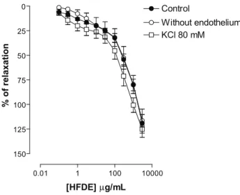
In intact rings of rat isolated superior mesenteric artery, HFDE (0.1, 0.3, 1, 3, 10, 30, 100, 300 and 1000
μg/mL, cumulatively) induced vasorelaxation in a
concentration dependent manner of tonus induced by 10
μM phenylephrine (Emax = 119±14%; n = 6) (Figure 1).
0.01 1 100 10000
0
25
50
75
100
125
150
Control
Without endothelium KCl 80 mM
[HFDE]µg/mL
% of relaxation
Figure 1. Concentration-response curves to HFDE (0.1; 0.3; 1; 3; 10; 30; 100; 300; 1000 and 3000 µg/mL) in rings of rat isolated superior mesenteric artery pre-contracted with 10 μM Phe (Control), without functional endothelium (Without endothelium) and rings without endothelium pre-contracted with K+-depolarizing solutions (KCl 80 mM). Values are expressed
as mean±SEM, n = 6. The data were analyzed with repeated measures two-way ANOVA followed by Bonferroni post-test.
It is well known that the endothelium is an important regulator of the vascular tone by releasing endothelium-derived relaxing factors (Moncada et al., 1991), mainly NO and COX-derived products, such as PGI2 (Moncada et al., 1991; Furchgott & Zawadzki, 1980). In order to investigate the participation of the endothelium in the vasorelaxant effect induced by HFDE, we performed experiments in the absence of functional endothelium. In these conditions, the vasorelaxant response induced by
HFDE was not signiicantly changed (Emax = 116±6%; n = 6)
(Figure 1). This suggests that the presence of endothelium is not essential for relaxant response expression and that an endothelium-independent pathway is probably implicated in this effect.
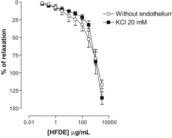
Potassium channels importantly contribute to the determination and regulation of the vascular tone (Nelson & Quayle, 1995; Jackson, 2000). The electrochemical gradient for K+ ions is such that the opening of K+ channels
results in the diffusion of this cation out of the cells with consequent hyperpolarization. This effect closes voltage-operated Ca2+ channels and leads to vasorelaxation
(Jackson, 2000). In order to investigate the involvement of K+ channels in the vasorelaxant effect of HFDE, we
performed experiments in the presence of 20 mM of K+.
This procedure partially prevents the eflux of K+ through
the membrane and, therefore inhibits the relaxations mediated by the opening of K+ channels (Campbell et al.,
1996). Thus, In rings without endothelium pre-contracted with Phe and incubated with KCl 20 mM, the
765
Rev. Bras. Farmacogn. Braz. J. Pharmacogn. 20(5): Out./Nov. 2010
(Emax = 135±9%; n = 6) (Figure 2), suggesting that K+
channels appears not to be involved in this response.
0.01 1 100 10000
0
25
50
75
100
125
150
Without endothelium KCl 20 mM
[HFDE]µg/mL
% of relaxation
Figure 2. Concentration-response curves to HFDE (0.1; 0.3; 1; 3; 10; 30; 100; 300; 1000 and 3000 µg/mL) in rings of rat isolated superior mesenteric artery without functional endothelium pre-contracted with 10 μM Phe (Without endothelium), and rings without endothelium pre-contracted with Phe and incubated with K+-depolarizing solutions (KCl 20 mM). Values are expressed as mean±SEM, n = 6. The data were analyzed with repeated measures two-way ANOVA followed by Bonferroni post-test.
Calcium is the primary regulator of tension in vascular smooth muscle (Gurney, 1994). It is well known that the maintenance of smooth muscle contraction depends on Ca2+ inlux from extracellular space through
voltage-and/or receptor-operated calcium channels (VOCCs and/ or ROCCs, respectively) (Karaki & Weiss, 1988). It is well reported that the increase of external K+ concentration
(KCl 80 mM) induces smooth muscle contraction through VOCCs activation and subsequent calcium release from the sarcoplasmic reticulum (Karaki & Weiss, 1988). The high K+-induced contraction is inhibited by Ca2+ channel
blockers or by removal of external Ca2+ and is, therefore,
entirely dependent of Ca2+ inlux (Karaki & Weiss, 1988).
Thus, we evaluated the HFDE effect on
endothelium-denuded rings pre-contracted with K+-depolarizing
solutions (KCl 80mM). This set of experiments revealed that HFDE induced vasorelaxations, which were not
signiicantly different of those observed in rings
pre-contracted with Phe (125±4%; n = 6) (Figure 1), suggesting
that the HFDE inhibits Ca2+ inlux through VOCCs.
In order to check the hypothesis above, we
constructed a concentration-response curve to CaCl2
(3 x 10⁻⁶, 10-5, 3 x 10⁻⁵, 10-4, 3 x 10⁻⁴, 10-3, 3 x 10⁻³, 10-2
and 3 x 10⁻² M) in presence of K+-depolarizing solution
(KCl 60 mM), before and after incubation with HFDE in doses of 300 and 1000 µg/mL. In these conditions, CaCl2 induced contractions in endothelium-denuded rings of rat mesenteric artery in a concentration-dependent manner
that were strongly inhibited after incubation with HFDE in concentrations of 300 and 1000 µg/mL (maximal inhibition = 25±7% and 95±1%; n = 6; respectively).
1 2 3 4 5 6 0 25 50 75 100
125 Control
After HFDE 1000µg/mL
*** *** *** *** ***
*** *
After HFDE 300µg/mL
* **
*
*
*** ***
- Log [CaCl2] M
% of contraction
Figure 3. Concentration-response curves to CaCl2 (3 x 10⁻⁶, 10-5,
3 x 10⁻⁵, 10-4, 3 x 10⁻⁴, 10-3, 3 x 10⁻³, 10-2 and 3 x 10⁻² M) in rings
of rat superior mesenteric artery, without endothelium before (control) and after pre-incubation with HFDE at concentrations of 300 and 1000 µg/mL, separately. Values are expressed as mean±SEM, n = 6. The data were analyzed with repeated measures two-way ANOVA followed by Bonferroni post-test. *p< 0.05, **p< 0.01 and ***p< 0.001 vs control.
As reported by Chan et al. (2000), nifedipine, a
L-type voltage-operated Ca2+ channel selective blocker,
also inhibited the concentration-response curve to CaCl2, suggesting strongly that HFDE could be acting possibly as a calcium channel blocker.
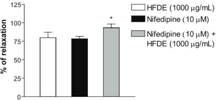
Finally, we performed experiments in that were observed the vasorelaxation response of HFDE (1000 µg/ mL) and 10 µM of nifedipine, separately, and the effect of HFDE (1000 µg/mL) after maximal vasorelaxant response induced by 10 µM of nifedipine. In this condition, HFDE
(1000 μg/mL) or nifedipine (10 μM) were capable of
inducing vasorelaxation of Phe tonus (Emax = 79±9 and 78±3%; n = 6, respectively), and HFDE (1000 µg/mL)
induced a small but signiicant additional vasorelaxation
effect on the maximal vasorelaxation of nifedipine (10 mM) (15±3%; n = 7), suggesting that HFDE appears to be acting in major part by the same pathway of the nifedipine.
However, the observation of an additional vasorelaxation after maximal response of nifedipine allow us to hypothesize that other pathway appears to be implicated in the HFDE-induced response, possibly involving intracellular calcium stores. However, further experiments are necessary to clearly elucidate this assumption.
endothelium-766 Rev. Bras. Farmacogn. Braz. J. Pharmacogn
20(5): Out./Nov. 2010 .
independent pathway, which appears to be due in major part to inhibition of the Ca2+ inlux through
voltage-operated Ca2+ channels.
0 25 50 75 100 125
Nifedipine(10µM)
Nifedipine(10µM) + HFDE (1000µg/mL) *
HFDE (1000µg/mL)
% of relaxation
Figure 4. Vasorelaxant effect of nifedipine (10 μM) before and after administration of HFDE (1000 µg/mL) in rings of rat isolated superior mesenteric artery without functional endothelium pre-contracted with 10 μM Phe. Values are expressed as mean±SEM, n = 6. The data were analyzed with one-way ANOVA followed by Bonferroni post-test. *p< 0.05 vs Nifedipine.
ACKNOWLEDGEMENTS
We thank technical assistance of Mr. Osvaldo Andrade Santos. This work was supported by grants from CNPq, CAPES, FAPITEC-SE, Ministério da Saúde, SES/ SE, Brazil.
REFERENCES
Akah PA, Nwambie AI 1993. Nigerian plants with anti-convulsant property. Fitoterapia 64: 42-44.
Altura BM, Altura BT 1970. Differential effects of substrate depletion on drug-induced contractions of rabbit aorta. Am J Physiol 219: 1698-1705.
Barbosa PPP, Ramos CP 1992. Studies on the antiulcerogenic activity of the essential oil of Hyptis mutabilis Briq in rats. Phytotherapy 6: 114-115.
Bispo MD, Mourão RHV, Franzotti EM, Bomim KBR, Arrigoni-Blank MF, Moreno MPN, Machioro M, Antoniolli AR 2001. Antinociceptive and antiedematogenic effects of the aqueous extract of Hyptis pectinata leaves in experimental animals. J Ethnopharmacol 76: 81-86. Campbell WB, Gebremedhin D, Prait PF, Herder DR
1996. Identiication of epoxyeicosatrienoic acids as endothelium-derived hyperpolarizing factors. Circ Res 78: 415-423.
Cândido EAF 2006. Caracterização parcial dos efeitos do extrato hidroalcoólico das folhas da Hyptis fruticosa no sistema nervoso central de camundongos. São Cristóvão, 75 p. Dissertação de Mestrado, Programa de Pós-graduação em Ciências da Saúde, Universidade Federal de Sergipe. Chan W, Yao X, Ko W, Huang Y 2000. Nitric oxide mediated
endothelium-dependent relaxation induced by glibenclamide in rat isolated aorta. Cardiovasc Res 46: 180-187.
Elizabetsky E 1986. New directions in ethnopharmacology. J Ethnobiol 6: 121-121.
Furchgott RF, Zawadzki JV 1980. The obligatory role of endothelial cells in the relaxation of arterial smooth muscle by acetylcholine. Nature 288: 373-376.
Goodfraind T, Miller R, Wibo M 1986. Calcium antagonism and calcium entry blockade. Pharmacol Rev 38: 321-416. Gurney AM 1994. Mechanisms of drug-induced vasodilatation. J
Pharm Pharmacol 46: 242-251.
Harley RM 1988. Revision of generic limits in Hyptis Jacq (Labiatae) and its allies. Bot J Linn Soc 98: 87-95. Jackson WF 2000. Ion channels and vascular tone. Hypertension
35: 173-178.
Karaki H, Weiss GB 1988. Calcium release in smooth muscle. Life Sci 42: 111-122.
Kuhnt M, Probstle A, Rimpler H, Bauer R, Heinrich M 1995. Biological and pharmacological activities and further constituents of Hyptis verticillata. Planta Med 61: 227-232.
Menezes IAC, Marques MS, Santos TC, Dias KS, Silva ABL, Mello ICM, Lisboa ACCD, Alves PB, Cavalcanti SCH, Marçal RM, Antoniolli AR 2007. Antinociceptive effect and acute toxicity of the essential oil of Hyptis fruticosa in mice. Fitoterapia 78: 192-195.
Moncada S, Palmer RMJ, Higgs EA 1991. Nitric Oxide: Physiology, pathophysiology and pharmacology. Pharmacol Rev 43: 109-142.
Nelson MT, Quayle JM 1995. Physiological roles and properties of potassium channels in arterial smooth muscle. Am J Physiol 268: C799-C822.
Santos MRV, Carvalho AA, Medeiros IA, Alves PB, Marchioro M, Antoniolli AR 2007. Cardiovascular effects of Hyptis fruticosa essential oil in rats. Fitoterapia 78: 186-191. Silva ABL, Dias KS, Marques MS, Menezes IAC, Santos TC,
Mello ICM, Lisboa ACCD Cavalcanti SCH, Marçal RM, Antoniolli AR 2006. Evaluation of the analgesic effect and acute toxicity of the aqueous extract of Hyptis fruticosa (Salmz. ex Benth.)”. RevBras J Pharmacogn 16: 475-479.