Rev Odontol UNESP. 2016 Sept-Oct; 45(5): 258-264 © 2016 - ISSN 1807-2577 ORIGINAL ARTICLE
Doi: http://dx.doi.org/10.1590/1807-2577.28415
Levobupivacaine induces vasodilatation, but
not vasoconstriction, in rat mesenteric artery
Levobupivacaína induz vasodilatação, mas não vasoconstrição em artéria mesentérica de rato
Liciane dos Santos MENEZES
a*, Liane Maciel de Almeida SOUZA
a,
Márcio Roberto Viana dos SANTOS
a, Patrícia Santos Cunha MENDONÇA
a,
Ítalo José Alves MOREIRA
a, Allan Carlos Araújo de OLIVEIRA
aaUFS – Universidade Federal de Sergipe, São Cristóvão, SE, Brasil
Resumo
Introdução: Levobupivacaína pode ser uma nova alternativa para analgesia por apresentar baixa toxicidade e vasoconstrição, permitindo sua utilização em pacientes que apresentam contra-indicação no uso de vasoconstritores. Objetivo: Avaliar os efeitos da levobupivacaína utilizando a técnica de reatividade vascular em artéria mesentérica isolada de rato e comparar este efeito à lidocaína. Material e método: Anéis foram obtidos da artéria mesentérica de ratos machos Wistar e foram mantidos em cubas. Para o registro das contrações isométricas, cada anel foi suspenso por linhas de algodão fixadas a um transdutor de força acoplado a um sistema de aquisição. Resultado: Tanto a lidocaína como a levobupivacaína não apresentaram efeito vasocontritor sobre o tônus basal em anéis com endotélio functional. No entanto, quando os anéis foram pré-contraídos com fenilefrina, ambas as drogas induziram um vasorrelaxamento concentração-dependente. O efeito vasorrelaxante causado pela levobupivacaína não foi diferente após a remoção do endotélio, ou com o tetraetilamônio (1mM), um bloqueador não seletivo dos canais para. K+. Em anéis sem endotélio
funcional e pré-contraídos com solução despolarizante de Tyrode (KCl 80mM), o vasorelaxamento induzido pela levobupivacaína não foi significativamente diferente daquele observado em anéis pré-contraídos com fenilefrina e não apresentou um efeito adicional significativo sobre o relaxamento máximo da nifedipina. Conclusão: Este estudo demonstrou que a levobupivacaína produz efeito vasorrelaxante em artéria mesentérica de rato, que é endotélio independente. Este efeito parece envolver os bloqueadores de canais para Ca2+ em célula muscular vascular lisa.
Descritores: Lidocaína; artéria mesentérica; vasodilatação.
Abstract
Introduction: Levobupivacaine (LEVO) can replace analgesia because it exhibits low toxicity and causes minor vasoconstriction, enabling its use in patients in whom vasoconstrictors are contraindicated. Objective: We aimed to evaluate the effects of LEVO in isolated rat superior mesenteric artery by using the vascular reactivity technique and compare its effect to that of lidocaine. Material and method: Arterial rings were obtained from the mesenteric artery of male Wistar rats and kept in organ baths. For recording isometric contractions, each ring was suspended by cotton threads from a force transducer, which was connected to a data acquisition system. Result: Both lidocaine and LEVO did not show a vasoconstrictor effect on the basal tone of the arterial rings with functional endothelium. However, when the rings were pre-contracted with phenylephrine, both drugs were able to induce concentration-dependent vasodilatation. The vasodilator effect induced by LEVO did not change after removal of the endothelium, or with the addition of tetraethylammonium (1 mM), a non-selective K+ channel blocker. In the rings without functional
endothelium, which were pre-contracted with depolarizing Tyrode’s solution (KCl 80 mM), LEVO-induced vasodilatation was not significantly different from that observed in the rings pre-contracted with phenylephrine. Moreover, it did not show a significant additional vasodilator effect compared to the maximal vasodilator effect of nifedipine. Conclusion: This study demonstrated that LEVO produces a vasodilator effect in the rat superior mesenteric artery in an endothelium-independent manner. This effect seems to be mediated via Ca2+ channel blockade in the vascular
smooth muscle cells.
INTRODUCTION
Advances in operation techniques and concerns about pain control have supported and sustained several pharmacological and clinical studies on local anesthetics1. Local anesthetics are widely
used in dentistry to minimize pain, in order to provide a safe and efective dental treatment2.
Local anesthetics are classiied according to their chemical structure into ester and amide-types, the latter being used more frequently in clinical settings. Amide-type anesthetics include prilocaine, lidocaine, mepivacaine, articaine, ropivacaine, and bupivacaine (BUPI)3. BUPI is a long-acting anesthetic, which can be used in
long surgeries without the need for anesthetic complementation. However, it exhibits a certain degree of cardiotoxicity, caused by the dextrorotatory enantiomer in the racemic mixture (S50-R50), which encouraged researchers to develop an anesthetic with a lower toxic potential4,5.
hus, levobupivacaine (LEVO), a local anesthetic with properties similar to those of BUPI but with less central and cardiac toxicity, was introduced. his diference is due to its predominant levorotatory enantiomer (S75-R25)5-7.
LEVO is considered as a new alternative to analgesia, which is associated with low toxicity when applied in the dental area and with minor vasoconstriction8-12. hus, it is a satisfactory option for
subjects in whom the use of vasoconstrictors is contraindicated13.
he present study aimed to evaluate the possible vasoconstrictor efect of LEVO in isolated rat superior mesenteric artery using the technique of vascular reactivity. he superior mesenteric artery is a resistant artery, which can relect some possible efects on the vascular reactivity in smaller blood vessels such as those present in the area treated by local anesthetics in dentistry. We also compared the efect of LEVO to that of lidocaine (LIDO).
MATERIAL AND METHOD
Animals
hirteen male Wistar normotensive rats (250-300 g) were obtained from the Department of Physiology in the Federal University of Sergipe, Sergipe, Brazil. hey were maintained in a large cage under controlled conditions of temperature and light (lights on: 06:00-18:00), and were fed with rodent diet and tap water
ad libitum. All procedures were approved by the Animal Research Ethics Committee of the Federal University of Sergipe (Protocol number 27/2012) and were carried out in compliance with the Guide for the Care and Use of Laboratory Animals published by the US National Institutes of Health (NIH publication 85-23, revised 1996).
Drugs
LEVO in enantiomeric excess 50% and lidocaine (LIDO) were purchased from Cristália and L-phenylephrine chloride (Phe), acetylcholine chloride (Ach), nifedipine (NIF), and tetraethylammonium (TEA) were purchased from Sigma-Aldrich.
Tissue Preparation
Rats were sacriiced by exsanguination under anesthesia and the superior mesenteric artery was removed, cleaned from the connective and fat tissues, and sectioned in rings (1-2 mm). hese rings were suspended in organ baths containing 10 mL of Tyrode’s solution, gassed with carbogen, and maintained at 37 °C under a resting tension of 0.75 g for 60 min (stabilization period). he isometric tension was recorded by a force transducer (Letica, Model TRI210, Barcelona, Spain) coupled to an ampliier-recorder (AECAD 0804, AVS, São Paulo – SP, Brasil). When necessary, the endothelium was removed with a ine steel wire and its functionality was assessed based on the ability of Ach (1 µM) to reduce more than 75% of the tone pre-induced by Phe (1 µM). he absence of Ach-induced relaxation was considered as evidence that the endothelium of the arterial rings became non-functional.
Characterization of the Efect of LIDO and LEVO on the
Basal Tone of the Arterial Rings
Ater conirming the presence of functional endothelium and the complete recovery of the basal tone, LIDO at concentrations of 3 x 10⁻⁷-3 x 10⁻⁴ M (n = 4) and LEVO at concentrations of
3 x 10⁻⁷-3 x 10⁻⁴ M (n = 9) were cumulatively and separately added
to the bath in order to construct a control concentration-response curve.
Characterization of the Efect of LIDO and LEVO on the
Pre-contracted Arterial Rings
Ater verifying the presence of functional endothelium, intact arterial rings were pre-contracted again with Phe (1 µM). hen, during the tonic phase of contraction, LIDO at concentrations of 3 x 10⁻⁷-3 x 10⁻⁴ M (n = 5) or LEVO at concentrations of
3 x 10⁻⁷-3 x 10⁻⁴ M (n = 6) was cumulatively added to the bath to
construct a concentration-response curve.
Assessment of the Role of the Vascular Endothelium in the
Responses Induced by LEVO
After verifying the absence of functional endothelium, the rings were pre-contracted with Phe (1 µM) and during tonic phase of contraction, increasing concentrations of LEVO (3 x 10⁻⁷-3 x 10⁻⁴ M; n = 4) were cumulatively added to the bath.
he concentration-response curve obtained from this experiment was compared with that obtained from the experiment conducted on arterial rings with functional endothelium.
Assessment of the Role of Ca
2+in the Responses Induced
by LEVO
he possible efect of LEVO on Ca2+ channels was investigated
using the concentration-response curve for LEVO (3 x 10⁻⁷-3 x 10⁻⁴ M;
n = 4) in arterial rings without endothelium in the presence of a high concentration of potassium. In this protocol, the normal Tyrode’s solution was replaced by a K+ depolarizing Tyrode’s solution
he involvement of dihydropyridine-sensitive voltage-operated calcium channels (Cavs) was assessed via testing the response of the rings without endothelium, pre-contracted with Phe (1 μM) to the anesthetic (3 x 10⁻⁴ M; n = 6) in the absence or presence of NIF
(10 μM), a selective blocker of the dihydropyridine-sensitive Cavs14.
Assessment of the Contribution of K
+Channels to the
Responses Induced by LEVO
he possible efect of LEVO on K+ channels was investigated using
the concentration-response curve for LEVO (3 x 10⁻⁷-3 x 10⁻⁴ M;
n = 7) in arterial rings without endothelium incubated with 1 mM of TEA for 30 min, which is a non-selective K+ channel blocker
at this concentration15. he concentration-response curve for this
experimental condition was compared with that obtained in the absence of the blocker.
Statistical Analysis
Values were expressed as mean ± S.E.M. he results were analyzed with one or two-way ANOVA followed by Bonferroni
post-hoc test. All analyses were performed using GraphPad Prism™ 5.0. A value of p<0.05 was considered signiicant.
RESULT
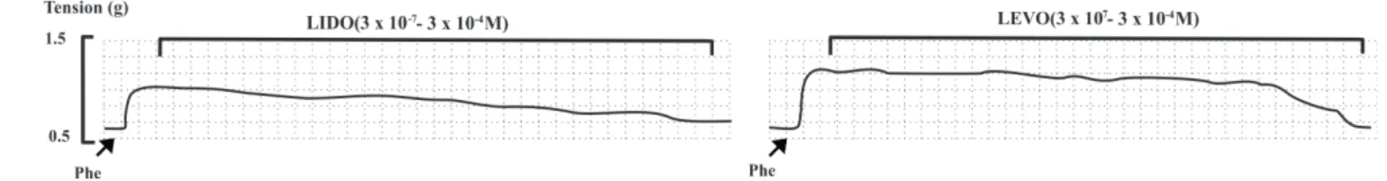
As shown in Figure 1A, both LIDO and LEVO were unable to induce vasoconstriction of the blood vessel during basal tone. However, in rings with functional endothelium, which was pre-contracted with phenylephrine, both LIDO and LEVO were capable of inducing concentration-dependent relaxation (Emax = 74.5 ± 11%; n = 5 and 91.13 ± 9.8%; n = 6, respectively) (Figures 1B and 2).
Figure 1. Original registration showing the efect of LIDO and LEVO in isolated arterial rings of the rat superior mesenteric artery on the basal tone (A) and in isolated arterial rings of the rat superior mesenteric artery pre-contracted with 1 µM of Phe (B).
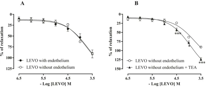
Figure 2. Concentration-response curves for LIDO (3 x 10⁻⁷-3 x 10⁻⁴ M; n = 9) and LEVO (3 x 10⁻⁷-3 x 10⁻⁴ M; n = 4) on the basal tone of
isolated rings of the rat superior mesenteric artery with functional endothelium (A) and in isolated rings of the rat superior mesenteric artery with
he concentration-dependent relaxation induced by LEVO in arterial rings without functional endothelium, which were pre-contracted with Phe (1 µM), was not signiicantly diferent from that obtained in rings with functional endothelium (Emax = 91.13 ± 9.8%; n = 6 and 90.4 ± 2.8%; n = 4, respectively) (Figure 3A).
he possible involvement of Ca2+ channels was investigated
using the curves, in which the rings were pre-contracted with depolarizing Tyrode’s solution (KCl 80 mM). LEVO was capable of
inducing vasodilatation similar to that achieved in the rings without functional endothelium, which were pre-contracted with Phe (1 µM) (Emax = 92.5 ± 1.0%; n = 4 and 90.4 ± 2.8%; n = 4, respectively), as shown in Figure 4A. LEVO (3 x 10⁻⁴ M) and NIF (10 µM) were
able to induce vasodilatation in the rings without endothelium, pre-contracted with Phe (89.3 ± 6.1%; n = 5 and 80.0 ± 4.6%; n = 6, respectively). However, LEVO did not induce a signiicant additional efect in the rings preincubated with NIF (97.0 ± 3.15%, n=6) (Figure 4B).
Figure 4. Concentration-response curves for LEVO (3 x 10⁻⁷- 3 x 10⁻⁵ M; n = 6) in isolated rings of the rat superior mesenteric artery without functional endothelium pre-contracted with Phe (n = 4) or KCL 80 mM (n = 4) (A), and the vasodilator efect of NIF (10 μM), LEVO (3 x 10⁻⁴ M), LEVO ater the maximum relaxation of NIF and NIF ater the maximum relaxation of LEVO in the rings of the rat mesenteric artery without endothelium pre-contracted with Phe (1 μM) (B). Values are expressed as mean ± S.E.M. he data were analyzed with one or two-way ANOVA followed by Bonferroni post-hoc test.
To assess the possible contribution of K+-channels to the vasodilator
efect induced by LEVO, rings without functional endothelium were pre-incubated with 1 mM of TEA. At this concentration, TEA was able to non-selectively block K+ channels15. In this protocol, the
vasodilator efect induced by LEVO was found to be signiicantly higher as compared to that induced by the drug in the absence of the inhibitor (Emax = 90.4 ± 2.8%; n = 4 vs 123.7 ± 3.6%; n = 7) (Figure 3B).
DISCUSSION
Studies showed that LEVO in 50% enantiomeric excess induced certain intrinsic vasoconstriction in vitro8-10,12,16 and in vivo17-19.
herefore, this study aimed to assess the vasoactivity of LEVO in
in vitro experiments using arterial rings prepared from isolated rat superior mesenteric artery, a resistant artery, which can relect the possible efects on the vascular reactivity in smaller blood vessels such as those present in the area treated by local anesthetic in clinical dentistry. Furthermore, this efect was compared to that of lidocaine, a standard local anesthetic used in dentistry.
In this study, it was shown that both LEVO and LIDO has no contractile efect on basal tone in the rat mesenteric artery. hese results corroborate the indings of Chang et al.20, who demonstrated
that LEVO did not exhibit a contractile efect on the rat tracheal smooth muscles. In contrast, Iida et al.21 and Mukozawa et al.9,12 have
shown that LEVO was able to induce vasoconstriction in studies on dog cerebral arterioles and rat thoracic aorta, respectively. hese controversial results may be attributed to the diferences in the structure of the chemical agents, the vascular bed, the experimental conditions, or the species used.
In addition, these opposite efects induced by LEVO can be justiied because some anesthetics, including LEVO and BUPI, exhibit a biphasic activity depending on the concentration used. It was observed that these anesthetics cause vasodilatation at high concentrations, and vasoconstriction at smaller concentrations, both in vivo and in vitro16-19.
Since no vasoconstrictor activity was observed on the basal tone, experiments were conducted to evaluate the possible vasodilator efect of LEVO and compare it to that of LIDO. he vasodilatation induced by LEVO was similar to that caused by the lidocaine.
Furthermore, we assessed the underlying mechanism responsible for the vasodilator efect of LEVO. Since the endothelium is an important regulator of the vascular tone via releasing endothelium-derived relaxing factors, mainly nitric oxide (NO) and products derived from the activation of cyclooxygenase (COX) enzyme, such as prostacyclins, experiments were carried out to assess the role of the endothelium in this response22. However the endothelium probably
does not contribute to vasodilatador efect of LEVO.
In accordance with the results of the present study, LEVO showed an endothelium-independent efect on the rat thoracic aorta12 as
well as on isolated human umbilical artery and vein11. However, it
was shown that the efect of LEVO on rat aorta was dependent on nitric oxide released by the endothelium8. hese indings reinforce
that the vascular efects of LEVO may difer depending on some variables, such as the vascular bed used.
We also evaluated whether the efects induced by LEVO are mediated via another endothelium-independent signaling pathway, such as Ca2+-channel blockade. It is known that increased
external K+ concentration induces smooth muscle contraction
through the activation of Cavs and subsequent release of calcium from the sarcoplasmic reticulum. Contractions induced by high concentrations of K+ are inhibited by Ca2+ -channel blockers or by
the removal of Ca2+ from the external environment because they
are totally dependent on Ca2+ inlux23.
LEVO induced vasodilatation on pre-contracted rings with depolarizing Tyrode’s solution (KCl 80 mM) and did not promote an additional vasodilator efect compared to the maximum efect induced by NIF, suggesting that LEVO may act mainly in a similar way as NIF, i.e., blocking dihydropyridine-sensitive Cavs.
Choi et al.8, Baik et al.10 and Mukozawa et al.12 demonstrated that
LEVO-induced vasoconstriction in the aorta is mediated mainly by the intracellular calcium and by Ca2+ inlux through Cavs8,10,12.
However, our results suggest that LEVO acts via inhibition of Ca2+
inlux through these channels. hese diferences might be related to the diferent vascular beds used and diferent experimental conditions. Nevertheless, additional studies are needed for a better understanding of the underlying mechanism of action.
Moreover, there are reports in the literature that K+ channels
also play an important role in the regulation of the vascular tone24.
K+ channel opening in the smooth muscle cell membrane causes K+
elux, resulting in hyperpolarization that closes the Cavs, resulting in decreased inlux of Ca2+ ions and thus, relaxation of the smooth
muscles25. LEVO was able to induce vasodilatation in rings without
functional endothelium with TEA, which suggests that these channels are not involved in LEVO-induced vasodilator efect.
CONCLUSION
he results of this study demonstrated that LEVO did not induce vasoconstriction of arterial rings prepared from rat superior mesenteric artery, however, it produced a vasodilator efect. his LEVO-induced vasodilatation was endothelium and K+ channel-independent, however, it could be mediated via the
blockade of Cavs in the vascular smooth muscle cells. We suggest that LEVO exhibits several vascular efects depending on the vascular bed used. Although LEVO did not display the expected vasoconstriction in our study, LEVO remains beneicial and can be widely used in dentistry as it is less cardiotoxic and less neurotoxic than BUPI, and it also provides long-lasting anesthesia.
ACKNOWLEDGEMENTS
REFERENCES
1. Carvalho RWF, Pereira CU, Anjos ED, Laureano JR Fo, Vasconcelos BCE. Anestésicos locais: como escolher e prevenir complicações sistêmicas. Rev Port Estomatol Med Dent Cir Maxilofac. 2010 Abr-Jun;51(2):113-20.
2. Vasconcelos RJH, Nogueira RVB, Leal AKR, Oliveira CTV, Bezerra JGB. Alterações sistêmicas decorrentes do uso da lidocaína e prilocaína na prática odontológica. Rev Cir Traumat Buco-Maxilo-Facial. 2002 Jan-Jun;1(2):13-9.
3. Ramacciato JC, Motta RHL, Groppo FC, Volpato MC, Ranali J. Anestésicos locais. Rev Assoc Paul Cir Dent. 2007;61(4):486-7.
4. Albright GA. Cardiac arrest following regional anesthesia with etidocaine or bupivacaine. Anesthesiology. 1979 Oct;51(4):285-7. http://dx.doi. org/10.1097/00000542-197910000-00001. PMid:484889.
5. Ohmura S, Kawada M, Ohta T, Yamamoto K, Kobayashi T. Systemic toxicity and resuscitation in bupivacaine-, levobupivacaine-, or ropivacaine-infused rats. Anesth Analg. 2001 Sep;93(3):743-8. http://dx.doi.org/10.1097/00000539-200109000-00039. PMid:11524350.
6. Fawcett JP, Kennedy JM, Kumar A, Ledger R, Kumara GM, Patel MJ, et al. Comparative efficacy and pharmacokinetics of racemic bupivacaine and S-bupivacaine in third molar surgery. J Pharm Pharm Sci. 2002 May-Aug;5(2):199-204. PMid:12207874.
7. Bajwa SJ, Kaur J. Clinical profile of levobupivacaine in regional anesthesia: a systematic review. J Anaesthesiol Clin Pharmacol. 2013 Oct;29(4):530-9. http://dx.doi.org/10.4103/0970-9185.119172. PMid:24249993.
8. Choi YS, Jeong YS, Ok SH, Shin IW, Lee SH, Park JY, et al. The direct effect of levobupivacaine in isolated rat aorta involves lipoxygenase pathway activation and endothelial nitric oxide release. Anesth Analg. 2010 Feb;110(2):341-9. http://dx.doi.org/10.1213/ANE.0b013e3181c76f52. PMid:19955508.
9. Mukozawa M, Takakura K, Mizogami M. Direct vasocontractile activities of bupivacaine enantiomers on the isolated rat thoracic aorta. Anesthesiol Res Pract. 2010;2010:820186. http://dx.doi.org/10.1155/2010/820186. PMid:20981258.
10. Baik JS, Sohn JT, Ok SH, Kim JG, Sung HJ, Park SS, et al. Levobupivacaine-induced contraction of isolated rat aorta is calcium dependent. Can J Physiol Pharmacol. 2011 Jul;89(7):467-76. http://dx.doi.org/10.1139/y11-046. PMid:21812525.
11. Kiliçaslan A, Duman A, Sahin AS. In vitro vasoactive effects of levobupivacaine and ropivacaine on the isolated human umbilical artery and vein. Balkan Med J. 2011;28(2):164-8.
12. Mukozawa M, Takakura K, Obata Y, Shimo K, Shigemi K. Levobupivacaine induces vasoconstriction via Ca2+ -dependent and -independent mechanisms in isolated rat thoracic aorta. Circulation Control in Medicine. 2013;34:71-7.
13. Barros EG, Marquez IM, Zanetta-Barbosa D. Avaliação comparativa da latência e da duração do cloridrato de levobupivacaína 0, 5 por cento sem e com vasoconstrictor em anestesia terminal infiltrativa. Rev Odontol UNESP. 2006 Abr-Jun;35(2):165-70.
14. Hagiwara S, Mitsui M, Karaki H. Effects of felodipine, nifedipine and verapamil on cytosolic Ca2+ and contraction in vascular smooth muscle. Eur J Pharmacol. 1993 Mar;234(1):1-7. http://dx.doi.org/10.1016/0014-2999(93)90698-H. PMid:7682512.
15. Cook NS. Effect of some potassium channel blockers on contractile responses of the rabbit aorta. J Cardiovasc Pharmacol. 1989 Feb;13(2):299-306. http://dx.doi.org/10.1097/00005344-198902000-00019. PMid:2468961.
16. Bouaziz H, Iohom G, Estèbe JP, Campana WM, Myers RR. Effects of levobupivacaine and ropivacaine on rat sciatic nerve blood flow. Br J Anaesth. 2005 Nov;95(5):696-700. http://dx.doi.org/10.1093/bja/aei242. PMid:16183680.
17. Aps C, Reynolds F. An intradermal study of the local anaesthetic and vascular effects of the isomers of bupivacaine. Br J Clin Pharmacol. 1978 Jul;6(1):63-8. http://dx.doi.org/10.1111/j.1365-2125.1978.tb01683.x. PMid:666948.
18. Newton DJ, McLeod GA, Khan F, Belch JJ. The effect of adjuvant epinephrine concentration on the vasoactivity of the local anesthetics bupivacaine and levobupivacaine in human skin. Reg Anesth Pain Med. 2004 Jul-Aug;29(4):307-11. http://dx.doi.org/10.1097/00115550-200407000-00002. PMid:15305248.
19. Ergil J, Akkaya T, Gozaydin O, Gunsoy B, Alicura S, Aladag E, et al. Vasoconstrictive and analgesic efficacy of locally infiltrated levobupivacaine in tonsillectomy patients. Int J Pediatr Otorhinolaryngol. 2012 Oct;76(10):1429-33. http://dx.doi.org/10.1016/j.ijporl.2012.06.016. PMid:22776810. 20. Chang HC, Chen SY, Huang YF, Liu FL, Cherng YG, Wang HW. Effects of levobupivacaine on isolated rat tracheal smooth muscle. J Anesth.
2015 Oct;29(5):809-12. http://dx.doi.org/10.1007/s00540-015-2026-8. PMid:25995060.
21. Iida H, Ohata H, Iida M, Nagase K, Uchida M, Dohi S. The differential effects of stereoisomers of ropivacaine and bupivacaine on cerebral pial arterioles in dogs. Anesth Analg. 2001 Dec;93(6):1552-6. http://dx.doi.org/10.1097/00000539-200112000-00046. PMid:11726442.
22. Furchgott RF, Zawadzki JV. The obligatory role of endothelial cells in the relaxation of arterial smooth muscle by acetylcholine. Nature. 1980 Nov;288(5789):373-6. http://dx.doi.org/10.1038/288373a0. PMid:6253831.
23. Karaki H, Weiss GB. Calcium release in smooth muscle. Life Sci. 1988;42(2):111-22. http://dx.doi.org/10.1016/0024-3205(88)90674-1. PMid:2447464.
24. Akata T. Cellular and molecular mechanisms regulating vascular tone. Part 2: regulatory mechanisms modulating Ca2+ mobilization and/ or myofilament Ca2+ sensitivity in vascular smooth muscle cells. J Anesth. 2007;21(2):232-42. http://dx.doi.org/10.1007/s00540-006-0488-4. PMid:17458653.
25. Ko EA, Han J, Jung ID, Park WS. Physiological roles of K+ channels in vascular smooth muscle cells. J Smooth Muscle Res. 2008 Apr;44(2):65-81. http://dx.doi.org/10.1540/jsmr.44.65. PMid:18552454.
CONFLICTS OF INTERESTS
*CORRESPONDING AUTHOR
Liciane dos Santos Menezes, Universidade Federal de Sergipe, Avenida Marechal Rondon, s/n, Jardim Rosa Elze, 49100-000 São Cristóvão - SE, Brasil, e-mail: [email protected]