RESUMO: “Triagem toxocológica do extrato de Mikania glomerata Spreng., Asteraceae, no sistema reprodutor, produção espermática e nível de testosterona em machos de ratos Wistar após tratamento crônico”. As plantas medicinais podem apresentar na sua constituição compostos capazes de causar efeitos adversos no organismo. Cumarina e lavonoides são substâncias encontradas em muitas espécies vegetais, cuja interferência na fertilidade de ratas e cadelas, respectivamente, foi evidenciada em estudos prévios. Mikania glomerata Spreng., Asteraceae, (guaco) é uma planta usada no tratamento de doenças respiratórias e em suas folhas foi detectada a presença de cumarina e lavonoides. Neste estudo, avaliou-se o efeito do extrato hidroalcoólico, preparado com partes aéreas de guaco, no sistema reprodutor de ratos submetidos a tratamento crônico. Ratos Wistar (trinta dias de idade) foram tratados com extrato hidroalcoólico de guaco na dose de 3,3 g/kg de peso corporal durante noventa dias. O peso corporal e de órgãos, a produção de espermatozoides, a concentração de testosterona plasmática e o consumo de ração foram avaliados. Não foi observada nenhuma alteração signiicativa das variáveis analisadas e o tratamento não afetou o consumo de ração. Estes dados sugerem que, na dose utilizada, o extrato hidroalcoólico de guaco não teve efeito tóxico e nem interferiu com a fertilidade de ratos Wistar submetidos a um tratamento de longa duração.
Unitermos: Mikania glomerata, órgãos reprodutivos, produção de espermatozoides, testosterona, rato Wistar.
ABSTRACT: Some compounds present in therapeutic plants may be responsible for the occurrence of adverse side effects. Coumarin and lavonoids are substances found in many plant species that showed antifertility activity in female rats and dogs, respectively. Mikania glomerata Spreng., Asteraceae, known as guaco in Brazil, is a plant largely used in folk medicine and its leaves are reported to have coumarin and lavonoids. This work analyzes the effect of chronic administration of M. glomerata on the reproductive system of male rats. Thirty-day-old Wistar rats were treated with M. glomerata hydroalcoholic extract at a dose of 3.3 g/kg of body weight for ninety days. Body and organ weights, gamete concentration on the epididymis cauda, serum testosterone level and food consumption were evaluated. No signiicant alteration was observed in any of the variables analyzed, suggesting the absence of toxic action or antifertility activity of the M. glomerata hydroalcoholic extract.
Keywords:Mikania glomerata, reproductive organs, sperm production, testosterone level, Wistar rat.
20(5): 718-723, Out./Nov. 2010
Article
Received 6 Aug 2009; Accepted 12 Nov 2009, Available online 16 Jul 2010.
Toxicological screening of
Mikania glomerata
Spreng., Asteraceae,
extract in male Wistar rats reproductive system, sperm
production and testosterone level after chronic treatment
Rita de Cássia da Silveira e Sá,
*,1Magda N. Leite,
2Reinaldo N. de Almeida
31Centro de Biologia da Reprodução, Departamento de Biologia, Universidade Federal de Juiz de Fora,
Juiz de Fora-MG, Brazil,
2Faculdade de Farmácia e Bioquímica, Universidade Federal de Juiz de Fora, 36036-330 Juiz de Fora-MG, Brazil, 3DFP/Laboratório de Tecnologia Farmacêutica, Universidade Federal da Paraíba, 58051-970 João Pessoa-PB,
Brazil.
INTRODUCTION
The genus Mikania contains more than four
hundred species (King & Robinson, 1987) Corrigir referência and is considered to be one of the largest genera of the Eupatorieae group. These species have been reported
to have constituents such as sesquiterpene lactones,
diterpenes, pimaradiene acids, steroids and coumarin
(Sarg & El-Dahmy, 1990; Cruz et al., 1996) and have extensively been used in folk medicine for their antiseptic, antitussive and expectorant properties (Cruz et al., 1996).
Mikania glomerata Spreng., Asteraceae, popularly known
& Sá, 1991), has been employed as an anti-snake venom, analgesic and anti-inlammatory agent (Ruppelt et al.,
1990). Phytochemical studies have revealed the presence
of several substances in M. glomerata, including kaurenoic
acid, cinnamoylgrandiloric acid, stigmaterol, lavonoids
and coumarin (Oliveira et al., 1993; Vilegas et al., 1997
a, b; Martins et al., 2000; Cabral et al., 2001), a natural substance reported to be the main active compound from
its leaves (Vilegas et al., 1997a).
The use of plants for therapeutic purposes has always been a common practice among the peoples of the world. In contemporary times special emphasis is being given to this subject in many ways, especially regarding
pharmacological and industrial approaches concerning plants of medicinal interest. Despite the importance of
this kind of work, close attention should be given to the
indiscriminate use of such plants and the occurrence of
adverse effects on the organism. The male reproductive
system and its associated endocrine system can undergo a
number of damaging alterations following exposures to a variety of chemical and physical agents that are capable of
interfering with the integrity of the reproductive function.
Among the chemical agents, there are many substances found in medicinal plants that are under current use by
the population for treating various types of diseases. For
instance, certain types of coumarin have been shown to
have antifertility activity in mature female rats and to cause glomerulocapsular adhesion and segmental fusion
in rats kidneys (Ulubelen et al., 1994). Flavonoids have been reported to have anti-snake activity (Nakagawa & Nakanisha, 1982), but are also known to produce
antiandrogenic activity and affect male fertility in dogs (Bhargava, 1989).
Taking into account the concern for reproductive
hazards and the fact that at least two potential antifertility
substances are present in M. glomerata, this study was aimed at investigating the effects of a hydroalcoholic
extract of this plant on the reproductive organs and some
vital organs, sperm production and serum testosterone
level of Wistar rats submitted to sub chronic treatment.
MATERIAL AND METHODS
Plant material
Mikania glomerata Spreng., Asteraceae, was
collected in the botanical garden of the Pharmacy and
Biochemistry Faculty, Universidade Federal de Juiz de
Fora (UFJF) and authenticated in the Herbarium Leopoldo
Krieger, Department of Botany, (UFJF), where a voucher
specimen registered under the number CESJ 34456 is deposited. The aerial parts were dried and powdered for the preparation of a hydroalcoholic extract. After extraction with 90% ethanol, the solvent was evaporated
in a rotavapor and the residue was dissolved in distilled water.
Animals and housing
Immature male Wistar rats (Rattus norvegicus
Berkenhout, 1769) of thirty days old and weighing around
70 g were obtained from the vivarium of UFJF, where they were born and bred. They were housed individually under standard laboratory conditions with a 12 h light/12 h dark photoperiod. They were fed on rat chow pellets and
received water ad libitum. Animal care and the experimental
protocol followed the principles and guidelines suggested
by the Brazilian College of Animal Experimentation (COBEA) and were approved by the Ethical Committee
of the Federal University of Juiz de Fora (UFJF) which
follows the international rules (EEC Directive-86/609/ EEC) (Protocol number: 013/2004-CEA).
Bioassay
The animals were randomly divided into control and treated groups, containing twenty animals each. The rats from the treated group received, by gavage and once
daily, M. glomerata hydroalcoholic extract of at a dose of
3.3 g/kg of body weight, administered for 90 d. The control group received 1 mL of distilled water following the same
protocol as the treated group.
During the experiment, the animals were inspected twice daily for detection of clinical signs of toxicity, such
as piloerection, and alterations in locomotor activity
(Mason & Kang, 1994). The animals were weighed before the beginning of treatment, once a week and at the end of
treatment. Food consumption was monitored daily. On the
ninety irst day, the animals were killed by an overdose
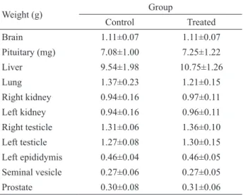
inhalation of anesthetic (halothane). Immediately after death, the following organs were dissected out and weighed: testes, left epididymis, seminal vesicle, prostate, liver,
kidneys, lungs, brain and the pituitary gland. The left testis and epididymis were then ixed in Bouin for histological examination. The tissues were dehydrated in a graded series of ethanol, embedded in parafin and sectioned at 3 µm thickness for routine haematoxylin and eosin staining, and light microscope examination (Humason, 1972).
Sperm were collected from the epididymal
secretion of the right epididymis cauda. The secretion was placed in a 0.3 mL drop of saline extract and later diluted in
distilled water. From this homogenate, a sample was taken
and the number of sperm counted using a hemocytometer with improved double Neubauer ruling (Moraes, 1994).
To analyze the serum testosterone level, a blood sample was withdrawn by cardiac puncture from control (n = 10) and treated (n = 10) rats. The blood was collected between 8 am and 1 pm and centrifuged at 17 oC, 250
Statistical analysis
The Student’s t test was applied for statistical
comparison of the differences in data between the test groups (α = 0.05) and the results were expressed by mean and standard deviation (SD). The serum testosterone level was analyzed using the Wilcoxon-Mann-Whitney test (α =
0.05) (Sokal & Rohlt, 1996).
RESULTS AND DISCUSSION
Plants have served as a natural source of
substances that may affect the development or normal
functioning of the reproductive system (Rajasekaran et al., 1988; Bidwai et al., 1990; Hiremath et al., 1997;
Montanari et al., 1998; Abdel-Magied et al., 2001). For
instance, coumarin is known to have antifertility activity
in female rats (Ulubelen et al., 1994) and lavonoids to
produce antiandrogenic activities and to affect male dog
fertility (Bhargava, 1989). Both substances have been reported to be among the active compounds of the aerial
parts of Mikania glomerata Spreng., Asteraceae, (Vilegas
et al., 1997 a, b; Martins et al., 2000) thus suggesting a possible toxic behavior of this plant on the reproductive
system.
In this work, immature male rats were treated with
M. glomerata hydroalcoholic extract at a dose level (3.3 g/
kg of body weight) that was six hundred times higher than the human dose for ninety consecutive days. The dose used was based on previous works in which the effect of M. glomerata extract administered during the spermatogenic
cycle of rats and its dominant lethality in male rats were
investigated (Sá et al., 2003, 2006). At the beginning of
treatment the reproductive organs of the rats were not
completely developed, but by the time the treatment was over, the animals were fully grown and sexually mature. The high dose and long-term administration of the extract did not seem to have caused any toxic effect in the rats as no death, no clinical signs of toxicity such as locomotor activity alterations and piloerection, and no signiicant body weight reduction (Figure 1) were detected. Daily food intake was not affected by the administration of the plant extract either (Figure 2). The treatment did not produce undesirable side effects on vital organs such as the liver, kidneys, lungs, brains and the pituitary gland, on the
organs of the reproductive system, and on the accessory
sex glands (Table 1), as evident by their weights which were not altered signiicantly. These results were similar to those obtained in male rats treated with M. glomerata
hydroalcoholic extract at two lower dose levels (1.1 and 2.2 g/kg of body weight) that were two hundred and four
hundred times higher than the human dose, respectively (data not shown).
The pituitary gland plays a major role on the
development and functioning of the male reproductive
system by secreting follicle-stimulating hormone (FSH) and
luteinizing hormone (LH) that act on the test is to produce sex steroid hormones and male gametes (Mahony & Hodgen, 1995). Although some sort of damage would be expected to occur because the animals used were still undergoing sexual maturation during the experimental procedure, no androgenic or anti-androgenic effects were detected in the treated animals corroborating the normal functioning of the reproductive organs, including the androgen-dependent accessory sex glands. Serum testosterone concentration in the adult male rat was measured on the ninety irst day and showed a large variation among the animals in both
control and treated groups, which ranged from a low 0.70
ng/mL to a high 19.3 ng/mL. The values obtained were not statistically different between the control and treated
animals (Control (C) = 4.20±5.00 ng/mL and Treated (T)
= 6.13±5.80 ng/mL) and were within the values reported in
previous studies (Coyotupa et al., 1973; Pujol et al., 1976;
Fahim et al., 1982; Viguier-Martinez et al., 1985; Linder et
al., 1995; Sá et al., 2003).
A comprehensive assessment of the effects of chemicals on male reproductive functioning requires study of effects on spermatogenesis and the quality of spermatozoa produced (Blazak et al., 1985). Despite the high dose used and the long duration of treatment, the
coumarin and lavonoids present in the M. glomerata
extract were not toxic to the testis, the left epididymis, the prostate and the seminal vesicle. No impairment of the spermatogenic cycle was observed and the sperm
concentration from the secretion of the right epididymis
cauda did not differ signiicantly between the groups:
(C) = 909.13±149.47 x 10⁶ sperm/mL (mean±SD) and
(T) = 1018.88±201.54 x 10⁶ sperm/mL. The sperm
morphology seemed unaffected by the treatment as the proportion of normal and abnormal spermatozoa was comparatively similar between the control and treated
groups. Spermatozoa without head or tail and with coiled
tail were the most common abnormalities present which also showed a similar concentration in both groups.
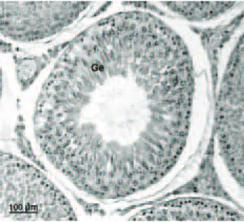
The histological examination revealed that there
was no structural alteration in the seminiferous epithelium
of the treated rats’ testis (Figure 3) when compared to the
controls. Similarly, the epididymis of control and treated
rats (Figure 4) showed a normal epithelium lining and presence of spermatozoa in all segments (head, body and
tail) of this organ.
In conclusion, the data showed in this work
points to the absence of toxicity of the M. glomerata
hydroalcoholic extract as a high dose and long-term treatment with this plant did not produce any toxic effect on important vital organs of the Wistar rat. The functioning
Figure 1. Body weight of control and Mikania glomerata extract treated Wistar rats (3.3 g/kg of body weight) submitted to subchronic treatment, with exposure of ninety days and death on the ninety irst day.
Figure 2. Daily food consumption of control and Mikania glomerata extract-treated Wistar rats (3.3 g/kg of body weight) submitted to subchronic treatment, with exposure of ninety days and death on the ninety irst day.
Figure 3. Cross section of the seminiferous tubule of M. glomerata extract-treated Wistar rats, showing the germinal epithelium (Ge) with normal morphology.
Table 1. Organ weights of control and Mikania glomerata extract-treated Wistar rats (3.3 g/kg of body weight) submitted to subchronic treatment, with exposure of ninety days and death on the ninety irst day.
Weight (g) Group
Control Treated
Brain 1.11±0.07 1.11±0.07
Pituitary (mg) 7.08±1.00 7.25±1.22
Liver 9.54±1.98 10.75±1.26
Lung 1.37±0.23 1.21±0.15
Right kidney 0.94±0.16 0.97±0.11
Left kidney 0.94±0.16 0.96±0.11
Right testicle 1.31±0.06 1.36±0.10
Left testicle 1.27±0.08 1.30±0.15
Left epididymis 0.46±0.04 0.46±0.05
Seminal vesicle 0.27±0.06 0.27±0.05
Prostate 0.30±0.08 0.31±0.06
Results expressed in mean±SD. N=12.
ACKNOWLEDGMENTS
The authors are grateful to Fernanda Lima Lopes and Maycon de Moura Reporedo for technical support and to Ms Sharon Lloyd for reviewing the English version of
this manuscript.
REFERENCES
Abdel-Magied EM, Abdel-Rahman HA, Harraz FM 2001. The effect of aqueous extract of Cynomorium coccineum and Withania somnifera on testicular development in immature Wistar rats. J Ethnopharmacol 75: 1-4. Bhargava SK 1989. Antiandrogenic effects of a lavonoid rich
fraction of Vitex negundo seeds: a histological and biochemical study in dogs. J Ethnopharmacol 27: 327-339.
Bidwai PP, Wangoo D, Bhullar N 1990. Antispermatogenic action of Celastrus paniculatus seed extract in the rat with reversible changes in the liver. J Ethnopharmacol 28: 293-303.
Blazak WF, Ernst TL, Stewart BE 1985. Potential indictors of reproductive toxicity: testicular sperm production and epididymal sperm number, transit time, and motility in Fischer 344 rats. Fund Appl Toxicol 5: 1097-1103. Cabral LM, Dos Santos TC, Alhaique F 2001. Development
of a proitable procedure for the extraction of 2-H-1-benzopyran-2-one (coumarin) from Mikania glomerata. Drug Dev Ind Pharm 27: 103-106.
Coyotupa J, Parlow AF, Kovacic N 1973. Serum testosterone and dihydrotestosterone levels following orchiectomy in the adult rat. Endocrinology 92: 1579-1581.
Cruz FG, Roque NF, Giesbrecht AM, Davino SC 1996. Antibiotic activity of diterpenes from Mikania triangularis.
Fitoterapia 67: 189-190.
Fahim MS, Fahim Z, Harman JM, Clevenger TE, Mullins W, Hafez SE 1982. Effect of Panax ginseng on testosterone level and prostate in male rats. Arch Andrology 8: 261-263.
Hiremath SP, Badami S, Swamy HKS, Patil SB, Londonkar RL 1997. Antiandrogenic effect of Striga orobanchioides. J Ethnopharmacol 56: 55-60.
Humason GL 1972. Animal Tissue Techniques (3rd edn). San Francisco: WH Freeman.
King RM, Robinson H 1987. The genera of the Eupatorieae (Asteraceae). Monogr Syst Bot 22: 1-581.
Linder RE, Klinefelter GR, Strader LF, Narotsky MG, Suarez JD, Roberts NL, Perreault SD 1995. Dibromoacetic acid affects reproductive competence and sperm quality in the male rat. Fund Appl Toxicol 28: 9-17.
Mahony MC, Hodgen GD 1995. Toxic effects on the hypothalamus-anterior pituitary-gonadal axis, control on the male and female reproductive system, and related issues. In: Witorsch RJ (Ed). Reproductive Toxicology. 2 ed. New York: Raven Press, p. 195-213.
Martins ER, Castro DM, Castellani DC, Dias JE 2000. Plantas Medicinais. Viçosa: Editora UFV.
Mason JM, Kang YJ 1994. Test methods for assessing female reproductive and developmental toxicology. In: Hayes AW (Ed). Principle and Methods of Toxicology. 3 ed. New York: Raven Press, p. 980-1037.
Montanari T, Carvalho JE, Dolder H 1998. Antispermatogenic effect of Achillea millefolium L. in mice. Contraception 58: 309-313.
Moraes GES 1994. Espermocitograma. Porto Alegre: Editora Médica Missau.
Nakagawa M, Nakanisha K 1982. Structures of cabenegrin I and II, potent anti-snake venoms. Tetrahedron Lett 23: 3855-3858.
Neves LJ, Sá MFA 1991. Contribuição ao estudo das plantas medicinais Mikania glomerata Spreng. Rev Bras Farmacogn 72: 42-47.
Oliveira F, Saito ML, Garcia LO 1993. Caracterização cromatográica em camada delgada do extrato luído de guaco Mikania glomerata Sprengel. Lecta 11: 43-56. Pujol A, Francis B, Louvet J-P, Boulard C 1976. Testosterone and
dihydrotestosterone concentrations in plasma, epididymal tissues, and seminal luid of adult rats. Endocrinology 98: 111-113.
Rajasekaran M, Bapna JS, Lakshmana S, Ramachandrannair AG, Veliath AJ, Panchanadam M 1988. Antifertility effect in male rats of oleanolic acid, a triterpene from Eugenia jambolana lowers. J Ethnopharmacol 24: 115-121. Ruppelt BM, Pereira EFR, Gonçalves LC, Pereira NA 1990.
Abordagem farmacológica de plantas recomendadas na medicina folclórica como antiofídicas. I-Atividades analgésica e antinlamatória. Rev Bras Farmacogn 71: 54-56.
extract on male Wistar rats’ reproductive organs, sperm production and testosterone level. Contraception 67: 327-331.
Sá RCS, Leite MN, Peters VM, Guerra MO, Almeida RN 2006. Absence of mutagenic effect of Mikania glomerata hydroalcoholic extrac on adult Wistar rats in vivo. Braz Arch Biol Technol 49: 599-604.
Sarg TM, El-Dahmy S 1990. A further new ent-labdane from Mikania alvimii. Fitoterapia 61: 160-161.
Sokal RR, Rohlf FJ 1996. Biometry. The Principles and Practice of Statistics in Biological Research. New York: WH Freeman and Co.
Ulubelen A, Ertugrul L, Birman H, Yigit R, Erseven G, Olgac V 1994. Antifertility effects of some coumarins isolated from Ruta chalepensis and R. chalepensis var. latifolia in rodents. Phytother Res 8: 233-236.
Viguier-Martinez M-C, De Reviers M-TH, Perreau C 1985. Effects of lutamide or of supplementation with testosterone in prepubertal male rats prenatally treated with bulsufan. Acta Endocrinol-Cop 109: 550-557.
Vilegas JHY, Marchi E, Lanças FM 1997a. Extraction of low-polarity compounds (with emphasis on coumarin and kaurenoic acid) from Mikania glomerata (“guaco”) leaves. Phytochem Analysis 8: 266-270.