A
KINESIOGRAPHIC STUDY OF MANDIBULAR
MOVEMENTS DURING FUNCTIONAL
ADAPTATION TO COMPLETE DENTURES
ESTUDO CINESIOGRÁFICO DOS MOVIMENTOS MANDIBULARES DURANTE
O PERÍODO DE ADAPTAÇÃO FUNCIONAL A PRÓTESES TOTAIS
Cláudio Rodrigues LELES
Professor, Department of Prevention and Oral Rehabilitation, School of Dentistry, Federal University of Goiás, Goiânia, Goiás, Brazil.
Marco Antonio COMPAGNONI
Professor, Department of Dental Materials and Prosthodontics, Araraquara Dental School, São Paulo State University, Araraquara, São Paulo, Brazil.
Raphael Freitas de SOUZA
Graduate student, Department of Dental Materials and Prosthodontics, Araraquara Dental School, São Paulo State University, Araraquara, São Paulo, Brazil.
Débora Barros BARBOSA
Graduate student, Department of Dental Materials and Prosthodontics, Araraquara Dental School, São Paulo State University, Araraquara, São Paulo, Brazil.
fter complete denture insertion, edentulous patients usually present transitory difficulties. This is one of the most critical stages of prosthetic treatment and it is strongly related to the prostheses acceptance. The aim of this study was to evaluate potential changes in the mandibular movement pattern related to insertion of the complete denture during the functional adaptation period. The kinesiograph instrument K6-I (Myotronics Research Inc., Seattle, WA) was used to evaluate eight edentulous patients that received conventional complete denture treatment. Recordings of opening and closure movement limits, movement velocity, postural rest position, chewing cycle and limits of eccentric movements were performed. Each patient was evaluated in four different stages: with the old dentures, immediately after insertion of the new dentures, and 30 days and 6 months after insertion of the dentures. The results showed that there were no remarkable changes in the mandibular motion patterns after insertion of the new dentures. It was suggested that the problems related to the process of utilization of new dentures are not strictly associated to the mandibular movement. Therefore, functional adaptation after denture insertion is not directly related to functional changes, but to the quality of the dentures and to individual features.
UNITERMS: Complete denture; Patients; Kinesiography.
INTRODUCTION
Denture insertion is the summit of all clinical procedures of denture construction and is eagerly awaited by both patient and clinician20. However, after denture insertion, a set of
adaptive difficulties can emerge. Transitory problems like masticatory impairment, mandibular incoordination, excessive salivary flow and phonetic troubles are common, even if the denture is well planned and constructed1. Pain
related to the denture-bearing tissues and faults in denture retention or stability are frequently observed, mainly due to occlusal imbalance, incorrect vertical dimension and occlusion, or improper contour of the denture base8. These
factors may reduce the patient’s comfort and satisfaction and influence the wearing of complete dentures on a daily basis4,19.
Fish5 observed that these initial difficulties are caused
by alterations in the tongue position relative to the oral mucosa, restriction of the space available for the tongue, load transmitted to the supporting tissues, changes in muscle proprioceptive impulses, and alteration of vertical facial height and mandibular rest position.
Several factors that influence this adaptation period were studied in the natural dentition, such as the vertical dimension3,6,13,14,22, oral sensory threshold12 and occlusal
status30. Research methods include clinical evaluation,
electromyography and kinesiography.
The study of mandibular movement dynamics is not new, but only in the 70’s Jankelson, et al.7 introduced the
kinesiograph as an instrument for diagnosis and clinical research in dentistry. This instrument was largely used for monitoring dentate individuals, although few studies were conducted for complete denture patients. Tallgren, et al.27,28
observed that patients without posterior dental support perform irregular movements and the insertion of immediate maxillary dentures opposed to mandibular removable partial dentures restored functional occlusion state and significantly improved the mandibular movement pattern. Additionally, a two-year longitudinal study showed that the range of movement is considerably reduced, probably due to the progressive loss of retention of the dentures28. Other
kinesiographic studies highlight the importance of the quality of the denture for restoration and maintenance of the chewing pattern and cyclic mandibular movements9,10,25.
The aim of the current study was to evaluate, by means of a kinesiographic instrument, the functional changes of mandibular movement parameters associated to insertion of new dentures, during different stages of adjustment of the patients to complete dentures.
MATERIAL AND METHODS
Completely edentulous patients who attended the Araraquara Dental School and were treated with complete dentures were randomly selected. Inclusion criteria included patients who needed new dentures, mentally receptive individuals, absence of dysfunctional disorders of the masticatory system, normal volume and resilience of the residual edentulous ridges, and absence of debilitating systemic diseases.
Patients were selected to receive upper and lower conventional complete dentures constructed according to a standardized protocol as described by Russi, et al.23 The
Ethics Committee of the school has approved the research project and an informed consent term was obtained from the patients before any experimental procedure.
Eight patients met the criteria and were selected for the study. Six were female and two were male, age ranging from 37 to 77 years (mean age: 61 years). The mean time of denture usage was 29 years.
After completion of clinical treatment, during the insertion visit, the quality of retention and stability, comfort and esthetics were tested. Points of occlusion were selectively ground and possible pressure spots were disclosed with a pressure indicator paste and relieved. After clinical adjustments, kinesiographic analysis was performed in the same appointment.
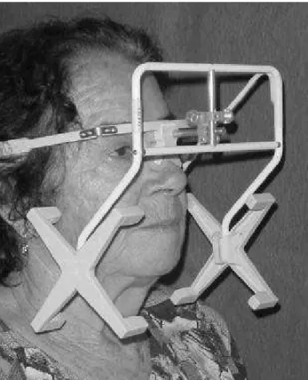
A kinesiographic instrument (K6-I Diagnostic System-Myotronics Research Inc., Seattle, WA, USA) connected to
a computerized system was used to graphically record the mandibular movements. This system has a sensor array (Figure 1) and a 12x6x3mm magnet tracking device (Figure 2) that records the spatial three-dimensional position of the mandible during functional movements.
Kinesiographic analysis was divided into three different periods of time: (1) immediate, (2) short-term and (3) long-term. Immediate period was evaluated with old and new dentures.
1. Immediate: during the denture insertion visit and after the intraoral adjustments. Assessment of transition between new and old dentures was recorded in the same visit. Measurement series were coded as STAGE I (old dentures), and STAGE II (new dentures). Stage I was considered the
FIGURE 1- Sensor array in position
pre-insertion pattern of mandibular movement.
2. Short-term (STAGE III): to avoid confounding factors, like sore points that might occur during the post-insertion period, recordings were made after the last follow-up visit, when complete adaptation to the dentures had been achieved. The estimated maximum time interval for this stage was 30 days after insertion of the denture.
3. Long-term (STAGE IV): about six months after insertion of the dentures.
The mean time interval elapsed between stage II (denture insertion) and stage III was 30 days (range: 18 – 32 days), and between stage II and IV was 170 days (range: 156 – 231 days).
Five tracing modes (scan) were selected for recordings. Description of mandibular movements and respective tracing modes are summarized in Table 1.
Recordings were performed according to the manufacturer’s instructions and three reproducible measurements were recorded for each scan mode.
Descriptive statistics were performed and comparative analysis was made using the non-parametric Wilcoxon signed ranks test, at a confidence level of 5%. Data analysis was done with the Statistical Package for the Social Sciences software (SPSS 10.0 for Windows, SPSS Inc., 1999).
RESULTS
The results of recordings are listed in Table 2-11, according to scan tracings.
DISCUSSION
Satisfactory function and comfort are related to a balanced occlusion, correct vertical dimension and proper base extension4. Regardless of general health condition and
psychological traits, the quality of the dentures and other local favorable aspects are good predictors of treatment success2. Nevertheless, functional adaptation to dentures has
multidimensional aspects and consequences that influence the patient’s satisfaction. Patients must be aware that, after insertion of the dentures, a new muscular behavior may develop in order to control the functional activities12,15,16.
Nearly 60% of denture wearers achieve satisfactory function within a week after insertion and about 20% within one month1.
Functional parameters to be considered after dentures’ insertion include patients’ subjective appraisal of dentures15,16, chewing efficiency16,29, masticatory frequency
and muscular coordination15,16, chewing cycle consistency15,
and occlusal perception11. Previous studies suggest that all
these parameters significantly improve after the post-insertion period because a compensatory neuromuscular
Scan Description
1 Normal opening and closure of mandibular movement
2 Speed of the mandible during opening and closure
3 Three-dimensional movement of the mandible from rest position to maximal occlusion
8 Three-dimensional movements of the mandible during chewing. Test food – a cube of 15x10x10mm cured polysulfide
(Permlastic Regular – Kerr Corporation, USA)
13 Sagittal and frontal range of motion during maximum opening, closure and lateral movements of the mandible. TABLE 1- Preprogrammed scan numbers corresponding to scan tracings
Patient Stage I Stage II Stage III Stage IV
1 56.7 57.8 56.9 57.3
2 56.8 60.3 59.5 57.8
3 41.4 35.8 45.6 42.1
4 27.6 27.2 35.5 42.6
5 66.0 50.6 55.6 54.1
6 41.2 47.5 49.3 47.7
7 47.0 38.2 48.4 45.9
8 54.2 54.9 54.2 59.2
modification is developed by the patients11,15,16,16,29. However,
these factors are not related to the mandibular coordination or extension of mandibular range of movement. It is better explained by the patients’ ability to cope with difficulties, learning capability and continuous reinforcement of
functional activities17.
Periodontal and temporomandibular joint receptors play an important role in controlling occlusal forces and mandibular movements, respectively17. Even if periodontal
proprioception is absent, as in the edentulous patient, the
Patient Stage I Stage II Stage III Stage IV
Max. Mean Max. Mean Max. Mean Max. Mean
1 177.1 91.6 152.5 76.3 134.6 75.9 143.8 84.2
2 191.6 107.2 260.8 151.1 276.7 159.1 188.0 182.9
3 120.4 60.7 129.6 76.2 86.3 48.8 135.9 65.7
4 103.8 60.7 83.8 53.3 127.5 74.6 208.4 120.4
5 140.0 79.9 117.5 77.9 120.4 76.1 112.5 69.2
6 230.5 138.1 180.9 108.8 137.5 80.3 131.3 71.7
7 65.5 36.5 82.5 43.8 75.4 54.5 113.4 70.0
8 133.8 88.0 161.4 77.4 150.1 72.8 210.0 185.0
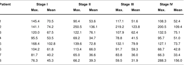
TABLE 4- Maximum and mean mandibular speed during opening movement (in millimeters per second)
Patient Stage I Stage II Stage III Stage IV
Max. Mean Max. Mean Max. Mean Max. Mean
1 145.4 70.5 90.4 53.6 117.1 51.6 108.3 52.4
2 141.1 74.2 250.5 136.1 219.2 123.8 200.5 109.4
3 120.0 67.5 122.1 76.1 107.9 62.4 132.5 75.1
4 95.5 53.5 69.2 34.7 78.8 41.5 95.7 51.0
5 168.4 102.8 139.6 72.8 132.1 79.9 127.1 73.7
6 104.2 61.8 113.4 66.0 91.7 59.3 66.7 42.8
7 81.7 40.2 65.0 36.6 83.8 36.0 66.3 33.4
8 76.3 45.3 66.2 39.3 59.5 31.9 288.3 156.0
TABLE 5- Maximum and mean mandibular speed during closure movement (in millimeters per second)
Patient Stage I Stage II Stage III Stage IV
Right Left Right Left Right Left Right Left
1 0.3 14.2 0.5 14.0 0.1 13.7 0.5 7.8
2 0.2 13.7 2.4 12.7 1.5 6.1 1.4 6.0
3 2.9 4.6 4.2 2.3 1.4 5.7 1.6 4.3
4 0.4 6.5 0.1 7.0 0.1 9.7 0.4 3.1
5 0.2 10.4 1.7 10.8 1.4 9.0 2.1 6.2
6 0.9 9.7 0.5 16.1 0.6 12.7 1.1 3.3
7 0.3 8.3 0.6 4.3 0.9 8.9 0.8 3.7
8 0.2 9.8 1.4 11.1 2.1 3.5 1.9 2.7
masticatory dynamics remains unaltered because chewing is controlled by a central neural generator that modulates rhythmic activities17,24.
The present study reveals that the insertion of dentures does not alter the pattern, speed and limits of nonfunctional movements. In contrast, Tallgren et al.28 observed
improvement in mastication and range of movement after
insertion of maxillary immediate denture and mandibular extension-base removable partial denture. This improvement can be affected by the retention of the dentures28.
Another factor that influences adaptation to new dentures is the change in occlusion. Investigations in dentate individuals show that modifications in occlusal guidance significantly alter lateral border movements21. Nevertheless,
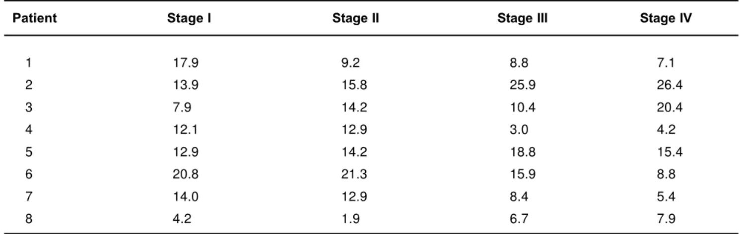
Patient Stage I Stage II Stage III Stage IV
1 17.9 9.2 8.8 7.1
2 13.9 15.8 25.9 26.4
3 7.9 14.2 10.4 20.4
4 12.1 12.9 3.0 4.2
5 12.9 14.2 18.8 15.4
6 20.8 21.3 15.9 8.8
7 14.0 12.9 8.4 5.4
8 4.2 1.9 6.7 7.9
TABLE 6- Speed of terminal contact during closure movement of mandible (in millimeters per second)
Patient Stage I Stage II Stage III Stage IV
V A-P L V A-P L V A-P L V A-P L
1 6.0 2.5 0.3 2.0 0.9 0.1 0.8 0.9 0.4 1.5 1.3 0.1
2 2.9 1.9 0.4 3.2 1.9 0.6 3.6 2.1 0.6 3.6 2.1 0.6
3 1.4 0.5 0.1 1.3 0.7 0.1 1.4 0.2 0.2 2.3 0.9 0.4
4 5.1 5.1 1.3 2.8 2.1 0.6 3.7 2.4 0.8 3.9 2.8 0.3
5 12.1 5.7 0.4 3.3 2.5 0.3 2.2 1.6 0.1 2.7 0.5 0.2
6 7.1 3.9 0.8 3.1 3.1 0.2 2.0 1.4 0.1 1.9 1.9 0.4
7 7.5 4.8 1.5 2.9 2.4 0.3 2.6 1.7 0.3 3.6 2.4 0.2
8 3.6 3.0 0.8 2.2 0.8 0.3 1.4 1.3 0.8 5.0 4.1 0.9
TABLE 7- Mean vertical (V), anteroposterior (AP) and lateral (L) movement between rest position and intercuspal position (in millimeters)
Patient Stage I Stage II Stage III Stage IV
1 7.2 9.2 6.2 8.8
2 11.0 9.2 13.5 13.4
3 6.0 4.8 6.3 13.1
4 11.0 12.6 9.9 11.4
5 14.2 10.4 8.6 11.3
6 7.5 11.0 9.0 6.7
7 8.7 7.2 6.9 7.0
8 9.9 10.6 7.1 8.3
it is not possible to reach any conclusion on the clinical significance of these changes if complaints are not detected and overall comfort, esthetic satisfaction and functional improvement are achieved.
The most significant modifications are related to the vertical dimension. The consequences are variations in muscular activity, rest position of the mandible and consequently in the interocclusal distance, observed in dentate6,13,14 and edentulous patients.18,26 However, it is well
recognized that minor or gradual moderate changes in vertical dimension do not cause deleterious problems if a stable and well distributed occlusion is achieved3.
It can be suggested that adaptation to new dentures is not closely related to functional changes. Intrinsic denture problems and individual subjective problems related to the patient may play a major role during the post-insertion process. Consequently, a careful appraisal of clinical and psychological conditions and a realistic outcome of the treatment are important aspects of patient management. A comprehensive treatment planning is essential for a satisfactory post-insertion period and long-term success of treatment.
CONCLUSION
Within the limitations of this study, it was concluded that the pattern of mandibular movement does not demonstrate significant changes between the pre and post-insertion stages of utilization of complete dentures. Functional transitory problems after insertion of the denture may not be associated to changes in the coordination of the mandibular movement.
ACKNOWLEDGMENTS
This research was supported by The State of Sao Paulo Research Foundation (Fapesp – Grant no. 99/06609-8).
RESUMO
Após a instalação de próteses totais, o paciente desdentado geralmente enfrenta dificuldades transitórias. Esse estágio é um dos mais críticos durante o tratamento protético desses pacientes, estando relacionado à aceitação das próteses. O presente estudo teve como objetivo avaliar as possíveis
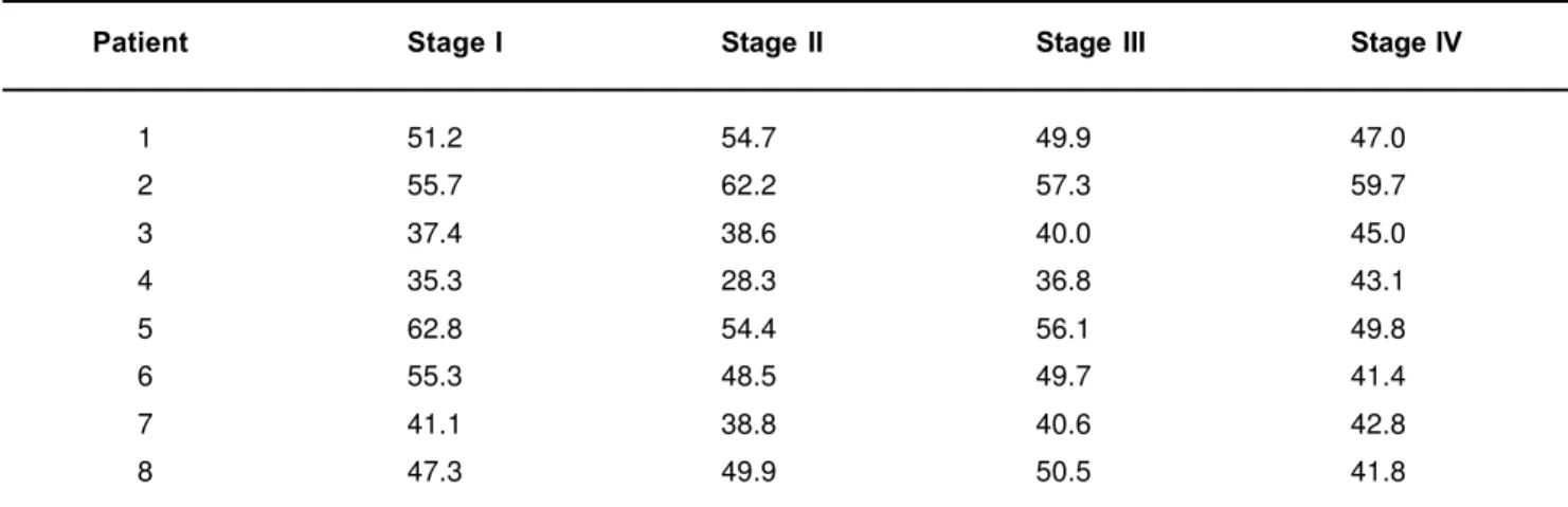
Patient Stage I Stage II Stage III Stage IV
1 51.2 54.7 49.9 47.0
2 55.7 62.2 57.3 59.7
3 37.4 38.6 40.0 45.0
4 35.3 28.3 36.8 43.1
5 62.8 54.4 56.1 49.8
6 55.3 48.5 49.7 41.4
7 41.1 38.8 40.6 42.8
8 47.3 49.9 50.5 41.8
TABLE 10- Mean distance (range of movement) between intercuspal position and maximum opening (in millimeters) Patient Stage I Stage II Stage III Stage IV
Ant. Right Left Ant. Right Left Ant. Right Left Ant. Right Left
1 8.1 9.8 6.6 6.7 7.3 8.3 4.4 10.5 8.8 7.2 10.2 9.7
2 3.5 10.6 10.2 2.9 13.8 6.9 2.6 12.1 9.5 2.0 12.4 9.7
3 2.9 3.4 2.2 2.0 6.7 1.7 7.0 4.8 2.4 10.0 8.8 7.7
4 3.2 4.7 5.9 6.7 4.1 9.1 7.1 1.0 8.4 7.1 5.8 7.5
5 4.8 7.9 4.0 9.8 10.3 4.3 11.5 9.7 5.8 9.4 11.5 4.7
6 5.9 5.8 1.2 5.0 5.6 0.4 6.8 5.0 2.3 5.4 5.9 4.9
7 6.5 5.6 7.2 7.3 8.9 6.7 1.8 9.9 4.4 7.9 10.3 8.2
8 4.3 5.5 5.8 4.1 5.8 4.3 9.1 8.0 7.6 7.0 7.1 8.1
alterações no padrão de movimentos mandibulares decorrentes da instalação de próteses totais em indivíduos desdentados, durante o período de adaptação funcional. O sistema de diagnóstico cinesiográfico K6-I (Myotronics Research Inc., Seattle, WA) foi empregado para avaliação de 08 pacientes que foram previamente tratados para confecção de próteses totais. Foram realizados registros da amplitude do movimento mandibular de abertura e fechamento, velocidade do movimento mandibular durante a abertura e fechamento, movimento da mandíbula entre a posição de repouso postural e a máxima intercuspidação, movimento da mandíbula durante a mastigação e registro do limite de movimento mandibular de abertura e fechamento, lateralidade e protrusão. Os pacientes foram avaliados com as próteses antigas, no período imediato à instalação das próteses e nos períodos de aproximadamente 30 dias e 6 meses pós-instalação. Os resultados demonstraram que o padrão dos movimentos mandibulares pouco se altera entre os períodos pré e pós-instalação e entre diferentes estágios após a instalação das próteses totais, o que sugere que as dificuldades funcionais após a instalação de próteses totais não estão relacionadas a alterações no padrão de coordenação dos movimentos mandibulares. Assim, a adaptação dos pacientes após a instalação de próteses totais não se relaciona propriamente a alterações funcionais do paciente, mas a características intrínsecas das próteses e a aspectos subjetivos individuais.
UNITERMOS: Dentadura completa; Pacientes; Cinesiografia.
REFERENCES
1- Berg E. Acceptance of full dentures. Int Dent J 1993; 43: 299-306.
2- Brunello D, Mandikos M. Construction faults, age, gender, and relative medical health: factors associated with complaints in complete denture patients. J Prosthet Dent 1998; 79: 545-54.
3- Carlsson G, Ingervall B, Kocak G. Effect of increasing vertical dimension on the masticatory system in subjects with natural teeth. J Prosthet Dent 1979; 41: 284-9.
4- Fenlon M, Sherriff M, Walter J. Association between the accuracy of intermaxillary relations and complete denture usage. J Prosthet Dent 1999; 81: 520-5.
5- Fish S. Adaptation and habituation to full dentures. Br Dent J 1969; 127: 19-26.
6- Garnick J, Ramfjord S. Rest position. An electromyographic and clinical investigation. J Prosthet Dent 1962; 12: 895-911.
7- Jankelson B, Swain C, Crane P, Radke J. Kinesiometric instrumentation: a new technology. J Am Dent Assoc 1975; 90: 834-40.
8- Jeganathan S, Payne J. Common faults in complete dentures: a review. Quintessence Int 1993; 24: 483-7.
Recordings Comparison between clinical stages
I-II I-III I-IV II-III II-IV III-IV
Range of opening movement
Right deviation during opening movement
Left deviation during opening movement * * *
Maximum speed during opening movement
Mean speed during opening movement
Maximum speed during closure movement
Mean speed during closure movement
Speed of terminal tooth contact
Vertical interocclusal space in rest position * *
Anteroposterior movement between rest position and intercuspal position * *
Lateral movement between rest position and intercuspal position
Vertical movement during chewing
Anterior movement limit
Right lateral movement limit *
Left lateral movement limit *
Range of movement between intercuspal position and maximum opening
9- Jemt T, Hedegård B, Wickberg K. Chewing patterns before and after treatment with complete maxillary and bilateral distal-extension mandibular removable partial dentures. J Prosthet Dent 1983; 50: 566-70.
10- Jemt T, Stålblad P. The effect of chewing movements on changing mandibular complete dentures to osseointegrated overdentures. J Prosthet Dent 1986; 55: 357-61.
11- Langer A, Michman J. Occlusal perception after placement of complete dentures. J Prosthet Dent 1968; 19: 246-51.
12- Manly R, Pfaffman C, Lathrop D, Keyser J. Oral sensory thresholds of persons with natural and artificial dentitions. J Dent Res 1952; 31: 305-12.
13- Manns A, Miralles R, Guerrero F. The changes in electrical activity of the postural muscles of the mandible upon varying the vertical dimension. J Prosthet Dent 1981; 45: 438-45.
14- Michelotti A, Farella M, Vollaro S, Martina R. Mandibular rest position and electrical activity of the masticatory muscles. J Prosthet Dent 1997; 78: 48-53.
15- Michman J, Langer A. Clinical and electromyographic observations during adjustment to complete dentures. J Prosthet Dent 1968; 19: 252-62.
16- Michman J, Langer A. Postinsertion changes in complete dentures. J Prosthet Dent 1975; 34: 125-34.
17- Mohl N. Neuromuscular mechanisms in mandibular function. Dent Clin North Am 1978; 22: 63-71.
18- Morimoto T, Abekura H, Tokuyama H, Hamada T. Alteration in the bite force and EMG activity with changes in the vertical dimension of edentulous subjects. J Oral Rehabil 1996; 23: 336-41.
19- Morstad A, Petersen A. Postinsertion denture problems. J Prosthet Dent 1968; 19: 126-32.
20- Nagle R. Postinsertion problems in complete denture prosthesis. J Am Dent Assoc 1958; 57: 183-7.
21- Nishigawa K, Nakano M, Bando E, Clark G. Effect of altered occlusal guidance on lateral border movement of the mandible. J Prosthet Dent 1992; 68: 965-9.
22- Rugh J, Drago C. Vertical dimension: a study of clinical rest position and jaw muscle activity. J Prosthet Dent 1981; 45: 670-75.
23- Russi S, Lombardo G, Compagnoni M, Nogueira S. Prótese Total. Manual de Laboratório (Teoria e Prática). Araraquara: Faculdade de Odontologia; 1982.
24- Schindler H, Stengel E, Spiess W. Feedback control during mastication of solid food textures - a clinical-experimental study. J Prosthet Dent 1998; 80: 330-6.
25- Shi C, Ouyang G, Guo T. A comparative study of mastication between complete denture wearers and dentate subjects. J Prosthet Dent 1991; 66: 505-9.
26- Tallgren A, Holden S, Lang B, Ash Jr M. Jaw muscle activity in complete denture wearers – a longitudinal electromyographic study. J Prosthet Dent 1980; 44: 123-32.
27- Tallgren A, Tryde G, Mizutani H. Changes in jaw relations and activity of masticatory muscles in patients with immediate complete upper dentures. J Oral Rehabil 1986; 13: 311-24.
28- Tallgren A, Mizutani H, Tryde G. A two-year kinesiographic study of mandibular movement patterns in denture wearers. J Prosthet Dent 1989; 62: 594-600.
29- Vinton P, Manly R. Masticatory efficiency during the period of adjustment to dentures. J Prosthet Dent 1955; 5: 477-80.
30- Wood W, Gelfant H, Hannam A. Effect of occlusal reconstruction on the reproducibility of chewing movements. J Prosthet Dent 1981; 46: 181-4.
Recebido para publicação em: 26/02/2003 Enviado para reformulações em: 26/06/2003 Pronto para publicação em: 28/07/2003
Adress: Cláudio R. Leles
Faculdade de Odontologia da UFG, Departamento de Prevenção e Reabilitação Oral
Praça Universitária, s/n, Setor Universitário CEP 74.605-220 Goiânia-GO