O
ri
gi
na
l
a
rt
ic
le
s
Effects of eight weeks of neuromuscular electrical stimulation
training on muscle activation / torque ratios in elderly women
with osteoarthritis
Fábio Juner Lanferdini1,2 Julio Cezar Lima da Silva1 Caroline Pieta Dias1,3 Alexandre Mayer1 Marco Aurélio Vaz1
1 Universidade Federal do Rio Grande do Sul, Escola de Educação Física, Laboratório de Pesquisa do
Exercício. Porto Alegre, RS, Brasil.
2 Universidade Regional Integrada do Alto Uruguai e das Missões, Curso de Educação Física. Santo
Ângelo, RS, Brasil.
3 Faculdade da Serra Gaúcha, Curso de Educação Física. Caxias do Sul, RS, Brasil.
Financial support: MCT/FINEP Call for Submissions – Cross Cutting Action – Assistive Technologies – 09/2005, register nº 2.265/05 and FAURGS Contract Nº 105074400.
Correspondence Fábio Juner Lanferdini
E-mail: [email protected]
Abstract
Introduction: Aging affects the musculoskeletal system, which can lead to osteoarthritis, causing degeneration of the articular cartilage and consequently resulting in functional impairment among elderly patients. However, neuromuscular electrical stimulation (NMES) training can be used as a mode of muscle strengthening. Objective: To investigate the effects of eight weeks of NMES training of the knee extensors on the RMS/torque ratio of elderly persons with osteoarthritis. Methods: Twenty-four elderly women were assigned into two groups: a healthy group (HE; n=12) and an osteoarthritis group (OA; n=12). The OA group was submitted to eight weeks of NMES training. Results: In the OA group, the RMS values increased from the pre-training to the post-training periods (p<0.05). The HE group did not differ from the OA group in the post-training period (p>0.05). Quadriceps torque was higher in the OA group in the post-training period at 90º of knee flexion (p<0.05) but the torque in the HE group remained higher than in the OA group for all the angles evaluated (p<0.05). The RMS/torque ratios increased in the post-training period at 60º, 75º and 90º of knee flexion (p<0.05), but did not differ between the HE and OA groups (p>0.05). Conclusion: Eight weeks of NMES training resulted in a significant increase in the RMS and torque values of the quadriceps, but these neural adaptations were not sufficient to improve the osteoarthritis group to levels similar to the healthy group.
Key words: Neuromuscular Electrical Stimulation; Osteoarthritis; Elderly; Root
IntroDuCtIon
Aging causes several changes in the musculoskeletal system, such as reduced muscle mass and the loss of muscle activation capacity, causing a decrease in force production capacity.1,2 This reduction, combined with other factors (changes in bone, cartilaginous and connective tissue) results in imbalances in the musculoskeletal structure, and may cause or aggravate degenerative processes in the same.3
Among the various degenerative diseases that affect the elderly, osteoarthritis (OA) is the most prevalent.4,5 OA is characterized as affecting the chondrocytes and cartilage matrix, resulting in structural and functional losses of cartilage. This process leads to a compensation of bone tissue, causing a remodeling of the same, which may aggravate the degenerative process.6,7
One of the joints most affected by OA is the knee joint. OA causes a loss in force production capacity and reduced activation of the muscles that make up the quadriceps, leading to a mechanical overload of the joint due to the inability of the knee extensors to absorb impact during activities of daily living.8
Various rehabilitation methods have been proposed to reverse the muscle weakness process. Among these, neuromuscular electrical stimulation (NMES) treatment has been used to reduce the risk factors associated with the development and aggravation of the degenerative processes of OA. Its main objective is to increase the musculature force that is inhibited due to joint pain, through the artificial generation of muscle contractions.9-14 However, there is little evidence describing the use of NMES and its relation to an improvement in neuromuscular and mechanical properties in elderly patients with OA. Similarly, the effects of NMES training on the root mean square (RMS)/torque ratio have not yet been fully explained.
The hypothesis of the present study was that NMES causes an improvement in the RMS/
torque ratio, resulting in values that are closer to those of healthy elderly persons and that are asymptomatic for OA. Therefore, the aim of this study was to evaluate the influence of eight weeks of NMES training on neuromuscular function, evaluated through the RMS/torque ratios of the knee extensor muscles in elderly women with OA of the knee.
MAterIALS AnD MetHoDS
Sample
A semi-experimental study was performed of 24 elderly patients, chosen from a hospital and two orthopedic clinics. The individuals were intentionally divided into two groups on a paired basis: the osteoarthritis group (OA), n = 12, body mass 76.17 kg (±11.82), height 155.71 cm (±8.00), age 60.17 years (±8.90); and the healthy group (HE, n=12, body mass 66.83 kg (±6.64), height 156.92 cm (±1.98), age 61.50 years (±5.96). The OA group underwent a process of eight weeks of NMES training of the extensor muscles of the knee. Osteoarthritis in the OA group was classified in accordance with Dejour et al.15 and diagnosed by a physician expert in the field of orthopedic traumatology through x-ray exam and clinical evaluation. Only patients with grade 2 (early arthritis with joint space narrowing on X-ray, subchondral condensation and presence of osteophytes) or 3 (osteoarthritis undergoing rapid evolution, in which joint imbalance can be identified due to the high degree of bone degeneration) osteoarthritis were included in the study. The exclusion criteria were: (1) medical contraindication for participation in maximum stress tests, (2) presence of any neurological, metabolic, cardiovascular or neoplastic disease.
The study was approved by the Ethics Research Committee at the Universidade Federal do Rio Grande do Sul (register nº 2007791/2008). All the participants signed a Term of Free and Informed Consent.
Examination procedure
The Western Ontario and McMaster Universities Osteoarthritis Index (WOMAC) questionnaire16 was used to evaluate the pain of OA subjects before and after NMES training.
Only the right lower limb of individuals from both groups was examined. A Biodex System 3 Pro brand isokinetic dynamometer (Biodex Medical Systems, Shirley - New York, USA) was used to assess the isometric torque of the knee extensors. Initially, subjects were seated in the chair of the dynamometer and fixed in position with Velcro strips, keeping the hip angle at approximately 90° flexion, and positioning the axis of the knee joint with the rotation axis of the dynamometer.16
Prior to performing the tests, a familiarization procedure was performed, which consisted of two series of three concentric repetitions (angular velocities of 120°/s and 60º/s) and an isometric contraction (at an angle of 60° of knee flexion). After familiarization, torque was measured using three maximal voluntary isometric contractions (MVICTs) at an angle of 60°, and an MVICT of the knee extensor muscles (randomized) at angles of 30°, 45°, 75° and 90°. A two-minute interval was provided between consecutive contractions to avoid possible effects of fatigue.
To obtain the electromyographic signal an eight channel electromyography system was used (AMT-8, Bortec Biomedical Ltda., Canada). The electromyographic signal (EMG) was acquired from the rectus femoris, vastus lateralis and vastus medialis muscles through pairs of passive surface electrodes (Meditrace - 100; Ag/AgCl; diameter of two centimeters) in bipolar configuration. The Dataq system (Dataq
Instruments Inc., Akron, USA) was used for data acquisition. The EMG signals and torque were digitized with a frequency of 2000 Hz per channel by a 720-DI analog-to-digital card (Dataq Instruments Inc., Akron, USA). The EMG signals were mathematically processed using a Matlab® routine. First, the signals were filtered with a third order Butterworth passband filter with a cutoff frequency of 20 to 500 Hz. After filtration, the root mean square (RMS) values of each portion of the quadriceps muscle were calculated. The average of the RMS values of the three portions of the quadriceps muscle was considered the activation of the knee extensors.
Muscle strengthening procedure
To identify the motor point an electrical stimulator with a stylus was used, employing faradic current of sufficient intensity and frequency to produce a tetanic contraction (with a frequency higher than 30 Hz) (EGF Carci, SP, Brazil). NMES was used for muscle strengthening, with a two-phase symmetrical current with frequency of 80 Hz and pulse width
of 500μs, using a portable electrical stimulator
Statistical analysis
Data was expressed as means and standard deviations of the RMS/torque ratio and muscle activation (mean RMS) of three portions of the quadriceps (rectus femoris, vastus lateralis and vastus medialis) at angles of 30°, 45°, 60°, 75° and 90° of knee flexion. The dependent t-test was used to compare the results of the OA group pre and post-training. To compare the OA group and the HE group the Levene test was used to test the homogeneity of the data, followed by an independent t-test. The SPSS 10.0 software for Windows program with a significance level of p<0.05 was used for statistical analysis.
reSuLtS
The results of the WOMAC16 questionnaire showed that there was a reduction in pain from the pre- (11.7±3.2) to the post-NMES treatment period (7.2±3.5); p<0.01.
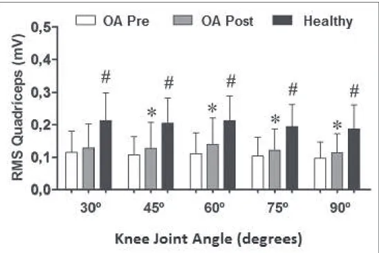
The RMS value of the quadriceps in the OA group increased after eight weeks of NMES training at angles of 45º (p=0.04), 60º (p<0.01), 75º (p=0.01) and 90º (p<0.01) before and after training. The HE group had a greater RMS value than the pre-training value of the OA group for all joint angles (p=0.03). No significant differences were observed during the post-training period (p>0.05). (Figure 1).
* Significant difference between the pre- and post-training period in the osteoarthritis group (OA) (p<0.05);
#Significant difference among the healthy group for pre-training group OA (p<0.05).
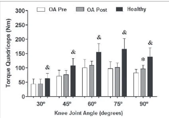
The torque produced by the knee extensors in the OA Group increased significantly at the 90° angle following the training period (p = 0.03).
The HE group had higher torque values than the OA group for all joint angles both pre- and post-training (p = 0.04). (Figure 2).
* Significant difference between pre and post-training in osteoarthritis group (OA) (p <0.05); & significant
difference between the healthy group and the pre and post-training OA group (p <0.05).
Figure 2. Maximum isometric torque of the knee extensors (quadriceps) at different angles. Porto Alegre, RS, 2012.
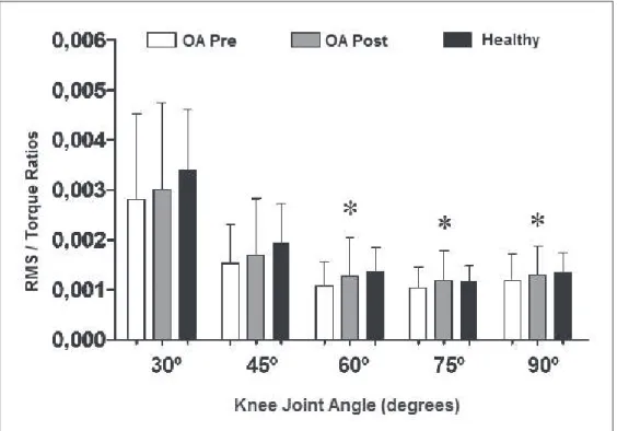
Regarding the RMS/torque ratio of the knee extensors, an increase in the ratio at the angles 60° (p = 0.03), 75° (p = 0.02) and 90° (p = 0.04) was observed in the OA group after
* Significant difference between pre and post-training period in the osteoarthritis group (OA) (p<0.05).
Figure 3. Root mean square (RMS)/torque ratios of the knee extensors at different angles. Porto Alegre, RS, 2012.
DISCuSSIon
Despite using older patients, the results of the present study agreed with the findings of Gondin et al.,17 which also observed an increase in the electrical activity of the muscles of younger individuals after eight weeks of training with artificial electrical stimulation. The HE group revealed greater electrical activation than the OA group during the pre-training period, but there was no difference between the groups in the post-training period, suggesting that training with NMES increases muscle electrical activity, achieving levels close to those of healthy individuals.
Bennell & Hinman18 suggested that aging causes specific adaptations, resulting in degeneration of the bone, cartilage, connective and muscular tissue. These degenerative factors (sarcopenia, loss of muscle strength and decreased muscle activation) are possible causes of OA, which in turn generates dysfunction, inhibition, and reduction of muscle force and activation.
of neuromuscular function, suggesting that the ratios also increase at angles close to the training angle. The absence of differences between the OA and HE groups suggests that the behavior of RMS/torque ratios was similar between the groups. However, it should be pointed out that muscle activation and the torque of the knee extensors were lower in the OA group than in the HE group.
Rosemffet et al.14 used a procedure with a short resting time (5 sec) and found an increase in force production after training with artificial electrical stimulation. The higher torque observed in the HE group may be explained by greater motor unit recruitment. Petterson et al.19 pointed out that subjects with OA have degeneration of the involved joint, causing joint instability, generating muscle inhibition and loss of force production capacity.
In addition, the results of this study partly corroborate those of Bruce-Brand et al.,20 which identified improved functional ability and reduced pain after eight weeks of training with artificial electrical stimulation. However, these authors found no significant increase in isometric torque after eight weeks of training, a finding which differs from the results of the present study, in which there was a significant increase in isometric torque at the training angle after NMES training. These results may be related to the lower training time per-session and maintenance of the same training parameters for every session, without changing volume and intensity, used by Bruce-Brand et al., 20 compared to the present study.16,21,22
In contrast, the present study conducted NMES training with only one knee flexion angle (90°), which resulted in increased torque after the training period solely for this angle, representing a possible limitation of the study. In order to optimize NMES training, it is suggested that in future studies such training is performed at more than one joint angle (such as 30°, 60° and 90° of knee flexion), facilitating gains in torque for the largest possible joint amplitude, hence improving the performance of daily living activities of individuals with OA. In addition, studies that seek to review functional parameters before and after training with NMES, such as the Timed Up and Go, Romberg and Sit and Raise tests, are also required, in order to evaluate the beneficial effects of such training on the functional capacity of older adults with osteoarthritis.20-23
ConCLuSIon
reFerenCeS
1. Doherty TJ. Invited review: aging and sarcopenia. J Appl Physiol 2003;95(4):1717-27.
2. Yu F, Hedstrom M, Cristea A, Dalen N, Larsson L. Effects of ageing and gender on contractile properties in human skeletal muscle and single fibres. Acta Physiol (Oxf) 2007;190(3):229-41.
3. Freemont AJ, Hoyland JA. Morphology, mechanisms and pathology of musculoskeletal ageing. J Pathol 2007;211(2):252-9.
4. Belo JN, Berger MY, Reijman M, Koes BW, Bierma-Zeinstra SM. Prognostic factors of progression of osteoarthritis of the knee: a systematic review of observational studies. Arthritis Rheum 2007;57(1):13-26.
5. Zhang Y, Jordan JM. Epidemiology of osteoarthritis. Rheum Dis Clin North América 2008;34(3):515-29.
6. Buckwalter JA. Osteoarthritis and articular cartilage use, disuse, and abuse: experimental studies. J Rheumatol Suppl 1995;43:13-5.
7. Gur H, Cakin N. Muscle mass, isokinetic torque, and functional capacity in women with osteoarthritis of the knee. Arch Phys Med Rehabil 2003;84(10):1534-41.
8. Esposito F, Cè E, Gobbo M, Veicsteinas A, Orizio C. Surface EMG and mechanomyogram disclose isokinetic training effects on quadriceps muscle in elderly people. Eur J Appl Physiol 2005;94(5-6):549-57.
9. Fitzgerald GK, Oatis C. Role of physical therapy in management of knee osteoarthritis. Curr Opin Rheumatol 2004;16(2):143-7.
10. Maurer BT, Stern AG, Kinossian B, Cook KD, Schumacher HR Jr. Osteoarthritis of the knee: isokinetic quadriceps exercise versus an educational intervention. Arch Phys Med Rehabil 1999;80(10):1293-9.
11. Mikesky AE, Mazzuca SA, Brandt KD, Perkins SM, Damush T, Lane KA. Effects of strength training on the incidence and progression of knee osteoarthritis. Arthritis Rheum 2006;55(5):690-9.
12. Gaines JM, Metter EJ, Talbot LA. The effect of neuromuscular electrical stimulation on arthritis knee pain in older adults with osteoarthritis of the knee. Appl Nurs Res 2004;17(3):201-6.
13. Laufer Y, Ries JD, Leininger PM, Alon G. Quadriceps femoris muscle torques and fatigue generated by neuromuscular electrical stimulation with three different waveforms. Phys Ther 2001;81(7):1307-16.
14. Rosemffet MG, Schneeberger EE, Citera G, Sgobba ME, Laiz C, Schmulevich H, et al. Effects of functional electrostimulation on pain, muscular strength, and functional capacity in patients with osteoarthritis of the knee. J Clin Rheumatol 2004;10(5):246-9.
15. Dejour H, Carret J, Walch G. Les gonarthroses. In: 7e Journées Lyonnaises de Chirurgie du Genou; 1991. [S.l.: s.n];1991. p. [775-9].
16. Vaz MA, Baroni BM, Geremia JM, Lanferdini FJ, Mayer A, Arampatzis A, et al. Neuromuscular electrical stimulation (NMES) reduces structural and functional losses of quadriceps muscle and improves health status in patients with knee osteoarthritis. J Orthop Res 2013;31(4):511-6.
17. Gondin J, Guette M, Ballay Y, Martin A. Neural and muscular changes to detraining after electrostimulation training. Eur J Appl Physiol 2006;97(2):165-73.
18. Bennell K, Hinman R. Exercise as a treatment for osteoarthritis. Curr Opin Rheumatol 2005;17(5):634-40.
19. Petterson SC, Barrance P, Buchanan T, Binder-Macleod S, Snyder-Mackler L. Mechanisms underlying quadriceps weakness in knee osteoarthritis. Med Sci Sports Exerc 2008;40(3):422-27.
Received: June 30, 2014 Revised: Dec 24, 2014 Accepted: May 13, 2015
21. Melo MO, Aragão FA, Vaz MA. Neuromuscular electrical stimulation for muscle strengthening in eldery with knee osteoarthritis: a systematic review. Complement Ther Clin Pract 2013;19(1):27-31.
22. Davis AM, MacKay C. Osteoarthritis year in review: outcome of rehabilitation. Osteoarthr Cartil 2013;21(10):1414-24.