A
RTIGO
C
IENTÍFICO
Revista Brasileira de FisioterapiaPeak torque and knee kinematics during gait
after eccentric isokinetic training of quadriceps
in healthy subjects
O pico de torque e a cinemática do joelho durante a marcha após treino
isocinético excêntrico do quadríceps em sujeitos saudáveis
Poletto PR1, Santos HH1, Salvini TF1, Coury HJCG1, Hansson GA2
Abstract
Objective: To evaluate the effects of eccentric isokinetic training on knee range of motion (ROM) of healthy subjects. Methods: The knee extensor and flexor isokinetic peak torques and ROM of flexion/extension and varus/valgus knee movements during gait of 18 healthy men (21.7r2.2 years; 1.73r0.10m; 68.7r9.4kg; body mass index: 22.6r2kg/m2) were analyzed, before and after six weeks of
bilateral eccentric isokinetic training of the knee extensors at 30o/s.Results: The knee extensor torque increased in both limbs (right,
from 229r54 to 304r53Nm; p<0.01; and left, from 228r59 to 311r63Nm; p<0.01), without any difference in torque gain between them. The knee flexor peak torque increased (from 114r30 to 123r22Nm; p<0.05), but the hamstrings/quadriceps (H/Q) ratio decreased (from 0.5r0.08 to 0.39r0.07; p<0.01) after the training. There were no differences in the flexion/extension and varus/valgus movements after the training, except for a small change (4°) in valgus for the left knee. Conclusions: The eccentric isokinetic training of the knee extensors increased the extensor torque and decreased the H/Q ratio, although the effect on the gait pattern seemed negligible in healthy subjects. Associated training for flexors, complementary to the extensor training, seems to be necessary for balance between knee agonists and antagonists.
Article registered in the Australian New Zealand Clinical Trials Registry (ANZCTR) under the number: 12607000590460.
Key words: electrogoniometer; gait; torque; knee; eccentric training.
Resumo
Objetivo: Avaliar os efeitos do treino isocinético excêntrico sobre a amplitude de movimento (ADM) do joelho em sujeitos saudáveis.
Métodos: Foram analisados os picos de torque isocinético dos extensores e flexores do joelho e a ADM de flexo/extensão e valgo/ varo, durante a marcha, de 18 homens saudáveis (21,7r2,2 anos; 1,73r0,10m; 68,7r9,4kg; índice de massa corpórea: 22,6r2kg/m2)
antes e após seis semanas de treino isocinético excêntrico bilateral dos extensores do joelho a 30o/s.Resultados: O torque extensor
do joelho aumentou em ambos os membros, direito (de 229r54 para 304r53Nm; p<0,01) e esquerdo (de 228r59 para 311r63Nm; p<0,01) sem diferença de ganho de torque entre eles. O pico de torque flexor aumentou (de 114r30 para 123r22Nm; p<0,05), mas a razão isquiotibiais/quadríceps (I/Q) diminuiu (de 0,5r0,08 para 0,39r0,07; p<0,01) após o treino. Não houve diferença para os movimentos de flexo/extensão e valgo/varo após o treino, exceto uma pequena mudança (4°) no valgo para o joelho esquerdo.
Conclusões: O treino isocinético excêntrico dos extensores do joelho aumentou o torque extensor e diminuiu a razão I/Q, entretanto o efeito sobre o padrão da marcha parece desprezível em sujeitos saudáveis. Um treino associado dos flexores, complementar ao treino dos extensores parece ser necessário para o equilíbrio entre agonistas e antagonistas do joelho.
Artigo registrado na Australian New Zealand Clinical Trials Registry (ANZCTR) sob o número: 12607000590460.
Palavras-chave: eletrogoniômetro; marcha; torque; joelho; treino excêntrico.
Recebido: 08/11/077 – Revisado: 23/04/08 – Aceito: 10/06/08
1Department of Physical Therapy, Universidade Federal de São Carlos (UFSCar) – São Carlos (SP), Brazil 2Department of Occupational and Environmental Medicine, University Hospital – Lund, Sweden
Corresponding to: Tania Fatima Salvini, Universidade Federal de São Carlos, Departamento de Fisioterapia, Rodovia Washington Luis, km 235, Caixa Postal 676, CEP 13565-905, São Carlos (SP), Brazil, e-mail: [email protected]
Introduction
Injuries and ligament reconstructions of the knee have been associated with changes in kinematic patterns during gait1-3. An altered gait, secondarily to ligament injury or reconstruction, may produce unfavorable loading of the cartilage of the knee joint4, and thus it may contribute to the development of arthritis5. Changes in gait pattern may occur as a consequence of joint tissue deran-gement, knee joint swelling, weakness of the quadriceps femoris muscle or muscle inhibition due to pain6. Atrophy of the extensor muscles is a common inding among patients undergoing ante-rior cruciate ligament (ACL) reconstruction7-9. herefore, recovery of knee extensor strength is essential for functional rehabilitation. Previous reports have shown that the functional outcome is po-sitively correlated with extensor strength, thus indicating that muscle strengthening is a precondition for functional recovery7,8.
It has been reported that training using eccentric contrac-tions is more efective for muscle recovery because it promotes greater changes in neural activation and muscle hypertrophy9-12. Both force generation and stretching are major factors in acti-vating protein synthesis, and the combination of these stimuli apparently has a pronounced additive efect13. Moreover, loa-ded eccentric exercise is a potent stimulus for hypertrophy14,15 and increases muscle strength16.
In a recent study17 in which we applied eccentric isokinetic training of the quadriceps muscles to subjects who had under-gone ACL reconstruction, the knee extensor torque and lexion/ extension range of motion (ROM) during gait increased signii-cantly after training. However, unexpectedly increased valgus, which was most pronounced during the swing phase, along with increased valgus/varus ROM. hese kinematic changes, which may imply adverse efects on the knee, were also observed in the ACL reconstructed knees when compared with the healthy untrained subjects. In this respect, it would be also important to examine the efect of isokinetic eccentric training on knee ROM in control groups (subjects with healthy knees).
hus, this study had the objective of evaluating the efects of eccentric isokinetic training on the strength of the extensor and lexor muscles of the knee, and the sagittal and coronal knee movements during gait, in healthy male subjects. In addi-tion, the present stride-based method for characterizing gait was compared with the method used in our previous study17.
Materials and methods
Subjects
Initially, the sample was 25 subjects, but only 18 completed the study. hese 18 healthy and active males ( four with right
dominance and 14 with left dominance) did not present any musculoskeletal injuries or symptoms or equilibrium disorders (age 21.7r2.2 years; height 1.73r0.10m; weight 68.7r9.4kg; body mass index: 22.6r2.0kg/m2). heir occupational and recreatio-nal activities did not change, and none of them was involved in any other training program to improve muscle strength, during the present study. his study was developed with approval from the Ethics Committee for Human Investigation of the Universi-dade Federal de Sao Carlos (UFSCar), number 144/2004 and all the subjects signed an informed consent statement.
Eccentric training
he training was performed twice a week for six consecu-tive weeks, totaling 12 sessions. he extensor muscles of both the right and left knees were trained during each session. To avoid any systematic diferences, the left knee was trained irst in one session and the right knee irst in the subsequent ses-sion. his procedure was repeated for the rest of the training. Eighteen subjects completed the training program, which had been developed in the Muscle Plasticity Unit of the Neuros-ciences Laboratory of UFSCar18.
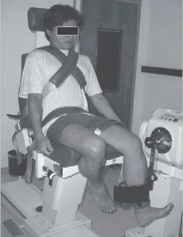
he subjects warmed up for ive minutes on a cycle ergome-ter (75W) and then the right and left quadriceps, hamstrings and calf muscles were stretched three times (30s of stretch with 30s rest). Next, the subjects were seated on the isokinetic dyna-mometer (Biodex Multi-Joint System 3, Biodex Medical Inc., Shirley, New York, USA) with the backrest reclined 5º from vertical and straps ixing the trunk, waist and distal thigh. he lateral femoral epicondyle was used as the body landmark for matching the rotation axes of the knee joint and the lever arm of the dynamometer. he dynamometer pad was then fastened around the leg 5cm proximally to the medial malleolus, and the subjects performed a series of three submaximal contractions for familiarization. he subjects then performed three series of ten consecutive maximal eccentric isokinetic contractions; the knee was moved by the dynamometer through the ROM from 20 to 90º of knee lexion at an angular velocity of 30º/s (Fi-gure 1). Each series was preceded by three minutes of resting, and there were no pauses between the ten contractions.
Knee extensor and flexor torque
Forty-eight hours before and after the training, the peak torque of the knees was assessed, during eccentric isokinetic contractions at 30°/s. he procedure and the equipment giving peak torque data for each contraction were the same as for the training (see above), except that only one session of ive con-tractions was performed. he peak torque was deined as the maximum value achieved during the ive contractions.
To assess knee functional ability and muscle balance, the hamstrings to quadriceps (H/Q) strength was derived as the ratio between the corresponding peak torques19-21.
Knee movements
Knee lexion/extension and valgus/varus movements were recorded bilaterally using biaxial lexible electrogoniometers and acquisition units (M110, DL1001 and Datalink 2.0, Biome-trics Ltd., Gwent, UK). One goniometer was ixed to the shaved lateral face of each knee. he center of the inter-joint line was considered to be the common reference for the leg and thigh. he center of the sensor springs was mounted so as to coincide with this line, and the two terminals were attached on the sagittal plane of the knee and aligned with the axis of the thigh (with the additional reference point of the greater trochanter of the femur) and the axis of the lower leg (with the external malleolus as the second reference point). he sampling rate was 100Hz. he refe-rence position (id estt 0° of lexion/extension and valgus/varus)
was derived as the mean value over a 16 seconds period, with the subject standing erect and relaxed. Positive angles denoted lexion and valgus. After familiarization with walking on a tread-mill at 5km/h, knee movements were recorded for 90 seconds.
Data analysis
Knee torque and eccentric training
he knee torque and movements and the efects of train-ing (id estt the post-training minus the pre-training value) were
calculated for both the left and right knees and evaluated by paired t-tests. he comparison between the right and left sides, both in the stride-based analysis and in the previous method used by Coury et al.17, also used paired t-Student test. he data
were also tested with regard to the normality of the distribu-tion (Shapiro-Wilk) and homogeneity of variance (Levene). All signiicance tests were performed at a predetermined alpha level of 0.05. Although no power calculation was made, the sample size was based on studies published in the literature (see Coury et al.17, Manal et al.22, Kurz et al.23, Lavcanska, Taylor
e Schache24).
Knee movements
From the central part of the electrogoniometric recording, 50 consecutive strides were identiied, independently for the right and left sides. From the lexion angles, the heel strikes were detected, deined as the irst minimum occurring after the maximum lexion25. he heel strike deined the beginning
of the strides and, for each stride, the data were normalized to the duration of the stride. During normal gait, as seen in the present study, the irst 60% of the stride represents the stance
phase and the subsequent 40%, the swing phase25. For each
stride, the maximum and minimum angles and the ROM (id est
the maximum minus the minimum angle) were derived for le-xion/extension and valgus/varus. he mean values of theses measurements for the 50 strides were used to characterize the knee movements for each subject. In addition, for each subject and knee, graphs of the mean values for the 50 strides were derived. his analysis was performed using software developed from MatLab 6.5 (MathWorks Inc., Natick, MA, USA).
Comparative analysis of the
electrogoniometric method
A comparative analysis was made between the present method and the previous method17 of electrogoniometric
re-cords. For this, the 1st and 99th percentiles, and the 99th minus
the 1st percentile of the angle distributions, were calculated
for 60s of the central part of the gait (the same method as used in our previous study17), for 81% of the recordings (29 out
of 36). he reference position was derived in the same way, and for the same time period, for both methods. We then compared this percentile analysis with the new method used in the present study.
Figure 1.Positioning of the subject in the isokinetic dynamometer during eccentric training of the knee extensor (note the isometric contraction of the flexor in the contralateral limb).
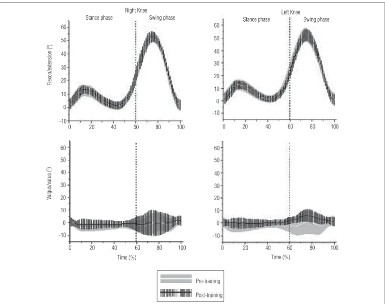
Figure 2. Mean amplitudes and standard deviations of flexion/extension and valgus/varus movements during 100% of consecutive strides, depicted in the stance and swing phases of gait for both knees before and after training, among 18 healthy male subjects. Positive angles denote flexion and valgus.
Right Knee
Stance phase Swing phase 60
Flexon/extension (º)
50 40 30 20 10 0 -10
0 20 40 60 80 100
Stance phase Swing phase Left Knee
60 50 40 30 20 10 0 -10
0 20 40 60 80 100
Time (%)
Valgus/varus (º)
60 50 40 30 20 10 0 -10
0 20 40 60 80 100
Time (%) 60
50 40 30 20 10 0 -10
0 20 40 60 80 100
Pre-training Post-training
Results
Peak torque
After the isokinetic eccentric training, both the right and the left limb presented increased knee extensor peak torque (38 and 41% greater, respectively). he right limb increased from 229r53 to 304r53Nm (p<0.01) and the left limb increased from 228r59 to 311r63Nm (p<0.01). Although, unexpectedly, the training increased the knee lexor peak torque by 8% ( from 114r30 to 123r22Nm, p<0.05), there was a natural decline in the H/Q ratio of 22% ( from 0.5r0.08 to 0.39r0.07, p<0.01).
Kinematic analysis
Means and standard deviations for the maximum, minimum and ROM angles are shown in Table 1, for before and after the trai-ning. Regarding the minimum lexion/extension values, there was
no noticeable hyperextension during walking, while the average maximum lexion/extension during the swing phase was between 53 and 54°, independent of knee and training. Figure 2, which pre-sents the mean ensemble curves with their standard deviations, for the 18 subjects, shows that the symmetry between the knees and the lack of any efect from the training on the lexion/exten-sion angles applied to all parts of the gait cycle.
here was no signiicant diference (right knee: p=0.2; left knee: p=0.54) in valgus/varus ROM between pre and post-trai-ning measurements, for both knees (Table 1). he maximum and minimum values, as presented in Figure 2, show that the valgus/varus angles, except for the left knee after training, were fairly symmetrically distributed around the reference position. After the training, the left knee displayed a general shift towards valgus, which was most pronounced during the swing phase (the average diference between the mean ensemble curves was 4.1°). he increased valgus is also shown by the increased minimum and maximum angles.
he standard deviations for the maximum and minimum valgus/varus angles were relatively large, compared with the standard deviations for the lexion/extension angles, thus indica-ting a higher inter-individual variation for valgus/varus than for lexion/extension (Table 1). he standard deviations in Figure 2 also show this relatively large inter-individual variation, and that it was most pronounced during the swing phase. It is interesting to observe that, for valgus/varus, the standard deviation decre-ased, id est the movement pattern of the subjects became more
uniform during the swing phase for the left knee after training. The kinematic analysis used in our previous study17 identified values that were almost identical to those from the present method. The differences in the results from the two methods (previous method minus present method) were: peak flexion 0.5° (IC95%=0.4°-0.6°), peak extension 0° (-0.5°-0.4°), range of flexion/extension 0.5° (0.1°-1°), peak valgus -0.5° (-0.7°–-0.4°), peak varus -0.5° (-0.8°–-0.3°) and valgus/varus range 0° (-0.3°-0.3°).
Discussion
he eccentric training increased both the peak extensor torque (by 40%) and the peak lexor torque (by 8%), but it de-creased the H/Q ratio ( from 0.5 to 0.39). hese changes had no signiicant efect on the gait kinematics of the knee, especially for the valgus/varus ROM, except for a small shift towards val-gus for the left knee after the training.
Methodological considerations
he training in the present study was similar to the training in our previous study17, except for shorter duration (six versus 12 weeks in the previous study). In spite of this, the torque gain was higher in the present study (39 versus 25% in the previous study). he lower eiciency in extensor torque gain during the training in the previous study17 was probably because the qua-driceps muscle had an abnormal pattern of motor unit recruit-ment after ACL reconstruction13.
One limitation of the present study was that the lexor peak torque was only measured for the nondominant limb, which was done for technical reasons. However, future studies that evaluate both the dominant and the nondominant limb are necessary, since a diference in valgus/varus during gait was found only on the left side.
he use of a stride-based analysis is more relevant for gait evaluation than the more general analysis of amplitude distributions that we have previously used. For technical rea-sons, only the 29 gait recordings that were recorded using the DL1001 data logger were used for comparing the methods. For
the derived measurement, the diferences between the two methods were surprisingly small, for both lexion/extension and valgus/varus. he very small and physiologically non-sig-niicant diferences enabled direct comparisons of the results obtained using the two methods. Moreover, when these me-thods are used for quantifying the efect of training, as in both the present study and our previous one, diferences between pairs of measurements made using the same method are cal-culated, thus virtually eliminating even the minute diferences between the methods. Hence, methodological considerations can be disregarded when comparing the results from the pre-sent study and our previous study.
Physiological effects
As expected, the eccentric training increased the exten-sor torque of the knees, similarly to what was found in the previous study using a similar training protocol for subjects who had undergone ACL reconstruction17. However, the unexpected gain in lexor torque was probably due to isome-tric contraction in the contralateral limb, performed by the subjects during eccentric actions of the extensors (Figure 1), since muscle strengthening of this group was not in the trai-ning program used. Among other factors, the increased lexor
Table 1. Means and mean differences of the flexion/extension and valgus/varus angles of the right and left knees before and after training, among 18 healthy male subjects during gait.
Movement Angle (º)
Maximum Minimum ROM Flexion/extension
Right side
Before training 52.6±3.7 -0.9±3.4 53.8±4.8 After training 53.5±4.2 0±2.7 53.4±4.4 Difference 0.9±3.3 0.9±3 -0.4±4.7
p-value 0.27 0.22 0.72
Left side
Before training 53±5.8 -0.7±2.8 53.6±5.9 After training 52.5±4.7 -1.4±2.6 53.9±5.4 Difference -0.5±3.3 -0.7±2.3 0.2±3.2
p-value 0.55 0.23 0.79
Valgus/varus Right side
Before training 5.5±3.9 -6±5.3 11.6±5.2 After training 6.4±6.2 -6.5±5 12.9±5.5 Difference 0.9±8.9 -0.5±7.9 1.4±4.3
p-value 0.68 0.8 0.2
Left side
Before training 5.9±4.9 -6.3±5.3 12.2±3.6 After training 8.3±4.1 -3.1±2.1 11.4±3.4 Difference 2.3±5 3.2±5.3 -0.8± 5.5
p-value 0.06 0.02 0.54
Note: Results are means±standard deviations; positive angles denote flexion and valgus; range of motion=ROM.
torque may be due to the hemispheric dominance of the task itself26, the size and uniformity of the sample, sex27, age28, func-tional asymmetry29, motor experience and type of strategies developed during practice26.
Nevertheless, the decline in H/Q ratio, after the training, indicates that the lexors presented reduced capacity for knee stabilization. his could be considered to be an increased risk of injury. Normal values for H/Q ratio of 0.4 to 0.5 have been reported based on peak moments, independent of contraction mode and velocity19,30. he approximate values for H/Q ratio (#0.3) suggest that the lexors muscles have a reduced capacity for dynamic knee joint stabilization during forceful knee lexion movements with simultaneous eccentric quadriceps muscle contraction31,32. his may relect predisposition to injury33.
he quadriceps muscle contraction may create signiicant anterior tibial translation or shear, especially at high contrac-tion forces with the knee towards full extension34-36, and this may produce substantial internal rotation of the tibia relative to the femur34,37. he co-activation of the hamstring muscles, in addition to the ACL tension, will signiicantly contribute towards counterbalancing the tibial shear32,37 or rotation30. herefore, the H/Q ratio may be used to indicate the extent to which the hamstring muscles are capable of counteracting the anterior tibial shear induced by maximal quadriceps muscle contraction19. he results from the present study show that when eccentric training is applied exclusively to the quadriceps muscles, it alters the forces involved in knee joint stabilization.
he decline in the H/Q ratio found in this study was a consequence of the higher torque gain of the knee extensor (about 40%), compared with the torque gain of the knee le-xor (8%). However, this result seems to only marginally afect the gait pattern in normal subjects. Speciically, no increase in valgus/varus ROM was observed, while such an increase was the main inding in our previous study on ACL reconstruc-tion patients. Such patients are probably more susceptible to alterations in gait kinematics due to eccentric training, and also present higher values in the H/Q ratio. If the decline in H/Q ratio is the same as in the present study, the mechanical
properties of the ligaments may be more sensitive to increased extensor torque, even when partly balanced by antagonistic torque of the lexors. hus, to prevent ACL reconstruction pa-tients from developing degenerative complications, secondary to the primary injury, rehabilitation that restores functional knee kinematics during gait seems important. Since eccentric exercise is an efective method for strengthening the knee ex-tensors, complementary eccentric training of the knee lexors may be required, in order to maintain a normal H/Q ratio and avoid possible abnormalities in the gait kinematics.
he comparison between the results from this study and our previous report17 indicates that some aspects of the efects of eccentric training on knee gait parameters in normal and ACL reconstruction subjects still remain to be considered in future studies. For example, it would be interesting to assess the efect of bilateral eccentric training of knee lexors and extensors on the gait pattern of normal and ACL reconstruc-tion subjects, and also the efects of diferent combinareconstruc-tions of frequency and duration of training.
Conclusions
Isokinetic eccentric training of knee extensors increased the torque of the knee extensor and decreased the H/Q ratio, but the efect on the gait pattern seems negligible in healthy subjects. Associated training of knee lexors, complementary to the training of the extensors, might be necessary in order to maintain the balance between knee agonists and antagonists.
Acknowledgements
Financial support from the Fundação de Amparo à Pesquisa do Estado de São Paulo, (Fapesp), and the Conselho Nacional de Desenvolvimento Cientiico e Tecnológico (CNPq). Patrícia Rios Poletto and Heleodório Honorato Santos hold PhD bur-saries from the Coordination Oice for the Coordenação de Aperfeiçoamento de Pessoal de Nível Superior (Capes).
336
Referências bibliográficas
1. Knoll Z, Kiss RM, Kocsis L. Gait adaptation in ACL deficient patients before and after anterior cruciate ligament reconstruction surgery. J Electromyogr Kinesiol. 2004;14(3):287-94.
2. Yoo JD, Papannagari R, Park SE, DeFrate LE, Gill TJ, Li G. The effect of anterior cruciate ligament reconstruction on knee joint kinematics under simulated muscle loads. Am J Sports Med. 2005;33(2):240-6.
3. Shelburne KB, Torry MR, Pandy MG. Effect of muscle compensation on knee instability during ACL-deficient gait. Med Sci Sports Exerc. 2005;37(4):642-8.
337
5. Kvist J, Gillquist J. Anterior positioning of tibia during motion after anterior cruciate ligament injury. Med Sci Sports Exerc. 2001;33(7):1063-72.
6. Ernst GP, Saliba E, Diduch DR, Hurwitz SR, Ball DW. Lower extremity compensations following anterior cruciate ligament reconstruction. Phys Ther. 2000;80(3):251-60.
7. Williams GN, Snyder-Mackler L, Barrance PJ, Buchanan TS. Quadriceps femoris muscle morphology and function after ACL injury: a differential response in coopers versus non-coopers. J Biomech. 2005;38(4):685-93.
8. McHugh MP, Tyler TF, Browne MG, Gleim GW, Nicholas SJ. Electromyographic predictors of residual quadriceps muscle weakness after anterior cruciate ligament reconstruction. Am J Sports Med. 2002;30(3):334-9.
9. Konishi Y, Fukubayashi T, Takeshita D. Possible mechanism of quadriceps femoris weakness in patients with ruptured anterior cruciate ligament. Med Sci Sports Exerc. 2002:34(9):1414-8.
10. Wilk KE, Romaniello WT, Soscia SM, Arrigo CA, Andrews JR. The relationship between subjective knee scores, isokinetic testing in the ACL-reconstructed knee. J Orthop Sports Phys Ther. 1994;20(2):60-73.
11. LaStayo PC, Woolf JM, Lewek MD, Snyder-Mackler L, Reich T, Lindstedt SL. Eccentric muscle contractions: their contribution to injury, prevention, rehabilitation, and sport. J Orthop Sports Phys Ther. 2003;33(10):557-71.
12. Miller LE, Pierson LM, Nickols-Richardson SM, Wootten DF, Selmon SE, Ramp WK et al. Knee extensor and flexor torque development with concentric and eccentric isokinetic training. Res Q Exec Sport. 2006;77(1):58-63.
13. Goldspink G. Molecular mechanisms involved in the determination of muscle fibre mass and phenotype. Adv Exerc Sports Physiol. 1999;5(2):27-39.
14. Gibala MJ, Interisano SA, Tarnopolsky MA, Roy BD, MacDonald JR, Yarasheski KE et al. Myofibrillar disruption following acute concentric and eccentric resistance exercise in strength-trained men. Can J Physiol Pharmacol. 2000;78(8):656-61.
15. McDonagh MJ, Davies CT. Adaptive response of mammalian skeletal muscle to exercise with high loads. Eur J Appl Physiol Occup Physiol. 1984;52(2):139-55.
16. Seger JY, Thorstensson A. Effects of eccentric versus concentric training on thigh muscle strength and EMG. Int J Sports Med. 2005;26(1):45-52.
17. Coury HJ, Brasileiro JS, Salvini TF, Poletto PR, Carnaz L, Hansson GA. Change in knee kinematics during gait after eccentric isokinetic training for quadriceps in subjects submitted to anterior cruciate ligament reconstruction. Gait Posture. 2006;24(3):370-4.
18. Brasileiro JS. Alterações funcionais e morfológicas do músculo quadríceps induzidas pelo treinamento excêntrico após reconstrução de LCA. (tese de Doutorado), São Carlos (SP): UFSCar; 2004.
19. Aagaard P, Simonsen EB, Trolle M, Bangsbo J, Klausen K. Isokinetic hamstring/quadriceps strength ratio: influence from joint angular velocity, gravity correction and contraction mode. Acta Physiol Scand. 1995;154(4):421-7.
20. Li RC, Maffulli N, Hsu YC, Chan KM. Isokinetic strength of the quadriceps and hamstrings and functional ability of anterior cruciate deficient knees in recreational athletes. Br J Sports Med. 1996;30(2):161-4.
21. Rosene JM, Forgarty TD, Mahaffey BL. Isokinetic Hamstrings: Quadriceps Ratios in Intercollegiate Athletes. J Athl Train. 2001;36(4):378-83.
22. Manal K, McClay Davis I, Galinat B, Stanhope S. The accuracy of estimating proximal tibial translation during natural cadence walking: bone vs. skin mounted targets. Clin Biomech (Bristol, Avon). 2003;18(2):26-31.
23. Kurz MJ, Stergiou N, Buzzi UH, Georgoulis AD. The effect of anterior cruciate ligament recontruction on lower extremity relative phase dynamics during walking and running. Knee Surg Sports Traumatol Arthrosc. 2005;13(2):107-15.
24. Lavcanska V, Taylor NF, Schache AG. Familiarization to treadmill running in young unimpaired adults. Human Mov Sci. 2005;24(4):544-57.
25. Kettelkamp DB, Johnson RJ, Smidt GL, Chao EY, Walker M. An electrogoniometric study of knee motion in normal gait. J Bone Joint Surg Am. 1970;52(4):775-90.
26. Schulze K, Lülders E, Jäncke L. Intermanual transfer in a simple motor task. Cortex. 2002;38(5):805-15.
27. Schmidt SL, Oliveira RM, Rocha FR, Abreu-Villaça Y. Influences of handedness and gender on the grooved pegboard test. Brain Cogn. 2000;44(3):445-54.
28. Uehara I. No transfer of visuomotor learning of button-pressing from right to left hands in right-handed four-year-olds. Percept Mot Skills. 1998;87(3 Pt 2):1427-40.
29. Stoddard L, Vaid J. Asymmetries in intermanual transfer of maze learning in right-and left-handed adults. Neuropsychologia. 1996;34(6):605-8.
30. Westing SH, Seger JY. Eccentric and concentric torque-velocity characteristics, torque output comparisons, and gravity effect torque corrections for the quadriceps and hamstring muscles in females. Int J Sports Med. 1989;10(3):175-80.
31. Aagaard P, Simonsen EB, Trolle M, Bangsbo J, Klausen K. Specificity of training velocity and training load on gains in isokinetic knee joint strength. Acta Physiol Scand. 1996;156(2):123-9.
32. Dvir Z. Isokinetics: Muscle testing, interpretation and clinical applications. 2nd ed. Edinburgh: Churchill Livingstone; 2004.
33. Baratta R, Solomonow M, Zhou BH, Letson D, Chuinard R, D’Ambrosia R. Muscular coactivation. The role of the antagonist musculature in maintaining knee stability. Am J Sports Med. 1988;16(2):113-22.
34. Hirokawa S, Solomonow M, Lu Y, Lou ZP, D’Ambrosia R. Anterior-posterior and rotational displacement of the tibia elicited by quadriceps contraction. Am J Sports Med. 1992;20(3):299-306.
35. Kaufman KR, An KN, Litchy WJ, Morrey BF, Chao EY. Dynamic joint forces during knee isokinetic exercise. Am J Sports Med. 1991;19(3):305-16.
36. Nisell R, Ericson MO, Németh G, Ekholom J. Tibiofemoral joint forces during isokinetic knee extension. Am J Sports Med. 1989;17(1):49-54.