ABSTRACT
Sealing ability of MTA, CPM, and MBPc as
root-Paulo Leal MEDEIROS1, Norberti BERNARDINELI1, Bruno Cavalini CAVENAGO1, Sérgio Aparecido TORRES2, Marco Antonio Hungaro DUARTE1, Clovis Monteiro BRAMANTE1, Marina Angélica MARCIANO1
1- Universidade de São Paulo, Faculdade de Odontologia de Bauru, Departamento de Dentística, Materiais Odontológicos e Endodontia, Bauru, SP, Brasil 2- Universidade de São Paulo, Faculdade de Odontologia de Bauru, Departamento de Microbiologia e Imunologia, Bauru, SP, Brasil
Corresponding address: Marina Angélica Marciano - Faculdade de Odontologia de Bauru, Universidade de São Paulo - Al. Octávio Pinheiro Brisolla, 9-75
- 17012-901 - Bauru - São Paulo - Brazil - e-mail: [email protected]
6XEPLWWHG-DQXDU\0RGL¿FDWLRQ$SULO$FFHSWHG-XQH
O
CPM, and MBPc) using an Ent er ococcus faecalis leakage model. Material and Methods: Seventy single-root extracted human teeth were instrumented and root-ends were resectedand MBPc cements. Ent er ococcus faecalis was coronally introduced and the apical portion was immersed in BHI culture medium with phenol red indicator. The bacterial leakage was monitored every 24 h for 4 weeks. The statistical analysis was performed using the
between the CPM and the other groups. Conclusions: The epoxy resin-based cement MBPc had lower bacterial leakage compared with the calcium silicate-based cements MTA and CPM.
Ke y w or ds: Endodontics. Dental pulp cavity. Epoxy resins.
I N TROD UCTI ON
Persistent microorganisms are the main factors associated with chronic apical periodontitis and consequent Endodontic failures10. One of the
indications for periradicular surgery is when the conventional treatment is not able to eliminate the microorganisms of the apical portion7. These
prevent bacterial leakage and the reinfection of the root canal system27. Therefore, it is crucial for the
cement used to obturate root-end cavities to be
Several materials were suggested for retrograde
and MTA2,32. The Mineral Trioxide Aggregate (MTA)
satisfactory physicochemical15,29 and biological
properties28,30. Other cements are studied as
alternatives for the MTA. In 2004, a novel
root-SA, Buenos Aires, Argentina) is composed of a powder mainly made of Portland cement and a liquid with distilled water in its composition. The CPM cement presents antimicrobial activity23,
satisfactory biological response18,20,and adequate
physicochemical properties4,24.
Epoxy-resin based cements have been widely studied for Endodontic procedures since 19578,19.
In 1984, Moraes and Berbert developed an epoxy resin-based cement containing calcium hydroxide to
13. The MBPc
is consisted of hydrophobic base paste/catalyst paste cement14. Promissory results regarding its
physical34, chemical14,33,and biological3,9 properties
were found. Nonetheless, this cement requires more investigation to be considered clinically useful.
There are several methods to evaluate the
34, radioisotope22, dye penetration6,14,
and bacterial leakage21. Radioisotope and dye
and dye particles smaller than bacteria31. On the
other hand, bacterial leakage is considered more clinically relevant31. This method is widely used
in Endodontic research to evaluate the sealing ability of root canal sealers12,26
materials1,11. A previous study has compared MTA,
CPM, and MBPc cements using a dye penetration model14. Considering the limitations of this method,
it is indispensable for us to investigate if there is
cements. Thus, the aim of the study was to evaluate
(white MTA, CPM, and MBPc) using an Ent erococcus
faecalis leakage model.
M ATERI AL AN D M ETH OD S
Seventy intact, caries-free, single-rooted permanent human teeth were selected. The ethics committee of the Bauru School of Dentistry approved the use of teeth for research purposes (CEP 049-2007). All teeth were autoclaved. The coronal portions were sectioned 1.0 mm below the dental-enamel junction using a 0.3 mm Isomet saw (Buehler, Lake Bluff, Illinois, USA). Roots were standardized at 15 mm and the working length (WL) was established 1.0 mm from the apical foramen. Root canals were prepared using the
crown-Ballaigues, Switzerland). Apical preparation was
The shaping procedure was completed with 2
Ballaigues, Switzerland). After the use of each instrument, we irrigated the canal with 2 mL of 2.5% sodium hypochlorite. The smear layer was removed with 2 mL of 17% EDTA (Biodinâmica, Ibiporã, Paraná, Brazil) for three minutes, then
each root was resected with a cylindrical diamond bur, perpendicularly to the long axis of the root. A root-end cavity with 3.0 mm of depth and 1.0 mm of diameter was prepared. A gutta-percha cone of size 40.02 was inserted into the root canal 3 mm
5RRWHQG¿OOLQJ
The specimens were randomly divided in three groups (n=20):
Brazil);
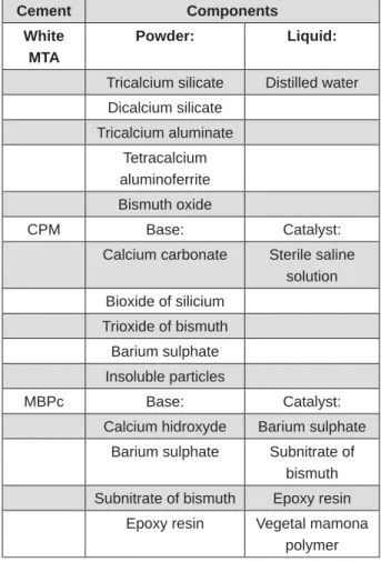
The composition of the evaluated cements is present in Figure 1. Cements were manipulated
by following the manufacturer’s instructions. They were incrementally inserted into the root-end cavities and vertically compacted using a hand plugger. Specimens were stored in 100% of humidity at 37°C for one week to allow the cements to set. For negative control (n=5), the entire root surface was coated with two layers of nail varnish (Colorama, L’Oréal, São Paulo, São Paulo, Brazil)
Ba ct e r ia l le a k a ge
The apparatus used to evaluate the leakage was prepared as previously described21
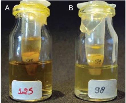
with rubber stoppers were adjusted to use. Using a high-speed handpiece, a hole was made through the centre of each rubber stopper in which each tooth was inserted under a pressure up to 4 mm from the apical portion. An epoxy resin-based varnish (Araldite, Brascola, São Paulo, São Paulo, Brazil) was used to seal the interface between tooth and rubber stopper. Cylinders prepared with 10 mL plastic syringes were adapted to the outer surface of the stoppers to create a chamber around the crown of the tooth. The apparatus was sterilized in ethylene oxide gas for a 4 h cycle at 56°C (Figure 2).
The standard bacterial strains of Ent er ococcus
faecalis (ATCC 29212) were used in the study.
Cement Components
White MTA
Powder: Liquid:
Tricalcium silicate Distilled water
Dicalcium silicate
Tricalcium aluminate
Tetracalcium aluminoferrite
Bismuth oxide
CPM Base: Catalyst:
Calcium carbonate Sterile saline solution
Bioxide of silicium
Trioxide of bismuth
Barium sulphate
Insoluble particles
MBPc Base: Catalyst:
Calcium hidroxyde Barium sulphate
Barium sulphate Subnitrate of bismuth
Subnitrate of bismuth Epoxy resin
Epoxy resin Vegetal mamona polymer
The Brain Heart Infusion Medium (BHI, Difco, BD Diagnostic Systems, Sparks, Maryland, USA) was used for bacterial growth. Ent er ococcus faecalis
was inoculated in tubes containing 5 mL of sterile BHI suspension and incubated at 37oC for 24 hours.
To adjust the experimental suspensions, turbidity
spectrophotometer (1105, Bel Photonics do Brasil Ltda., Osasco, Brazil) at 540 nm, bacterial cells were resuspended according to the MacFarland scale to
8 colony-forming
units (CFU)/mL. The purity of the cultures was
morphology of the colony.
Sterile pipettes were used to place 400 μl of bacterial inoculum in each access cavity found in the syringe apparatuses (in the upper region, removing the gauze stop). The 4 mm of root-ends were immersed in sterile BHI. The apparatus was incubated at 37°C for 120 days and checked daily for turbidity in the BHI broth. Bacterial leakage was
considered when turbidity was observed (Table 1).
St a t ist ica l a n a ly sis
Statistical analysis was performed using the
groups (p<0.05).
RESULTS
The criteria evaluated with experimental periods are shown in Figure 3. All cements showed bacterial leakage after 24 hours, except for the negative control group. The MTA showed a higher number of specimens that leaked compared with the MBPc group (p<0.05). Statistical similarities for bacterial leakage were found between the CPM and the MBPc and also the MTA (p>0.05). After 31 days until the
Cement Total of
samples
GD\V GD\V GD\V GD\V Total
White MTA 20 7 0 0 0 7
CPM 20 4 0 0 0 4
MBPc 20 1 0 1 0 2
Positive control 5 5 0 0 5 5
Negative control
5 0 0 0 0 0
Table 1- Number of samples with positive bacterial leakage every 30 days
D I SCUSSI ON
properties of the cement used17. The aim of root-end
conditions to periapical healing32. The sealing ability
of a cement can be determined using leakage methods12,14, confocal microscopy32, scanning
electron microscopy25, and more recently
micro-computed tomography analysis35. The bacterial
leakage method has the advantage of providing a clinically relevant adaptability date of materials for root canal walls31.
The Ent er ococcus faecalis was chosen for this
study model because it is commonly present in secondary infections from Endodontic treatment failures. This microorganism is a gram-positive coccus and is highly resistant to alkaline pH such as the one present in the calcium hydroxide
canals and their drug resistance, the E. faecalis
has been proposed as an Endodontic pathogen16.
Furthermore, this microorganism is easily arranged and interpreted from the study data11. Under the
conditions of this study, this microorganism was able to leak in all experimental cements. On the other hand, Jacobovitz, et al.5 (2009), using another
model of in vit r o microleakage analysis with the same microorganism, found that the MTA showed no microbial growth after 30 days.
The results found in the present study showed that the bacterial leakage occurred after 24 hours in at least one specimen for MTA, CPM, and MBPc
cements. During the experimental period, all groups showed decrease in the number of leaked specimens. A possible explanation is that the cements expanded into the cavities during the
in the present study are according to that previously found by Orosco, et al.35(2008), although they have
used the dye penetration method.
Although no statistical differences were found between MBPc and CPM cements (p>0.05), the
with the MTA (p<0.05). In a previous study, the MBPc showed satisfactory results for marginal adaptation and leakage14. Vasconcelos, et al.34
(2011) reported low leakage ranges for epoxy-resin sealers MBPc and AH Plus compared with the
presence of epoxy-resin improved the sealing of MBPc and AH Plus in dentin walls.
calcium carbonate, silicon dioxide, bismuth trioxide, and barium sulphate33. Probably, the presence of
calcium carbonate could be the responsible for offering a great calcium ions release, which may promote the adhesion to dentinal canal walls, thus improving sealing properties33. According to the
results, this cement has not been able to inhibit bacterial leakage completely. The absence of studies
regarding its sealing ability.
Further studies should use the confocal laser scanning (CLSM) microscopy to detect and quantify bacterial viability in void spaces or gaps between cavity walls. Moreover, prospective clinical
studies evaluating the success rate of Endodontic surgeries using the tested sealers might prove to be informative.
CON CLUSI ON
The epoxy resin-based cementMBPc had lower bacterial leakage compared with the calcium silicate-based cements MTA and CPM.
REFEREN CES
1- Adamo HL, Buruiana R, Schertzer L, Boylan RJ. A comparison materials using a bacterial microleakage model. Int Endod J. 1999;32(3):197-203.
2- Beltes P, Zervas P, Lambrianidis T, Molyvdas I. I n vit r o study of Traumatol. 1988;4(2):82-4.
and MBPC: microscopic analysis of implants in alveolar bone of rats. J Endod. 2006;32(6):556-9.
Filho M. Radiopacity evaluation of root canal sealers containing calcium hydroxide and MTA. Braz Oral Res. 2009;23(1):119-23. 5- Jacobovitz M, Vianna ME, Pandolfelli VC, Oliveira IR, Rossetto aggregates: an in vit r o analysis of bacterial microleakage. Oral Surg Oral Med Oral Pathol Oral Radiol Endod. 2009;108(1):140-4. Barcellos DC. Assessment of the apical seal of root canals using 7- Lopes HP, Siqueira JF Junior. Endodontics: biology and techniques. Rio de Janeiro: MEDSI; 1999.
8- Marciano MA, Ordinola-Zapata R, Cunha TV, Duarte MA,
Int Endod J. 2011;44(4):321-9.
9- Miranda RB, Fidel SR, Boller MA. L929 cell response to root perforation repair cements: an in vit r o cytotoxicity assay. Braz Dent J. 2009;20(1):22-6.
human teeth with therapy-resistant periapical lesions: a long-term light and electron microscopic follow-up study. J Endod. 1990;16(12):580-8.
A comparative evaluation of the sealing ability of 2 root-end
Ent er ococcus f aecalis. Oral Surg Oral Med Oral Pathol Oral Radiol Endod. 2011;112(2):e74-7.
12- Oliveira ACM, Tanomaru JM, Faria-Junior N, Tanomaru-Filho MTA-based sealers. Int Endod J. 2011;44(4):370-5.
as apical plugs. J Appl Oral Sci. 2008;16(1):50-4.
2010;18(2):127-34.
15- Parirokh M, Torabinejad M. Mineral trioxide aggregate: a comprehensive literature review--Part I: chemical, physical, and antibacterial properties. J Endod. 2010;36(1):16-27.
with periapical lesions. Int Endod J. 2003;36(1):1-11.
of tissue reactions to a new endodontic material. J Endod. 2010;36(7):1174-8.
Filho M, Cerri PS. Histological and histomorphometrical evaluation J. 2011;44(2):100-10.
20- Schroeder A. Endodontics - science and practice: a textbook for student and practioner. Chicago: Quintessence; 1981.
bacterial leakage. J Endod. 2001;27(11):673-5.
22- Szeremeta-Browar TL, VanCura JE, Zaki AE. A comparison of the sealing properties of different retrograde techniques: an autoradiographic study. Oral Surg Oral Med Oral Pathol. 1985;59(1):82-7.
23- Tanomaru JM, Tanomaru-Filho M, Hotta J, Watanabe E, Ito IY. Antimicrobial activity of endodontic sealers based on calcium hydroxide and MTA. Acta Odontol Latinoam. 2008;21(2):147-51. 24- Tanomaru-Filho M, Faleiros FB, Sacaki JN, Duarte MA,
mineral trioxide aggregate. J Endod. 2009;35(10):1418-21. 25- Tanzilli JP, Raphael D, Moodnik RM. A comparison of the marginal adaptation of retrograde techniques: a scanning electron microscopic study. Oral Surg Oral Med Oral Pathol. 1980;50(1):74-80.
26- Tasdemir T, Yildirim T, Buruk K, Celik D, Cora S, Tahan E, et in canals shaped with two different rotary systems: a bacterial leakage study. Oral Surg Oral Med Oral Pathol Oral Radiol Endod. 2009 [cited 2016 Jan 20];108(3):e129-34. Available from: http:// dx.doi.org/ 10.1016/j.tripleo.2009.05.007.
27- Torabinejad M, Higa RK, McKendry DJ, Pitt Ford TR. Dye leakage of four root end filling materials: effects of blood contamination. J Endod. 1994;20(4):159-63.
28- Torabinejad M, Hong CU, Lee SJ, Monsef M, Pitt Ford TR. dogs. J Endod. 1995;21(12):603-8.
29- Torabinejad M, Hong CU, McDonald F, Pitt Ford TR. Physical 1995;21(7):349-53.
30- Torabinejad M, Parirokh M. Mineral trioxide aggregate: a comprehensive literature review--part II: leakage and biocompatibility investigations. J Endod. 2010;36(2):190-202. 31- Torabinejad M, Rastegar AF, Kettering JD, Pitt Ford TR. Bacterial J Endod. 1995;21(3):109-12.
32- Torabinejad M, Watson TF, Pitt Ford TR. Sealing ability of a J Endod. 1993;19(12):591-5.
33- Vasconcelos BC, Bernardes RA, Cruz SM, Duarte MA, Padilha PM, Bernardineli N, et al. Evaluation of pH and calcium ion release Oral Radiol Endod. 2009;108(1):135-9.
34- Vasconcelos BC, Bernardes RA, Duarte MA, Bramante CM,
Sci. 2011;19(4):324-8.
35- Zakizadeh P, Marshall SJ, Hoover CI, Peters OA, Noblett WC,