Teresa BERLINCK
Justine Monteiro Monnerat TINOCO
Fernanda Leal Fonseca de CARVALHO
Luciana Moura SASSONE Eduardo Muniz Barretto TINOCO
Universidade do Estado do Rio de Janeiro – UERJ, School of Dentistry, Departamento de Procedimentos Clínicos Integrados – PROCLIN, Rio de Janeiro, RJ, Brazil.
Epidemiological evaluation of apical
periodontitis prevalence in an urban
Brazilian population
Abstract: The present study aimed to assess the prevalence of apical periodontitis (AP) in an urban Brazilian population according to gender, age group and tooth type. Data were collected from clinical iles containing the medical and dental histories and periapical radiographs of 1,126 patients treated at the School of Dentistry at Universidade do Estado do Rio de Janeiro between March 2000 and December 2010. A total of 15,724 periapical radiographs were evaluated. All the radiographs were evaluated by two independent, previously calibrated endodontists (kappa = 0.88). Periapical areas on the radiographs were classiied as N (normal) or AR (apical radiolucency). The frequency of AP and the 95% Conidence Interval (95%CI) were calculated according to gender, age group and tooth type. Differences between groups were calculated using the Z-test at a signiicance level of 5% (p < 0.05). AP was present in 7.87% of the samples, with 16.70% occurring on previously endodontically treated teeth and 44.65% occurring on teeth referred for endodontic treatment (TR-RCT). The frequency of AP was higher among females (64%) than among males (35%). The central and lateral maxillary incisors were the most frequently affected teeth. The frequency of AP was higher among individuals between 30 and 49 years of age. In this population, AP was more prevalent among females and among individuals between 30 and 49 years of age, and the central and lateral maxillary incisors were the most frequently affected teeth.
Keywords: Periapical Periodontitis; Periodontitis; Radiography.
Introduction
Apical periodontitis (AP) is a complex multifactorial condition originating from an infected necrotic pulp or a failed endodontic treatment. AP often develops asymptomatically, and the prognosis is typically less favorable when it is detected late. Epidemiological studies on the prevalence of AP in different countries have revealed that AP is a widespread oral health problem that may jeopardize systemic health.1 AP affects a signiicant proportion of people in different populations, and several authors have reported frequencies in Kosovo (46.3%), Turkey (67.9%), Belgium (40%), Denmark (52%), Lithuania (39%), Canada (44% and 51%), Germany (61%), Scotland (51%), Spain (64.5%), and the United States (39%).2,3,4
Results from cross-sectional studies have often associated AP with inadequate root-illings,5 and the quality of the root illing has been
Declaration of Interests: The authors certify that they have no commercial or associative interest that represents a conflict of interest in connection with the manuscript.
Corresponding Author:
Justine Monteiro Monnerat Tinoco E-mail: [email protected]
DOI: 10.1590/1807-3107BOR-2015.vol29.0051
Submitted: Sep 02, 2014
extensively documented as the most important determinant of endodontic success.5,6,7,8,9,10,11,12,13,14 Certain authors have indicated that age, gender, social class, education, occupation and use of dental services are among the risk indicators that may be associated with the presence of apical periodontitis.10 Other descriptive epidemiological studies have suggested that the presence of an apical lesion is dependent on tooth type.11 The present study aimed to assess the prevalence of apical periodontitis in an urban Brazilian population according to gender, age group and tooth type.
Methodology
Sample selection
Data were collected from 1,126 clinical files containing medical and dental histories, dental exams and periapical radiographs of patients treated by undergraduate students at the Endodontics Clinic at the School of Dentistry at Universidade do Estado do Rio de Janeiro – UERJ between March 2000 and December 2010. The mean patient age was 37.1 ± 16.4 years, and the cohort contained 738 females (65.55%) and 388 males (34.45%). The study was approved by the Ethics Committee of the Hospital Universitário Pedro Ernesto (CEP-HUPE 2032-2008).
Radiographic examination
Radiographic evaluation was used to assess the number of endodontically treated teeth, endodontically treated teeth with apical radiolucency, teeth referred for endodontic treatment and teeth referred for endodontic treatment with apical radiolucency. A total of 15,724 periapical radiographs were evaluated. All teeth referred for endodontic treatment were treated at the Endodontics Clinic at the School of Dentistry at UERJ. The operatory radiographs of these teeth were taken using an Odontomax 70/7P X-ray unit (Odontomax, Astex, São Paulo, Brazil) with the paralleling technique and Kodak Intraoral E-speed ilm (Eastman Kodak, Rochester, USA). All the radiographs were evaluated by two independent, previously calibrated endodontists (kappa = 0.88). Radiographs with cuts, distortion, poor it of the apical region, veiling or signiicant radiographic density alteration and images suggestive of
endo-periodontal disease or root-end surgery were excluded (n = 664). The periapical status of each tooth was assessed using the Periapical Index (PAI)15 as follows:
• N (normal): the periodontal ligament space and the surrounding bone show no alteration (PAI scores 1 and 2);
• AR (apical radiolucency): the presence of api -cal radiolucency or widening of the periapi-cal ligament space to greater than twice the normal width (PAI scores 3, 4 and 5).
The same environmental conditions were maintained at all times during the radiograph analysis. A ilm-observer distance of 20 cm was maintained throughout the analysis. The room was darkened, and a black mask was used around the radiograph to limit light emission from the illuminating viewing box.
Statistical analysis
The frequency of apical radiolucency and the 95% Conidence Interval (95%CI) were calculated according to gender, age group and tooth type. The Z-test for proportions was used to determine statistically significant differences between proportions. The signiicance level was set at 5% (p < 0.05). The prevalence of apical radiolucency was assigned as a dependent variable, and age group, gender and tooth type were included as independent variables. The Statistical Package for the Social Sciences (SPSS 8.0 for Windows, Tokyo, Japan) was used for data management.
Results
monthly income of 1 minimum wage. Table 1 shows the distribution of patients by age group.
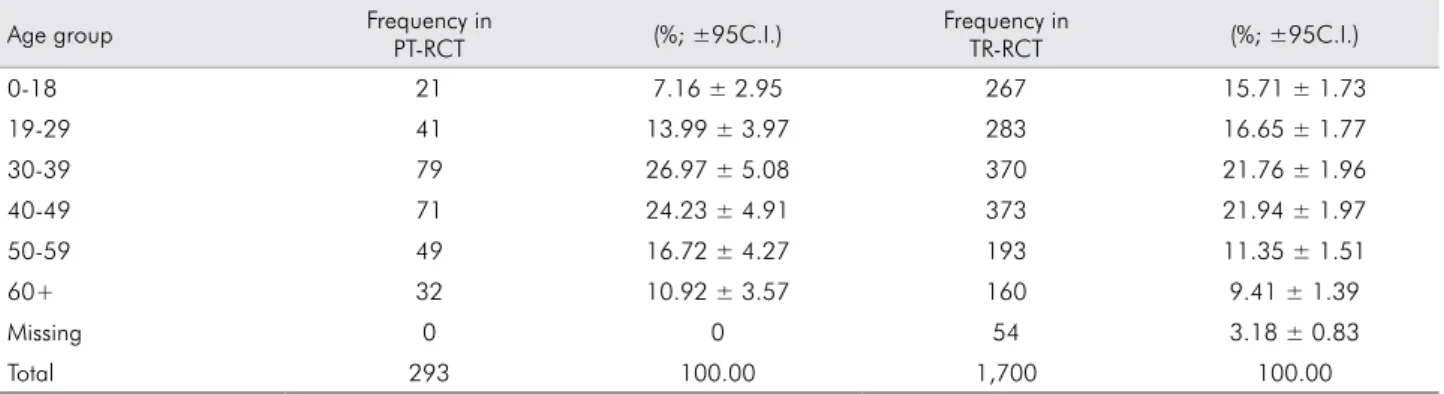
Apical radiolucency was present in 1993/25292 (7.87%) of the examined radiographs. Among previously endodontically treated teeth (PT-RCT), 293/1754 (16.70%) showed apical radiolucency, and 1700/3807 (44.65%) of the teeth referred for endodontic treatment (TR-RCT) showed apical radiolucency. The distribution and prevalence of apical radiolucency by age group is shown in Table 2.
The frequency of apical radiolucency was higher among females (62.41% in TR-RCT; 66.21% in PT-RCT) than among males (37.59% in TR-RCT; 33.79% in PT-RCT), although women were overrepresented in this sample (Table 3).
The central and lateral maxillary incisors were the most frequently affected teeth (10.87% and 7.70%, respectively), followed by the mandibular second premolars and irst molars (3.60% and 3.44%, respectively), among those teeth referred for endodontic
treatment. The central and lateral maxillary incisors were the most frequently affected teeth (13.13% and 17.76%, respectively), followed by the mandibular irst molar and the second and irst maxillary premolars (21.77%, 13.77% and 12.57, respectively), among the previously endodontically treated teeth (Table 4).
Discussion
This cross-sectional observational study aimed to describe the prevalence of periapical lesions in an urban Brazilian population using periapical radiographs and dental and medical histories. Although most studies are based on radiographic evaluation, certain authors have used other variables, such as clinical notes and interviews.9 Previously, periapical radiography was the only imaging method available to diagnose post-treatment apical periodontitis.16 Periapical radiographs were chosen to assess periapical radiolucency because they have a higher sensitivity in detecting periapical osteolytic lesions than panoramic radiographs, especially in the anterior region.17 Muhammed et al.18 found no statistically signiicant difference between panoramic radiographs and full-mouth surveys in detecting periapical lesions.
The population described in the present study represents a convenience sample from the School of Dentistry at UERJ and is not representative of the entire Brazilian population. Therefore, comparisons with other populations should be performed cautiously. Differences in health care system, age, and methodology are likely to contribute to discrepancies.19,20
Table 1. Age distribution and percentage of the total sample (n = 1,126)
Age group N (%; ±95C.I.)
0-18 125 11.1 ± 1.83
19-29 215 19.09 ± 2.3*
30-39 213 18.92 ± 2.29*
40-49 265 23.53 ± 2.48*
50-59 178 1.60 ± 0.73
60+ 115 10.21 ± 1.77
Total 1,126 100.0
*Statistically significant (p < 0.05)
Table 2. Frequency and distribution of apical radiolucency among teeth with previous endodontic treatment (PT-RCT) and among teeth referred for endodontic treatment (TR-RCT) by age group (n =1,993)
Age group Frequency in
PT-RCT (%; ±95C.I.)
Frequency in
TR-RCT (%; ±95C.I.)
0-18 21 7.16 ± 2.95 267 15.71 ± 1.73
19-29 41 13.99 ± 3.97 283 16.65 ± 1.77
30-39 79 26.97 ± 5.08 370 21.76 ± 1.96
40-49 71 24.23 ± 4.91 373 21.94 ± 1.97
50-59 49 16.72 ± 4.27 193 11.35 ± 1.51
60+ 32 10.92 ± 3.57 160 9.41 ± 1.39
Missing 0 0 54 3.18 ± 0.83
The number of individuals aged 40-49 years was signiicantly higher than that for the other age groups, with the exception of the 30-39 age group, suggesting that individuals of this age more frequently seek dental service. The mean age of the population described in the present study (37.1 ± 16.4 years) is similar to that described by Eriksen et al.7 but is lower than those reported by Tsuneishi et al.13 (50.8 years of age) and Georgopoulou et al.20 (48.0 years of age).
Certain authors have shown an increase in both the prevalence of root-illed teeth and the presence of apical periodontitis with increasing patient age.7, 13,19,20,21 The results presented herein revealed that the prevalence of apical periodontitis among teeth with previous endodontic treatment or teeth referred for endodontic treatment was highest in the 40-49 age group, followed by the 30-39 age group. Both the 50-59 and the 60+ age groups contained fewer patients with AP. Other studies Table 3. Distribution of teeth with apical radiolucency among teeth with previous endodontic treatment (PT-RCT) and among teeth referred for endodontic treatment (TR-RCT) by gender (n = 1,993)
Gender Frequency in PT-RCT (%±95 C.I.) Frequency in TR-RCT (%±95 C.I.)
Female 194 66.21 ± 5.42 1,061 62.41 ± 2.3
Male 99 37.79 ± 5.55 639 37.59 ± 2.3
Total 293 100.0 1,700 100.0
Table 4. Prevalence of apical radiolucency (AP) according the tooth type among teeth with previous RCT (TP-RCT) and among teeth referred for RCT (TR-RCT) (percentage and 95%CI).
Tooth type TP- RCT AP (% of Total; 95%C.I.) TR-RCT AP (% of Total; 95%C.I.) Maxilla
Central incisors 259 37 13.13 + 4.11 811 414 10.87 ± 0.99*
Lateral incisors 214 41 17.76 ± 5.12 541 293 7.70 ± 0.85*
Canines 146 21 12.32 ± 5.33 299 124 3.26 ± 0.56
First premolars 183 26 12.57 ± 4.8 330 126 3.31 ± 0.57
Second premolars 167 26 13.77 ± 5.23 367 121 3.18 ± 0.56
First molars 139 15 8.63 ± 4.67 88 27 0.71 ± 0.27
Second molars 50 7 6 ± 6.58 21 2 0.05 ± 0.07
Third molars 5 0 0 ± 0 0 0 0.0
Subtotal 1,163 173 4.38 ± 4.18 2,457 1,107 29.08±1.44**
Mandible
Central incisors 22 14 63.59±20.89 95 70 1.84±0.43
Lateral incisors 26 8 19.23±15.15 81 52 1.37±0.37
Canines 47 9 12.76±9.54 119 55 1.44±0.38
First premolars 112 20 15.17±6.64 195 85 2.23±0.47
Second premolars 117 16 11.11±5.69 342 137 3.60±0.59
First molars 147 34 21.77±6.67 324 131 3.44±0.58
Second molars 107 19 14.95±6.76 149 54 1.42±0.38
Third molars 13 0 0±0 14 3 0.08±0.09
Subtotal 591 120 16.92±3.02 1,319 587 15.42±1.15**
Missing values 0 0 0 18 6 0.16±0.13
Unclassified teeth 0 0 0 13 0 0.0
Total 1,754 293 14.31±1.64 3,807 1,700 44.65±1.58
have reported the same skewed age distribution.4,22 One possible explanation is that younger individuals seek dental treatment at dental schools more often than older individuals due to the lower treatment cost at dental schools. Additionally, one cannot eliminate the possibility that older persons may undergo more tooth extractions rather than root canal treatments. Nevertheless, there was no statistically signiicant difference in the prevalence of AP between the age groups (p = 0.068).
The sample in our study contained more females (63.98%) than males (36.02%). Previous surveys that have reported similar results explained this gender discrepancy as occurring because women have a greater interest in receiving dental care.19, 20 The present study
showed a signiicant difference in AP between men (37.59%) and women (62.41%) (p < 0.05), which may be explained by the smaller number of males in this sample.
Most studies have shown that molars and premolars are the most common root-illed teeth13, 20 with associated AP.8,11,21 The present results indicated that the central and
lateral maxillary incisors were the most frequently affected teeth, followed by the premolars and the mandibular first molars. This difference could be explained by the socioeconomic status of our sample population; this population probably chose tooth extraction rather than root canal treatment followed by costly prosthetic treatment. In addition, central and lateral maxillary incisors are the teeth most frequently affected by trauma, which is often associated with AP lesions.
Indeed, socioeconomic variables have been frequently associated with AP. In our sample, 70.33% of the patients had only an elementary education degree, and 93.61% had a monthly income of 1 minimum wage. These variables provide a broad overview of the social class, living conditions and general health of the patients, although these variables may only indirectly affect disease occurrence. Other studies have indicated that regular dental care and caries experiences are strong disease determinants.21
Our study sample consisted of teeth that had been endodontically treated or had been referred for endodontic treatment. In the present study, 16.7% of the cases that had previous endodontic treatment also presented with apical periodontitis. This prevalence was similar to those previously reported.13,23 In cases
that were referred for endodontic treatment, 44.65% of the patients presented with apical periodontitis; this prevalence is similar to that reported in other countries (16%-65%).4,6,21,22,24,25 Certain studies have described a higher prevalence of apical periodontitis (60%-67.5%).19,20 According to Khabbaz et al.,26 poorer
dental health and inappropriate techniques may contribute to this higher prevalence.
Kirkevang and Wenzel10 have classified risk indicators for apical periodontitis, and they concluded that the most important risk indicator for apical periodontitis was radiographic evidence of root illings. This inding is supported by several epidemiological studies.4,6,7,11,19,20,22,24,26,27,28,29,30,31 Ng et al.32 demonstrated the
need to improve the technical quality of root illings performed by undergraduate students, especially in molars. The technical quality of the root canal treatment and coronal restoration has a signiicant impact on the periapical health of root-illed teeth.
Apical periodontitis (AP) is a common sequela of pulp infection,33 and the local inlammatory process aims to
conine and limit the spread of infectious elements.34 However, in AP, pulpal and periapical infection can potentially spread throughout the body, this being the most serious endodontic pathological condition from a general health view.35 The association between
inlammation of endodontic origin and systemic health has not been adequately reported in the scientific literature.36,37,38 The available scientiic evidence is limited, but it is consistent in suggesting that AP may contribute to a systemic immune response that is not conined to the localized lesion, potentially leading to increased systemic inlammation.39 Chronic apical periodontitis has been independently associated with coronary heart disease (CHD),38 and surgical endodontic treatment has
been demonstrated to reduce systemic inlammation.40 These studies highlight the relevance of AP and the need for more studies on the epidemiology, etiology and risk factors of this important oral condition.
Conclusion
1. Segura-Egea J, Castellanos-Cosano L, Machuca G, Lopez-Lopez J, Martin-Gonzalez J, Velasco-Ortega E, et al. Diabetes mellitus, periapical inflammation and endodontic treatment outcome. Med Oral Patol Oral Cir Bucal. 2012 Mar 1;17(2):e356-61.
2. Eriksen HM, Berset GP, Hansen BF, Bjertness E. Changes in endodontic status 1973-1993 among 35-year-olds in Oslo, Norway. Int Endod J. 1995 May;28(3):129-32.
3. Weiger R, Hitzler S, Hermle G, Löst C. Periapical status, quality of root canal fillings and estimated endodontic treatment needs in an urban German population. Endod Dent Traumatol. 1997 Apr;13(2):69-74.
4. De Moor RJG, Hommez GMG, De Boever JG, Delme KIM, Martens GEI. Periapical health related to the quality of root canal treatment in a Belgian population. Int Endod J. 2000 Mar;33(2):113-20.
5. Kirkevang LL, Vaeth M, Hörsted-Bindslev P, Wenzel A. Longitudinal study of periapical and endodontic status in a Danish population. Int Endod J. 2006 Feb;39(2):100-7. 6. Marques MD, Moreira B, Eriksen HM. Prevalence of apical
periodontitis and results of endodontic treatment in an adult, Portuguese population. Int Endod J. 1998 May;31(3):161-5. 7. Eriksen HM, Kirkevang LL, Petersson K. Endodontic
epidemiology and treatment outcome: general considerations. Endod Topics. 2002 July;2(1):1-9.
8. Lupi-Pegurier L, Bertrand MF, Muller-Bolla M, Rocca JP, Bolla M. Periapical status, prevalence and quality of endodontic treatment in an adult French population. Int Endod J. 2002 Aug;35(8):690-7.
9. Dugas NN, Lawrence HP, Teplitsky PE, Pharoah MJ, Friedman S. Periapical health and treatment quality assessment of root filled teeth in two Canadian populations. Int Endod J. 2003 Mar;36(3):181-92.
10. Kirkevang LL, Wenzel A. Risk indicators for apical periodont it is. Com mun it y Dent Oral Epidem iol. 2003 Feb;31(1):59-67.
11. Loftus JJ, Keating AP, McCartan BE. Periapical status and quality of endodontic treatment in an adult Irish population. Int Endod J. 2005 Feb;38(2):81-6.
12. Siqueira Junior JF, Rôças IN, Alves FRF, Campos LC. Periradicular status related to the quality of coronal restorations and root canal fillings in Brazilian population. Oral Surg Oral Med Oral Pathol Oral Radiol Endod. 2005 Sep;100(3):369-74. 13. Tsuneishi M, Yamamoto T, Yamanaka R, Tamaki N, Sakamoto
T, Tsuji K, et al. Radiographic evaluation of periapical status and prevalence of endodontic treatment in an adult Japanese population. Oral Surg Oral Med Oral Pathol Oral Radiol. 2005 Nov;100(5):631-5.
14. Imura N, Pinheiro ET, Gomes B, Zaia AA, Ferraz C, Souza-Filho F. The outcome of endodontic treatment: a retrospective study of 2000 cases performed by a specialist. J Endod. 2007 Nov;33(11):1278-82.
15. Ørstavik D, Kerekes K, Eriksen HM. The Periapical index: a scoring system for radiographic assessment of apical periodontitis. Endod Dent Traumatol. 1986 Feb;2(1): 20–34. 16. Wu MK, Shemesh H, Wesselink PR. Limitations of previously
published systematic reviews evaluating the outcome of endodontic treatment. Int Endod J. 2009 Aug;42(8):656-66. 17. Molander B, Ahlqwist M, Gröndahl HG. Panoramic and
restrictive intraoral radiography in comprehensive oral radiographic diagnosis. Eur J Oral Sci. 1995 Aug;103(4):191-8. 18. Muhammed AH, Manson-Hing LR, Ala B. A comparison of
panoramic and intraoral radiographic surveys in evaluating a dental clinic population. Oral Surg Oral Med Oral Pathol. 1982 Jul;54(1):108-17.
19. Jiménez-Pinzón A, Segura-Egea JJ, Poyato-Ferrera M, Velasco-Ortega E, Ríos-Santos JV. Prevalence of apical periodontitis and frequency of root-filled teeth in aldult Spanish population. Int Endod J. 2004 Mar;37(3):167-73. 20. Georgopoulou MK, Spanaki-Voreadi AP, Pantazis N,
Kontakiotis EG. Frequency and distribution of root filled teeth and apical periodontitis in a Greek population. Int Endod J. 2005 Feb;38(2):105-11.
21. Kirkevang LL, Horsted-Bindslev P, Orstavik D, Wenzel A. Frequency and distribution of endodontically treated teeth and apical periodontitis in an urban Danish population. Int Endod J. 2001 Apr;34(3):198-205.
22. De Cleen MJ, Schuurs AH, Wesselink PR, Wu MK. Periapical status and prevalence of endodontic treatment in an adult Dutch population. Int Endod J. 1993 Mar;26(2):112-9. 23. Eckerbom M, Flygare L, Magnusson T. A 20-year follow-up
study of endodontic variables and apical status in a Swedish population. Int Endod J. 2007 Dec;40(12):940-8.
24. Buckley M, Spangberg LS. The prevalence and technical quality of endodontic treatment in an American subpopulation. Oral Surg Oral Med Oral Pathol Oral Radiol Endod. 1995 Jan;79(1):92-100.
25. Tronstad L, Asbjørnsen K, Døving L, Pedersen I, Eriksen HM. Influence of coronal restorations on the periapical health of endodontically treated teeth. Endod Dent Traumatol. 2000 Oct;16(5):218-21.
26. Khabbaz MG, Protogerou E, Douka E. Radiographic quality of root fillings performed by undergraduate students. Int Endod J. 2010 Jun;43(6):499-508.
27. Eriksen HM, Bjertness E. Prevalence of apical periodontitis and results of endodontic treatment in middle-aged adults in Norway. Endod Dent Traumatol. 1991 Feb;7(1):1-4. 28. Figdor D. Apical periodontitis: a very prevalent problem.
Oral Surg Oral Med Oral Pathol Oral Radiol Endod. 2002 Dec;94(6):651-2.
29. Santos SMC, Soares JA, Costa GM, Brito-Júnior M, Moreira AN, Magalhães CS. Radiographic parameters of quality of root canal fillings and periapical status: a retrospective cohort study. J Endod. 2010 Dec;36(12):1932-7.
30. Boltacz-Rzepkowska E, Pawlicka H. Radiographic features and outcome of root canal treatment carried out in the Lodz region of Poland. Int Endod J. 2003 Jan;36(1):27-32.
31. Barrieshi-Nusair KM, Al-Omari MA, Al-Hiyasat AS. Radiographic technical quality of root canal treatment performed by dental students at the Dental Teaching Center in Jordan. J Dent. 2004 May;32(4):301-7.
32. Ng Y-L, Mann V, Gulabivala K. Outcome of secondary root canal treatment: a systematic review of the literature. Int Endod J. 2008 Dec;41(12):1026-46.
33. Caplan DJ. Epidemiologic issues in studies of association between apical periodontitis and systemic health. Endod Topics. 2004 Jul;8(1):15-35.
34. Segura-Egea JJ, Castellanos-Cosano L, Machuca G, López-López J, Martín-González J, Velasco-Ortega E, et al. Diabetes mellitus, periapical inflammation and endodontic treatment outcome. Med Oral Patol Oral Cir Bucal. 2012 Mar 1;17(2):e356-61.
35. Márton IJ, Bergenholtz G. The periapical inflammatory process – systemic and local manifestations: introduction. Endod Topics. 2004 Jul;8(1):1-2.
36. Buttke TM, Shipper G, Delano EO, Trope M. C-reactive protein and serum amyloid A in a canine model of chronic apical periodontitis. J Endod. 2005 Oct;31(10):728-32. 37. Joshipura KJ, Pitiphat W, Hung HC, Willett WC, Colditz
GA, Douglass CW. Pulpal inflammation and incidence of coronary heart disease. J Endod. 2006 Feb;32(2):99-103. 38. Costa TH, Figueiredo Neto JA, Oliveira AE, Maia MFL,
Almeida AL. Association between chronic apical periodontitis and coronary artery disease. J Endod. 2014 Feb;40(2):164-7. 39. Gomes MS, Blattner TC, Sant'Ana Filho M, Grecca FS, Hugo
FN, Fouad AF, et al. Can apical periodontitis modify systemic levels of inflammatory markers? A systematic review and meta-analysis. J Endod. 2013 Oct;39(10):1205-17.