Periradicular lesions in HIV-infected patients
attend-ing the faculty of dentistry: clinical findattend-ings,
socio-demographics status, habits and laboratory data
- seeking an association
Tatiana Vasconcellos Fontes,ISonia Maria Soares Ferreira,IIArley Silva-Ju´nior,IIIPatrı´cia dos Santos Marotta,ICesar Werneck Noce,III Dennis de Carvalho Ferreira,IV,V,VI,VIIILucio Souza Gonc¸alvesI,VII
IEsta´cio de Sa´ University, Dental School, Rio de Janeiro/RJ, Brazil.IIEducational Foundation Jayme de Altavila (FEJAL/CESMAC), Dental School, Maceio´/AL,
Brazil.IIIFluminense Federal University, Oral Pathology, Rio de Janeiro/RJ, Brazil.IVCAPES Foundation, Bolsista da Coordenac¸a˜o de Aperfeic¸oamento de Pessoal de Nı´vel Superior (CAPES), Proc. n˚BEX 9203, Brası´lia/DF, Brazil.VUniversity of Groningen, Faculty of Mathematics and Natural Science, Microbial Ecology - Centre for Ecological and Evolutionary Studies, The Netherlands.VIVeiga de Almeida University, Dental School, Department of Oral Medicine, Rio de Janeiro/RJ, Brazil.VIIFederal University of Rio de Janeiro, Paulo de Go´es Institute of Microbiology, Rio de Janeiro/RJ, Brazil.VIIIFaculdade de Farma´cia e Enfermagem (UNIABEU), Disciplina de Microbiologia e Epidemiologia, Rio de Janeiro/RJ. Brazil.
OBJECTIVE:The purpose of this study was to estimate the prevalence of periradicular lesions in HIV-infected Brazilian patients and to assess the correlation of several factors with the periradicular status.
METHOD: One hundred full-mouth periapical radiographs were evaluated. A total of 2,214 teeth were evaluated for the presence of periradicular lesions, caries lesions, coronal restorations, pulp cavity exposure and endodontic treatment.
RESULTS: The prevalence of periradicular lesions was 46%. There were no significant differences between individuals with or without periradicular lesions with respect to their socio-demographic status, habits, laboratory data and route of HIV infection. However, the presence of a periradicular lesion was statistically correlated with the number of teeth with endodontic treatment (p= 0.018), inadequate endodontic treatment
(p= 0.025), images suggesting pulp cavity exposure (p= 0.002) and caries lesions (p= 0.001).
CONCLUSIONS:The prevalence of periradicular lesions in HIV-infected individuals was 46% and was not related to HIV infection.
KEYWORDS: HIV; Root Canal Filling; Endodontic Treatment; Periradicular Status; Apical Periodontitis.
Fontes TV, Ferreira SM, Silva-Ju´nior A, Marotta PS, Noce CW, Ferreira DC, et al. Periradicular lesions in HIV-infected patients attending the faculty of dentistry: clinical findings, socio-demographics status, habits and laboratory data - seeking an association. Clinics. 2014;69(9):627-633.
Received for publication onApril 4, 2014;First review completed onApril 27, 2014;Accepted for publication onApril 27, 2014 E-mail: [email protected]
Tel.: 55 21 2502-3238
& INTRODUCTION
Recently, the human immunodeficiency virus (HIV) and AIDS disease, which were characterized by a rapidly progressive course of immunodeficiency leading to death, evolved to a chronic pattern of disease as a consequence of the new therapeutic regimen that was introduced in 1996 (1). This new therapy, known as highly active antiretroviral therapy (HAART), decreased the viral load, which resulted in the absence of detectable virus as well as in reduced
morbidity and mortality related to AIDS. Additionally, after the introduction of HAART, there was a significant decrease in the rate of opportunistic infections in HIV-infected patients, such as oral candidiasis (2) and herpesviruses (3,4). With respect to periodontal disease, HIV-infected patients have increased chances of developing a more aggressive form of chronic periodontitis (5). Recently, Gonc¸alves et al. (6) demonstrated that the long-term use of HAART in Brazilian HIV-infected individuals resulted in a decrease in the severity of chronic periodontitis.
The human immunodeficiency virus (HIV) can lead to severe depression of the immune system; therefore, HIV infection may be a factor that modifies pathological changes and interferes in the development/repair of periradicular lesions of endodontic origin. In this type of infection, the microorganisms that are in contact with the periradicular tissues stimulate, either directly or indirectly, an inflamma-tory response, leading to several forms of apical periodontitis
Copyrightß2014CLINICS– This is an Open Access article distributed under
the terms of the Creative Commons Attribution Non-Commercial License (http:// creativecommons.org/licenses/by-nc/3.0/) which permits unrestricted non-commercial use, distribution, and reproduction in any medium, provided the original work is properly cited.
No potential conflict of interest was reported.
that spread outward from the infected root canal system (7). Chronic apical periodontitis occurs with persistent exposure to irritants in the root canal and is characterized by a loss of hard tissue and a radiolucent area around the tooth apex (8). Interaction among several immunocompetent cells deter-mines the structural changes in the periradicular bone (9).
Despite our extensive understanding of the role of bacteria and their products in the etiology and persistence of periradicular lesions (10), studies have shown that certain systemic conditions, such as diabetes, herpesvirus infections and genetic polymorphisms, as well as systemic changes caused by smoking, stress and depression, can compromise the host immune response and avoid or delay the healing of periradicular lesions. Thus, the above-mentioned conditions and factors are considered risk factors or disease modifiers (11). In this context, HIV infection is one of these modifying factors that can vary from individual to individual, resulting in different disease progression and having repercussions in the oral cavity.
The impact of HIV infection on the characteristics of periradicular lesions is poorly understood. The histological findings from a case report on the extraction of teeth with periradicular lesions in HIV/AIDS patients showed lesions that were almost devoid of TCD4+lymphocytes but with an abundance of TCD8+ lymphocytes (12). Additionally, HIV can be detected in the vital dental pulp of these patients, but the pulp tissue’s immune response has not yet been determined (12,13). Immunohistological studies, case reports and the principles of basic immunology have shown that treating periradicular lesions could have an unfavor-able prognosis in immunocompromised patients, such as those infected with HIV. According to these authors, T cells, which are affected by HIV, play an important role in both the pathogenesis (the balance of protective and tissue-destructive processes) and repair of periradicular lesions (14-17).
Therefore, because there is limited information on periradicular lesions in HIV-infected patients, studies to clarify this issue are warranted. The aim of this study was to estimate the prevalence of periradicular lesions in Brazilian HIV-infected patients and to assess the correlation of several factors with the periradicular status.
& MATERIAL AND METHODS
Subject Population
A cross-sectional study was conducted on a convenience sample of subjects who were recruited from a pool of patients from Clementino Fraga Filho University Hospital of the Federal University of Rio de Janeiro, Brazil, from 1997 to 2004. All subjects were informed about the aims, risks and benefits of the study and all signed a consent form. The study protocol was approved by the Review Committee for Human Subjects of the Clementino Fraga Filho University Hospital/UFRJ. Patients were included in the study if they had available full-mouth periapical radiographs. These radiographs were selected according to the required informa-tion related to age, gender, educainforma-tion, smoking, socio-economic level, family income, CD4+ lymphocyte level, viral load, medications used, AIDS-defining diseases (tuber-culosis, pneumocystosis, toxoplasmosis, cytomegalovirus and Kaposi’s sarcoma), alcohol consumption, drug use and full-mouth periapical radiographs taken a maximum of five months before or after the laboratory tests in patients who
had at least 5 teeth in their mouths. The patients included in this study were all undergoing highly active antiretroviral therapy (HAART).
Radiographic evaluation
Periradicular lesions were detected by evaluating of the full-mouth periapical radiographs for each individual. The quality of the root canal filling and the coronal restorations were analyzed according to criteria of the Tronstad et al. (18) with a slight modification, as described below:
Endodontic Treatment
a) Adequate restoration: all canals were filled; lack of empty spaces; root canal fillings ended within 0-2 mm of the radiographic apex.
b) Inadequate restoration: root canal filling ended more than 2 mm from the radiographic apex, or the apex was overfilled; root canal filling showed empty spaces, inade-quate density, unfilled canals and/or poor condensation.
Coronal Restoration
a) Adequate: any permanent restoration that appeared well adapted to the edge of the tooth upon radiographic examination;
b) Inadequate: any overhanging permanent restoration, any restoration poorly adapted to the edge; presence of recurrent caries or of temporary coronal restoration upon radiographic examination; teeth that appeared to be in need of restoration.
The endodontic treatment was categorized according to the Strindberg (19) criteria as follows:
Success: normal width of the periodontal ligament space
and normal appearance of the surrounding bone.
Failure: periradicular radiolucency.
Caries lesion
A caries lesion was defined as a radiolucency identified in the coronal tooth surface.
Image suggesting pulp exposure
Pulp exposure was defined as a radiolucency identified in the coronal tooth surface in contact with the pulp cavity in a tooth with or without root canal filling.
Two independent endodontic specialists, using an endo-dontic illuminator with an attached magnifier (4x) (Endo
Gold line), analyzed all radiographs. There was no
disagree-ment between the observers; however, had there been disagreement, a third endodontic specialist would have been consulted. The two observers were previously cali-brated and the kappa coefficient was used to analyze agreement regarding the presence/absence of a periradi-cular lesion (k= 0.89).
Data analysis
the studied variables using the Mann-Whitney test for continuous data and the Chi-squared test or Fisher’s exact test for categorical data. The threshold of statistical significance was set at 5% (p#0.05).
& RESULTS
In the current study, we enrolled 100 patients; most were male (64%). The prevalent age range was from 45 to 49 years (67.7%). Considering the household income and education the lowest levels in most individuals, who reported 0-2 minimum wage (61.1%) and 50% who had a maximum of a high school education. Most of the individuals did not smoke (65.3%), 83.9% did not consume alcohol and 91.8% were not drug users. The comparison between individuals with and without periradicular lesions with respect to socio-demographic and health-related behavior data did not reveal statistically significant differences for any of the studied variables (p.0.05; Chi-squared test and Fisher’s exact test) (Table 1).
The immunological data for the HIV-infected individuals indicated a population with advanced immunodeficiency with average lymphocyte TCD4+ counts ,300 cells/mm3 and a high prevalence of individuals with a viral load
.10,000 copies/mL (40.7%). The most frequent route of HIV transmission was sexual (84.7%). With regard to the oral and systemic manifestations studied, herpes simplex (41.7%) and herpes zoster (28%) were the most common systemic manifestations, and pseudomembranous candidia-sis (14%) and erythematous candidiacandidia-sis (11%) were the most common oral manifestations (data not shown). All indivi-duals included in this study were receiving HAART; zidovudine was the most frequently used drug (62.0%), followed by stavudine (46%), didanosine (37%), lamivudine (23%) and indinavir (23%) (data not shown). There was no statistically significant difference between individuals with
or without periradicular lesions when the laboratory data and the route of HIV infection (Table 2) were compared; the same was true for all oral and systemic manifestations and antiretrovirals (data not shown) (p.0.05; Mann-Whitney test and Fisher’s exact test).
Table 1 -Bivariate analysis between periradicular lesions and the socio-demographic/health-related characteristics of the study population.
VARIABLE PERIRADICULAR LESIONS p-value
Yes NO
N (%) N (%)
Gender*
Male 26 (56.5) 38 (70.4) 0.150
Female 20 (43.5) 16 (29.6)
Education"
Illiterate 3 (6.5) 3 (5.3) 0.562
Family income (wages)"
0-2 24 (55.8) 34 (65.4) 0.297
3-4 10 (23.3) 13 (25.0)
$5 9 (20.9) 5 (9.6)
Smoking (cigarettes)
No 29 (65.9) 33 (64.7) 0.446
1-20 12 (27.3) 17 (33.3)
.20 3 (6.8) 1 (2.0)
Alcohol consumption"
No 34 (73.9) 39 (72.2) 0.941
1-2 times/week 6 (13.0) 6 (11.1)
$3 times/week 1 (2.2) 1 (1.9)
*Chi-squared test. "
Fisher’s exact test.
Table 2 -Bivariate analysis between periradicular lesions and the laboratory data of the study population.
VARIABLE PERIRADICULAR LESIONS p-value
Laboratory Data Yes No
TCD4 lymphocytes1 -Mean (sd)
281.7 (191.1) 315.7 (267.3) 0.997
Total leukocytes1 -Mean (sd)
5369.8 (2010.5) 6942 (9743.2) 0.521
Neutrophils1
- Mean (sd) 2639.3 (1487.4) 2827.3 (1488.8) 0.405 Total lymphocytes1
-Mean (sd)
1658.1 (762.6) 1976.2 (1052.9) 0.177
Viral load"
- N (%) 0.319
0-1000 10 (29.4) 20 (42.6)
1001-10000 10 (29.4) 8 (17)
.10000 14 (41.2) 19 (40.4)
Route of HIV infection-N (%)
0.222
Transfusion 1 (2.3) 4 (7.8)
Sexual 38 (86.4) 45 (88.2)
Intravenous drug user 0 1 (2.0)
Mother to child transmission
1 (2.3) 0
Do not know 4 (9.0) 1 (2.0)
1
Mann-Whitney test. "Fisher’s exact test.
sd (standard deviation).
Table 3 -Radiographic characteristics of the study population.
VARIABLE RESULTS
Total number of studied teeth 2214 Mean (¡dp) of teeth/individual 22.1¡6.4
Periradicular lesion-N (%)
Yes 46 (46.0)
No 54 (54.0)
N (%) of individuals with:
Number of periradicular lesions
0 49 (49.0)
1-2 39 (39.0)
$3 12 (12.0)
Caries lesions
0 8 (8.0)
1-5 lesions 66 (66.0)
.5 lesions 26 (26.0)
Inadequate coronal restorations
No 38 (38.0)
1-4 restorations 51 (51.0)
.4 restorations 11 (11.0)
Images suggesting pulp cavity exposure
0 20 (20.0)
1-3 teeth 56 (56.0)
.3 teeth 24 (24.0)
Endodontic treatment
0 68 (68.0)
1-2 26 (26.0)
$3 6 (6.0)
Inadequate endodontic treatment
0 72 (72.0)
1 17 (17.0)
Radiographic characteristics
Table 3 presents the radiographic features of the study sample (100 individuals and 2,214 teeth). The prevalence of periradicular lesions was 46%. More than 50% of the individuals had 1-4 inadequate restorations and 66% and 56% had 1-5 caries lesions and 1-3 teeth with images suggestive of pulp cavity exposure, respectively. Approximately 70% of the study participants had not undergone any endodontic treatment and only 12% had more than 1 tooth with inadequate endodontic treatment.
There were no statistically significant differences between individuals with or without periradicular lesions when the radiographic characteristics were compared (p.0.05; Chi-squared test and Fisher’s exact test) (Table 4). Logistic
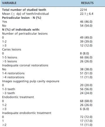
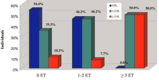
regression showed no significant associations between periradicular lesions and all radiographic variables consid-ered in this study. Nevertheless, periradicular lesions, categorized as ‘‘no lesion’’, ‘‘1-2 lesions’’ and ‘‘$3 lesions’’, were statistically correlated with the number of teeth with endodontic treatment (ET) (p= 0.018) (Figure 1), the number of teeth with inadequate endodontic treatment (IET) (p= 0.025) (Figure 2), images suggesting pulp cavity exposure (PCE) (p= 0.002) (Figure 3) and caries lesions (CL) (data not shown) (p= 0.001, Fisher’s exact test). The number of teeth with inadequate restorations was not statistically correlated with periradicular lesions (p.0.05) (data not shown).
& DISCUSSION
Since the beginning of the HIV epidemic, few studies have evaluated the possible relationship between HIV and endodontic infections. HIV infection is a transient sympto-matic disease characterized by high HIV replication and severe immune response suppression (20). The main targets of HIV-infection are TCD4+ cells, leading to a rapid depletion of the T cell repertoire and to functional damage of TCD4+cells in the weeks following infection. These cells work as a marker of immunodeficiency (21). In the current study, the participants demonstrated signs of advanced immunodeficiency with mean TCD4+ levels
,300 cells/ mm3 and most had a viral load .10,000 copies/ml. The laboratory data was not significantly different for indivi-duals with and without periradicular lesions. Suchina et al. (1) reported that a mean TCD4+cell count = 240 cells/mm3 does not significantly influence the success of endodontic treatment. Nevertheless, de Brito et al. (21) reported that 79.2% of individuals in need of endodontic treatment had a TCD4+cell level,500 cell/mm3
even though most of them were receiving HAART, suggesting poor compliance with the antiretroviral treatment.
When first receiving antiretroviral therapy, most patients develop inflammatory or cellular proliferative diseases because of T and B cell depletion or because of residual dysfunction in association with an opportunistic infection
Table 4 -Bivariate analysis between periradicular lesions and the radiographic characteristics of the study population.
VARIABLE PERIRADICULAR LESIONS p-value
Yes No
Caries lesions 2 (4.3) 6 (11.1) 0.117
0 28 (60.9) 38 (70.4)
1-5 lesions 16 (34.8) 10 (18.5)
.5 lesions
Inadequate coronal restorations 0.749
0 16 (34.8) 22 (40.7)
1-4 restorations 24 (52.2) 27 (50.0)
.4 restorations 6 (13.0) 5 (9.3)
Images suggesting pulp cavity exposure
0.245
0 6 (13) 14 (25.9)
1-3 teeth 27 (58.7) 29 (53.7)
.3 teeth 13 (28.3) 11 (20.4)
Endodontic treatment 0.124
0 28 (60.9) 40 (74.1)
1-2 13 (28.3) 13 (24.1)
$3 5 (10.9) 1 (1.8)
Inadequate endodontic treatment 0.156
No 30 (65.2) 42 (77.8)
1 8 (17.4) 9 (16.7)
$2 8 (17.4) 3 (5.5)
p-values refer to the Fisher’s exact test.
(22,23). However, no previous reports exist regarding whether patients requiring endodontic therapy have peri-radicular lesions. HAART can decrease the viral load to undetectable levels (,50 copies/mL) (24) and can decrease the morbidity and mortality associated with HIV; this treatment has also improved the quality of life and has restored and preserved the immunological functions of HIV-infected patients (25). Additionally, HAART has reduced the incidence of periodontitis in the HIV-infected population (26). It is also worth noting that the few studies on HAART in the current era that have compared successful endodontic treatments between HIV-infected and non-HIV infected patients have reported no statistically significant differences (17,27-30).
However, the risk of caries lesions is likely increased because some antiretroviral medications may induce xer-ostomia (26). In the present study, a high prevalence of caries was observed: 66% of 100 individuals (all undergoing
HAART) had 1 to 5 caries lesions. Zidovudine (AZT) was the most frequently prescribed antiretroviral for patients undergoing HAART (62.0%); this was also true in a study by de Brito et al. (21) (50.6%), whereas Gonc¸alves et al. (31) reported that lamivudine (3TC) was the most frequently prescribed drug (45.9%), followed by zidovudine (36.5%).
In the current study, radiographic evaluation was used as a criterion for determining the presence of periradicular lesions in HIV-infected patients. We evaluated 2,214 teeth and 46% of the study participants had periradicular lesions. The presence of a periradicular lesion was correlated with the number of teeth with endodontic treatment, the number of teeth with possible pulp exposure and the number of teeth with caries lesions as well as with inadequate endodontic treatment. The number of inadequate coronal restorations did not have a significant correlation with the presence of periradicular lesions. Several studies have
Figure 2 -Bivariate analysis between the number of teeth with inadequate endodontic treatment (IET) (categorized as 0, 1 and$2) and the frequency (%) of individuals with periradicular lesions (PL) (categorized as 0, 1-2 and$3) (p= 0.025; Fisher’s exact test).
Figure 3 -Bivariate analysis between the number of teeth with images suggesting pulp cavity exposure (PCE) (categorized as 0, 1-3 and
demonstrated the influence of endodontic treatment quality on periradicular tissue (10,32-36). In fact, inadequate endodontic treatment quality may be the main factor responsible for the high prevalence of poor outcomes (12,37-40).
The prevalence of periradicular lesions (46%) in the present study was as high as that observed in other countries, such as Belgium (40%) (32), Denmark (52%) (33), Canada (44% to 51%) (34), Spain (64.5%) (35), Lithuania (39%) (41), Germany (61%) (42), Scotland (51%) (43) and the United States (39%) (44). However, it is important to note that the present study has a cross-sectional design and some limitations should be considered. For example, the data were obtained in the absence of longitudinal follow-up and the sample size is not representative of the HIV-infected adult population in Brazil. Radiolucent images associated with teeth that underwent endodontic treatment were classified as persistent periradicular lesions; however, they might be considered periradicular lesions in the healing phase (45). Another limitation of this study is that the information was based only on radiographs, increasing the possibility of an incomplete diagnosis. In some cases, periradicular lesions are restricted to the cancellous bone; as a result and they would not be detectable by conventional radiographic examination (46). Additionally, radiographs do not completely show the quality of the canal seal (47). However, a cross-sectional study has a reduced chance of researcher bias compared with a longitudinal study.
In conclusion, the prevalence of periradicular lesions (46%) in HIV-infected individuals that was observed in this study is similar to that reported in studies of HIV-seronegative subjects. These lesions were not correlated with the HIV infection data (laboratory exams, routes of infection, antiretrovirals and systemic/oral manifestations) or with the number of inadequate coronal restorations. However, the lesions were correlated with the number of teeth that underwent endodontic treatment, the number of teeth with possible pulp exposure and the number of teeth with caries lesions as well as with inadequate endodontic treatment. Further studies should be performed to test additional relevant hypotheses and to address specific questions.
& AUTHOR CONTRIBUTIONS
Fontes TV, Ferreira SM, Noce CW and Gonc¸alves LS conceived and designed the study. Fontes TV, Marotta PS and Noce CW collected the data. Marotta PS, Noce CW, Ferreira SM, Silva Jr A, Ferreira DC and Gonc¸alves LS analyzed the data. Fontes TV, Ferreira SM, Silva Jr A, Ferreira DC and Gonc¸alves LS wrote the paper. Ferreira SM, Silva Jr A, Ferreira DC and Gonc¸alves LS reviewed the manuscript.
& REFERENCES
1. Suchina JA, Levine D, Flaitz CM, Nichols CM, Hicks MJ. Retrospective clinical and radiologic evaluation of nonsurgical endodontic treatment in Human Immunodeficiency Virus (HIV) infection. J Contemp Dent Pract. 2006;7(1):1-8.
2. Palella FJ Jr, Delaney KM, Moorman AC, Loveless MO, Fuhrer J, Satten GA, et al. Declining morbidity and mortality among patients with advanced human immunodeficiency virus infection. HIV outpatients study investigators. N Engl J Med. 1998;338(13):853-60.
3. dos Reis HL, Cavalcante FS, dos Santos KR, Passos MR, Ferreira Dde C. Herpes zoster as a sign of AIDS and nonadherence to antiretroviral therapy: a case report. Clinics. 2011; 66(12):2179-81, http://dx.doi.org/ 10.1590/S1807-59322011001200028.
4. Pinheiro Rdos S, Ferreira Dde C, No´brega F, Santos NS, Souza IP, Castro GF. Current status of herpesvirus identification in the oral cavity of HIV-infected children. Rev Soc Bras Med Trop. 2013;46(1):15-9, http://dx.doi. org/10.1590/0037-868217172013.
5. McKaig RG, Patton LL, Thomas JC, Strauss RP, Slade GD, Beck JD. Factors associated with periodontitis in an HIV-infected southeast USA study. Oral Dis. 2000;6(3):158-65.
6. Gonc¸alves LS, Ferreira SM, Souza CO, Souto R, Colombo AP. Clinical and Microbiological Profiles of Human Immunodeficiency Virus (HIV)-Seropositive Brazilians Undergoing Highly Active Antiretroviral Therapy and HIV-Seronegative Brazilians With Chronic Periodontitis. J Periodontol. 2007;78(1):87-96, http://dx.doi.org/10.1902/jop.2007. 060040.
7. Siqueira JF Jr. Microbiology of apical periodontitis. In: Orstavik D, Pitt Ford (eds) Essential endodontology, 2nd edn. Oxford, UK; 2007. pp. 135-196.
8. Gutmann JL. Clinical radiographic and histologic perspectives on success and failure in endodontics. Dent Clin North Am. 1992; 36(2):379-92.
9. Milia E, Campus G, Bandiera P, Pirino A. Activation of the immune system in periapical infection. 1. Lympho-monocytoid and plasma cellular elements of reactive soft tissue cells. Minerva Stomatol. 1996;45(1-2):37-48.
10. Sjogren U, Figdor D, Person S, Sundqvist G. Influence of infection at the time of root filling on the outcome of endodontic treatment of teeth with periapical periodontitis. Int Endod J. 1997;30(5):297-306, http://dx.doi. org/10.1111/j.1365-2591.1997.tb00714.x.
11. Loss BG, John RP, Laine ML. Identification of genetic risk factors for periodontitis and possible mechanisms of action. J Clin Periodontol. 2005;32(Suppl. 6):159-79, http://dx.doi.org/10.1111/j.1600-051X.2005. 00806.x.
12. Gerner NW, Hurlen B, Doblong J, Brandzag P. Endodontic treatment and immunopathology of periapical granuloma in AIDS patient. Endod Dent Traumatol. 1988;4(3):127-31, http://dx.doi.org/10.1111/j.1600-9657.1988. tb00310.x.
13. Glick M, Trope M, Pliskin ME. Detection of HIV in the dental pulp of a patient with AIDS. J Am Dent Assoc 1989;119(5):649-50.
14. Pulver WH, Taubman MA, Smith DJ. Immune components in human dental periapical lesions. Arch Oral Biol. 1978;23(6):435-43, http://dx. doi.org/10.1016/0003-9969(78)90074-2.
15. Torabinejad M, Kettering JD. Identification and relative concentration of B and T lymphocytes inhuman chronic periapical. J Endod. 1985;11(3): 122-5.
16. Marton IJ, Kiss C. Protective and destructive immune reactions in apical periodontitis. Oral Microbiol Immunol. 2000;15(3):139-50, http://dx.doi. org/10.1034/j.1399-302x.2000.150301.x.
17. Quesnell BT, Alves M, Hawkinson RW Jr, Johnson BR, Wenckus CS, BeGole EA. The effect of human immunodeficiency virus on endodontic treatment outcome. J Endod. 2005;31(9):633-6.
18. Tronstad L, Asbjornsen K, Doving L, Pedersen I, Eriksen HM. Influence of coronal restorations on the periapical health of endodontically treated teeth. Endod Dent Traumatol. 2000;16(5):218-21, http://dx.doi.org/10. 1034/j.1600-9657.2000.016005218.x.
19. Strindberg LZ. The dependence of the results of pulp therapy on certain factors: an analytic study based on radiographic and clinical follow-up examinations. Thesis, Royal School of Dentistry in Stockholm. 1956. 20. Kahn JO, Walker BD. Acute human immunodeficiency virus type 1
infection. N Engl J Med. 1998;339(1):33-9.
21. de Brito LC, da Rosa MA, Lopes VS, Ferreira EF, Vieira LQ, Sobrinho AP. Brazilian HIV-infected population: assessment of the needs of endodon-tic treatment in the post-highly active antiretroviral therapy era. J Endod. 2009;35(9):1178-81.
22. French MA. Disorders of immune reconstitution in patients with HIV infection responding to antiretroviral therapy. Curr HIV/AIDS Rep. 2007;4(1):16-21, http://dx.doi.org/10.1007/s11904-007-0003-z. 23. French M, Colebunders R. Immune restoration disease. Curr Opin HIV
AIDS. 2008;3(4):417-8, http://dx.doi.org/10.1097/COH.0b013e32830341fc. 24. Kaplan SS, Mounzer KC. Antiretroviral therapy in HIV infected patients with multidrug-resistant vı´rus: applying the guidelines to pratice. AIDS Patient Care STDS. 2008;22(12):931-94, http://dx.doi.org/10.1089/apc. 2008.0021.
25. Department of Health and Human Services. Guideline for the use of antiretroviral agents in HIV-1 infected adults and adolescents. 2006;1-121.
26. Flint SR, Tappuni A, Leigh J, Schmidt-Westhausen AM, MacPhail L. Markers of immunodeficiency and mechanisms of HAART therapy on oral lesions. Adv Dent Res. 2006;19(1):146-51, http://dx.doi.org/10. 1177/154407370601900126.
27. Shetty K, Garcia J, Leigh J. Success of root canal therapy in HIV-positive patients. Gen Dent. 2006;54(6):397-402.
28. Campo J, Cano J, del Romero J, Hernando V, Rodrı´guez C, Bascones A. Oral complication risks after invasive and non-invasive dental proce-dures in HIV-positive patients. Oral Dis. 2007;13(1):110-6, http://dx.doi. org/10.1111/j.1601-0825.2006.01262.x.
30. Tootla S, Owen CP. A comparison of endodontic treatment outcomes between HIV-positive and HIV-negative patients. SADJ. 2012;67(7): 322-5.
31. Gonc¸alves LdeS, Ferreira SM, Silva A Jr, Villoria GE, Costinha LH, Colombo AP. Association of T CD4 lymphocyte levels and chronic periodontitis in HIV-infected brazilian patients undergoing highly active anti-retroviral therapy: clinical results. J Periodontol. 2005;76(6):915-22, http://dx.doi.org/10.1902/jop.2005.76.6.915.
32. De Moor RJ, Hommez GM, De Boever JG, Delme KL, Martens GE. Periapical health related to the quality of root canal treatment in a Belgian population. Int Endod J. 2000;33(2):113-20, http://dx.doi.org/10. 1046/j.1365-2591.2000.00295.x.
33. Kirkevang LL, Orstavick D, Horsted-Bindslev P, Wenzel A. Periapical status and quality of root fillings and coronal restorations in a Danish population. Int Endod J. 2000;33(6):509-15, http://dx.doi.org/10.1046/j. 1365-2591.2000.00381.x.
34. Dugas NN, Lawrence HP, Teplitsky PE, Pharoah MJ, Friedman S. Periapical health and treatment quality assessment of root-filled teeth in two Canadian population. Int Endod J. 2003;36(3):181-92, http://dx.doi. org/10.1046/j.1365-2591.2003.00640.x.
35. Segura-Egea JJ, Jimenez-Pinzon A, Poyato-Ferrera M, Velasco-ortega E, Rios-Santos JV. Periapical status and quality of root fillings and coronal restorations in an adult Spanish population. Int Endod J. 2004;37(8):525-30, http://dx.doi.org/10.1111/j.1365-2591.2004.00826.x.
36. Moreno JO, Alves FR, Gonc¸alves LS, Martinez AM, Roˆc¸as IN, Siqueira JF Jr. Periradicular status and quality of root canal fillings and coronal restorations in an urban Colombian population. J Endod. 2013;39(5): 600-4.
37. Siqueira JF Jr, Roˆc¸as IN, Alves FR, Campos LC. Periradicular status related to the quality of coronal restorations and root canal fillings in a Brazilian population. Oral Surg Oral Med Oral Pathol Oral Radiol Endod. 2005;100(3):369-74, http://dx.doi.org/10.1016/j.tripleo.2005.03.029.
38. Georgopoulou MK, Spanaki-Voreadi AP, Pantazis N, Kontakiotis EG, Morfis AS. Periapical status and quality of root canal fillings and coronal restorations in a Greek population. Quintessence Int. 2008;9(2):e85-92. 39. Tavares PB, Bonte E, Boukpessi T, Siqueira JF Jr, Lasfargues JJ.
Prevalence of apical periodontitis in root canal-treated teeth from an urban French population: influence of the quality of root canal fillings and coronal restorations. J Endod. 2009;35(6):810-3.
40. Pak JG, Fayazi S, White SN. Prevalence of periapical radiolucency and root canal treatment: a systematic review of cross-sectional studies. J Endod. 2012;38(9):1170-6.
41. Sidaravicius B, Aleksejuniene J, Eriksen HM. Endodontic treatment and prevalence of apical periodontitis in an adult population of Vilnius, Lithuania. Endod Dent Traumatol. 1999;15(5):210-5, http://dx.doi.org/ 10.1111/j.1600-9657.1999.tb00776.x.
42. Weiger R, Hitzler S, Hermle G, Lost C. Periapical status, quality of root canal fillings and estimated endodontic treatment needs in an urban German population. Endod Dent Traumatol 1997;13(2):69-74.
43. Saunders WP, Saunders EM. Prevalence of periraricular periodontitis associated with crowned teeth in an adult Scottish subpopulation. Br Dent J. 1998;185(3):137-40, http://dx.doi.org/10.1038/sj.bdj.4809750. 44. Ray HA, Trope M. Periapical status of endodontically treated teeth in
relation to the technical quality of the root filling and the coronal restoration. Int Endod J. 1995;28(1):12-8, http://dx.doi.org/10.1111/j. 1365-2591.1995.tb00150.x.
45. Friedman S. Treatment outcome and prognosis of endodontic therapy. In: Orstavick D, Pitt Ford TR (eds) Essential Endodontology. Prevention and treatment of apical periodontitis, London, 1998. pp. 367-401. 46. Bender IB. Factors influencing the radiographic appearance of bony
lesions. J Endod. 1982;8(4):161-70.