Bruna Feltrin ANTONIAZZI(a)
Carine Weber PIRES(a)
Carmela Rampazzo BRESOLIN(a)
Rita Niederauer WEISS(b)
Juliana Rodrigues PRAETZEL(a)
(a)Universidade Federal de Santa Maria – UFSM, School of Dentistry, Department of Stomatology, Santa Maria, RS, Brazil.
(b)Universidade Federal de Santa Maria – UFSM, Department of Microbiology, Santa Maria, RS, Brazil.
Antimicrobial activity of different filling
pastes for deciduous tooth treatment
Abstract: Guedes-Pinto paste is the illing material most employed in
Brazil for endodontic treatment of deciduous teeth; however, the Rifocort®
ointment has been removed. Thus, the aim of this study was to investigate the antimicrobial potential of illing pastes, by proposing three new pharmacological associations to replace Rifocort® ointment with drugs
of already established antimicrobial power: Nebacetin® ointment, 2%
Chlorhexidine Gluconate gel, and Maxitrol® ointment. A paste composed
of Iodoform, Rifocort® ointment and Camphorated Paramonochlorophenol
(CPC) was employed as the gold standard (G1). The other associations were:
Iodoform, Nebacetin® ointment and CPC (G2); Iodoform, 2% Chlorhexidine
Digluconate gel and CPC (G3); Iodoform, Maxitrol® ointment and CPC
(G4). The associations were tested for Staphylococcus aureus (S. aureus), Streptococcus mutans (S. mutans), Streptococcus oralis (S. oralis), Enterococcus faecalis (E. faecalis), Escherichia coli (E. coli), and Bacillus subtilis (B. subtilis),
using the methods of dilution on solid medium – oriice agar – and broth dilution. The results were tested using statistical analysis ANOVA and Kruskal-Wallis. They showed that all the pastes had a bacteriostatic effect on all the microorganisms, without any statistically signiicant difference, compared with G1. S. aureus was statistically signiicant (multiple
comparison test of Tukey), insofar as G2 and G3 presented the worst and the best performance, respectively. All associations were bactericidal for
E. coli, S. aureus, S. mutans and S. oralis. Only G3 and G4 were bactericidal
for E. faecalis, whereas no product was bactericidal for B. subtilis. Thus, the
tested pastes have antimicrobial potential and have proved acceptable for endodontic treatment of primary teeth.
Keywords: Tooth, Deciduous; Root Canal Obturation; Microbial
Sensitivity Tests.
Introduction
One of the purposes of the pediatric dental clinic is to maintain primary teeth in anatomical and functional condition until their normal exfoliation, insofar as these teeth perform important roles, such as aesthetic, phonetic
and morphological functions.1 Oftentimes, dental traumas or extensive
caries lesions affect the pulp of deciduous teeth, making the maintenance of these on the arc dependent on endodontic treatment.
Use of the pulpectomy technique on deciduous dentition has several obstacles, such as molar root curvature, root canal complexity and physiological resorption.2 In addition to these dificulties, there is the
Declaration of Interests: The authors certify that they have no commercial or associative interest that represents a conflict of interest in connection with the manuscript.
Corresponding Author: Bruna Feltrin Antoniazzi
E-mail: [email protected]
DOI: 10.1590/1807-3107BOR-2015.vol29.0005
Submitted: Jan 21, 2014
infection of root canals, of polymicrobial origin.3,4 For all of these reasons, the chemical-mechanical preparation of these root canals becomes complex, requiring the reduction or elimination of any bacteria with antimicrobial irrigants and, especially, with an antimicrobial illing material of vast microbial spectrum, good diffusion and resorbability.
The illing materials most commonly employed for pulp therapy in deciduous teeth are zinc oxide paste (OZE), iodoform-based paste (IBP) and calcium
hydroxide (HCa), among others.5,6 In addition, in
Brazil, the most widely used illing paste is
Guedes-Pinto paste,7,8 a drug combination composed of
Iodoform, Rifocort® ointment and Camphorated
Paramonochlorophenol (CPC),9 which has shown
great potential as a antimicrobial and biocompatible illing material for primary teeth.10,11 However, one of its components – Rifocort® ointment – composed of an
association of antibiotic (Sodium Rifamycin SV) with corticosteroid (21-prednisolone acetate) was removed from the market, restricting current use of this paste.
Therefore, this study aimed at investigating the antimicrobial potential of illing pastes, by proposing three new pharmacological associations to replace Rifocort® ointment with drugs of already established
antimicrobial power.
The null hypothesis is that all the tested pastes have the same antimicrobial potential.
Methodology
The antimicrobial activity of different pastes was tested against standard strains of microorganisms obtained from a reference collection of the National Institute of Quality Control in Health (NIQCH) of the Osvaldo Cruz Foundation (FIOCRUZ, Rio de Janeiro, Brazil).
Specimens of S. aureus (ATCC 29213), S. mutans
(Atcc 25175), S. oralis (ATCC 10557), E. (ATCC 29212), E. coli (ATCC 25922) and B. subtilis (ATCC 6633)were used.
The association composed of Iodoform
(Biodinâmica, Ibiporã, Brazil) (0.30 g), Rifocort®
(Medley S.A., Campinas, Brazil) (0.25 g) and CPC (Biodinâmica, Ibiporã, Brazil) 3:7 (0.1 mL), was employed as the gold standard (G1). In obtaining
the test paste, Riforcort® was replaced, using the
same amount of Nebacetin® (Nycomed Pharma,
Santo Amaro, Brazil) ointment (G2), 2% Chlorhexidine Digluconate gel (Nova Derme, Santa Maria, Brazil) (G3) and Maxitrol® (Alcon, Sao Paulo, Brazil) ointment (G4).
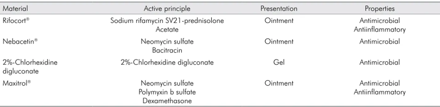
The proportions of all the groups were distributed according to a digital analytical scale (Marte AS2000. Marte®, Sao Paulo, Brazil). The different properties
of each material can be found in Table 1.
The method to evaluate the bacteriostatic activity
was Dilution on Solid Medium – Oriice Agar.12 The
analyses were performed in triplicate. Each microbial strain suspension was adjusted to the turbidity level, corresponding to tube #1 of the McFarland scale, for an approximate concentration of 3 x 10⁸ cells per mL. Seventy-eight plates were prepared, 18 for G1 paste (6 microorganisms in triplicate); 18 for G2 paste; 18 for G3 paste; and 18 for G4 paste. Six control plates were prepared without the addition of pastes, to conirm the bacterial growth on the pour-plate, at 37°C, for 24/48 hours.
After the Petri plates were prepared with Brain
Hearth Infusion medium (BHI - HiMedia®, Mumbai,
India) + 2% of pure Agar (Merck, Darmstadt, Germany) [plus 5% deibrinated sheep blood (Laborclin, Pinhais, Brazil), for S. mutans and S. oralis], the new cultures
were separated (18/24 hours) on BHI broth, into
Table 1. Different properties of each material.
Material Active principle Presentation Properties
Rifocort® Sodium rifamycin SV21-prednisolone Acetate
Ointment Antimicrobial
Antiinflammatory
Nebacetin® Neomycin sulfate
Bacitracin
Ointment Antimicrobial
2%-Chlorhexidine digluconate
2%-Chlorhexidine digluconate Gel Antimicrobial
Maxitrol® Neomycin sulfate
Polymyxin b sulfate Dexamethasone
Ointment Antimicrobial
standard strains of bacteria, which were used to indicate the antimicrobial activity of the pastes.
The procedure for pour-plate preparation consisted of mixing 0.5 mL of recent culture with 4.5 mL of melted, sterilized agar. This mixture was poured into a Petri plate, onto the particular medium, using circular motions until it illed the plate and solidiied. A hole was made in the center of each plate to add the materials. The hole made in the agar for placement of the pastes was performed with a sterile tube speciically for this purpose. The hole depth and diameter were 3 mm and 15 mm, respectively. The bottom of the hole was sealed with melted BHI agar to avoid leakage of the pastes.
The procedure for paste preparation consisted of placing the components in ceramic mortar and mixing them with a pestle, using circular motions until the paste appeared smooth and homogeneous. The mixture was placed in a 3 mL disposable sterile syringe (PlastipakTM,Curitiba, Brazil) to facilitate accurate application.
The plates were then identiied and 0.2 mL of each paste were dispensed into previously prepared holes. They were left undisturbed for 15 minutes to allow for product diffusion, and were incubated at 37°C for 24/48 hours.
The reading of the plates was performed in 24/48 hours to observe bacteriostatic activity. A transparent and smooth track around the oriice, corresponding to inhibition of bacterial growth, was observed with the naked eye on an illuminated background to obtain the results. The measurement was performed using a millimeter ruler from the
edge of the opening in the agar to the end of the area observed. The agar in the area where the product does not act appeared opaque from bacterial growth.
A sample of the inhibition range of each paste tested was collected with a bacteriological loop to investigate the bactericidal activity. Each sample was placed in nutrient broth for 24/48 hours to determine bacterial growth, which was observed as turbidity of the medium. Since the medium did not become turbid, the bactericidal action was conirmed. When there was bacterial growth, the test organism was conirmed by chiming in nutrient agar.
The data were analyzed with the SPSS program for Windows 15.0 (SPSS Inc., Chicago, USA). Data normality was tested with the Shapiro-Wilk test. ANOVA was used for variables that represent the microorganisms, and showed normal distribution, whereas Kruskal-Wallis was used for non-normal variables. A signiicance level of 5% was considered.
Results
The analytical statistics of data on measures of central tendency (mean or median) and dispersion (1Q3 or standard deviation) of the growth inhibition tracks of microorganisms tested are shown in Table 2.
Analyzing the pastes in regard to bacteriostatic capacity, it was observed that all the pastes, including G1 paste, were bacteriostatic against all the organisms tested.
Pastes G2, G3 e G4 performed similarly in regard to bacteriostatic activity, when comparing their ability to control E. faecalis, E. coli, B. subtilis, S. oralis and S. mutans
microorganisms. However, an analysis of Table 2 shows that there was a statistically signiicant difference for
Table 2. Measures of central tendency and dispersion of the growth inhibition tracks of microorganisms tested and significance (*).
Microorganism Median (1Q3) p value
G1 G2 G3 G4
E. Faecalis 10 (8-11)a 11 (11-12)a 12 (11-13)a 9 (8-12)a 0.193
E. Coli 7 (7-11)b 7 (7-7)b 6 (6-7)b 8 (7-8)b 0.108
B. Subtilis 34 (34-34)c 34 (22-34)c 34 (34-34)c 27 (25-30)c 0.082
S. Oralis 22 (20-25)d 18 (18-20)d 15 (14-23)d 21 (19-25)d 0.247
Mean (sd)
S. Aureus 6 (± 1.000)e 5 (± 0.000)f 10 (± 2.000)g 7.33 (± 2.51)e 0.01*
S. Mutans 16 (± 3.60)h 16 (± 2.646)h 16 (± 2.646)h 17 (± 1.000)h 0.954
S. aureus, as tested by ANOVA. This difference was
observed by applying the Tukey multiple comparison test, according to which the difference was found to be between G2 and G3, which showed the worst and the best performance, respectively.
Evaluating the pastes for bactericidal capacity, it was observed that the E. coli, S. aureus, S. oralis and S. mutans
microorganisms were sensitive to their bactericidal effect in all the groups. Only groups G3 and G4 showed bactericidal activity for E. faecalis, and no group was
bactericidal for the B. subtilis microorganism.
Discussion
This study was developed because of the need to ind a illing material to treat deciduous teeth, having optimal antimicrobial potential and good diffusion, and being resorbable. The results showed that the null hypothesis was accepted, since all the pastes tested showed similar antimicrobial activity, compared with Guedes-Pinto paste.
The OZE is widely used in the endodontic treatment of primary teeth. However, despite high success rates, there are disadvantages involving the difference between the degree of tooth resorption
and the paste.13 Moreover, if the paste is forced
through the radicular apex, there is a risk of causing a deviation of the eruption trajectory of the permanent successor tooth.14 The HCa paste is another optional illing material, due its favorable properties, such as biocompatibility, as well as bactericidal, resorbable
and radiopaque features.15 However, its use is
associated with internal resorption.16
The IBP have long been applied in dentistry.17
Guedes-Pinto paste is one such paste, and has demonstrated high rates of clinical and radiographic success in pulpotomies18 and pulpectomies.19 However, the fact that one of the components of the original paste is no longer available on the market has required that alternative components be found and researched to make a similar paste.
An important consideration in choosing the materials to be used was that 2% Chlorhexidine Digluconate gel is a potent antimicrobial agent widely used in dentistry for disinfecting root canals, and has a very high therapeutic index
in endodontics.20 This agent delivers excellent
action against gram-positive and gram-negative bacteria, offers 12 weeks of dentine substantivity,
and is biocompatible with the periapical tissues.21
However, studies suggest that chlorhexidine in high concentrations, as that used in this study, poses a potential systemic risk by producing reactive oxygen species (ROS), causing oxidative damage to deoxyribonucleic acid (DNA), and the release of para-chloroaniline, a cytotoxic agent
with carcinogenic potential in humans.22,23
Although Nebacetin® ointment and Maxitrol®
ointment are not used in dental treatment, they have already established antimicrobial power in their
respective uses. Maxitrol® ophthalmic ointment has
a composition similar to that of Rifocort®, insofar
as it is composed of associations of corticosteroids (Dexamethasone) and antibiotics (Neomycin Sulfate and Polymyxin B Sulfate), and is used in ocular
inlammatory conditions from bacterial infection.24
Nebacetin® ointment consists of the association
of two antibiotics – Bacitracin and Neomycin Sulfate.
It provides bactericidal action against both gram positive and gram negative bacteria, and is used to treat infections of the skin and mucosa.25 These drugs were selected not only for these characteristics, but also because they have no contraindication regarding drug interaction with the other components of the paste.
However, the antimicrobial activity of associations is only the first step, since a balance needs to be reached between this feature and cytocompatibility. Thus, after the antimicrobial effect has been proven, the biocompatibility of the pastes must be tested by many parameters, such as genotoxicity, mutagenicity, carcinogenicity, cytotoxicity, histocompatibility and microbial effects.26
In spite of the proven presence of strictly anaerobic microorganisms in pathogenic pulp lora, pure aerobic and facultative anaerobic cultures were chosen, because these microorganisms are most commonly used in studies that test the antimicrobial activity of materials used in endodontic therapy.4,27 Furthermore, studies show that the microbiota of root canals with endodontic failure is usually composed of facultative anaerobic gram-positive bacteria.3,4,28
determining its potential. Regarding bacteriostatic power, G1 paste demonstrated this potential in all
the microorganisms tested, as already shown.11,27
Similarly, all paste tests showed this property for all microorganisms. When G2 and G3 pastes were
tested against S. aureus, they were statistically
different, compared with the control paste, i.e., G2
underperformed regarding inhibition halos and G3 had the best performance. The superiority of G3 against S. aureus could be related to its proven eficacy
as an antimicrobial agent in infected root canals,29
and its diffusion power. Because Nebacetin® ointment
is a drug that meets practically all the criteria for an ideal antibiotic, considering that it has broad spectrum bactericidal activity (including gram-positive and gram-negative action), the inferiority of the G2 paste can be explained by its consistency, which could have hindered the diffusion of this paste in the medium.
In relation to bactericidal activity, all the pastes demonstrated this characteristic against S. aureus, S. mutans, S. oralis and E. coli. Only G3 and G4 pastes
were bactericidal for E. faecallis, and no paste showed
bactericidal power against the B. subtilis microorganism.
These results corroborate the indings of previous studies, where the G1 paste was not bactericidal for
E. faecallis and B. subtilis microorganisms.27
Because B. subtilis is not a microorganism found
in pathogenic oral flora, the above result has no
clinical relevance in the endodontic treatment of primary teeth. As already described, this bacterium was used because it served as a quality parameter in sensitivity tests of antimicrobial agents, which
demonstrated excellent action.30
The resistance of the E. faecalis microorganism
to antimicrobial agents, as already demonstrated,4,28 was confirmed in this study. This finding is clinically relevant for the filling of deciduous teeth, because this organism has been found in infected root canals.3
The aim of achieving pharmacological associations of filling materials for primary teeth, having antimicrobial power able to act on endodontic pathogenic lora, was achieved in its irst stage.
Conclusion
The antimicrobial potential evidenced in tested pharmacological associations showed that they are capable of being used in the endodontic treatment of primary teeth. However, further studies are required to evaluate the tissue compatibility of associations, so as to allow their clinical use.
Acknowledgements
We would like to thank the Stomatology and Microbiology departments of the Universidade Federal de Santa Maria – UFSM for their support in this study.
1. Imparato JCP, Duarte DA, Manfro ARG. Odontopediatria: Prática de saúde baseada em evidências. 1st ed. São Paulo: Elsevier; 2012. 2. Cleghorn BM, Boorberg NB, Christie WH. Primary human teeth
and their root canal systems. Endod Topics. 2010 Sep;23(1):63-33. 3. Ruviére DB, Leonardo MR, Silva LAB, Ito IY, Nelson-Filho
P. Assessment of the microbiota in root canals of human primary teeth by checkerboard DNA-DNA hybridization. J Dent Child (Chic). 2007 May-Aug;74(2):118-23.
4. Silva LAB, Nelson-Filho P, Faria G, Souza-Gugelmin MCM, Ito IY. Bacterial profile in primary teeth with necrotic pulp and periapical lesions. Braz Dent J. 2006 Apr-Jun;17(2):144-8. 5. Barja-Fidalgo F, Moutinho-Ribeiro M, Oliveira MA, de Oliveira BH. A systematic review of root canal filling materials for deciduous teeth: is there an alternative for zinc oxide-eugenol?. ISRN Dent. 2011 Jan;2011:367318. DOI: 10.5402/2011/367318.
6. Barcelos R, Santos MP, Primo LG, Luiz RR, Maia LC. ZOE paste pulpectomies outcome in primary teeth: a systematic review. J Clin Pediatr Dent. 2011 Spring;35(3):241-8. 7. Bergoli AD, Primosch RE, Araujo FB, Ardenghi TM, Casagrande
L. Pulp therapy in primary teeth--profile of teaching in Brazilian dental schools. J Clin Pediatr Dent. 2010 Winter;35(2):191-5. 8. Kramer PF, Faraco-Júnior IM, Feldens CA. [Current status
of pulp therapy in Brazilian Universities, pulpotomy and pulpectomy in primary teeth]. J Bras Odontopediatr Odontol Bebe. 2000 May-Jun;3(3):222-30. Portuguese.
9. Guedes-Pinto AC, Paiva JG; Bozzola JR. [Endodontic treatment of primary teeth with pulp necrosis]. Rev Assoc Paul Cir Dent. 1981;35(3):240-45. Portuguese.
10. Amorim LF, Toledo OA, Estrela CRA, Decurcio DA, Estrela C. Antimicrobial analysis of different root canal filling pastes used in pediatric dentistry by two experimental methods. Braz Dent J. 2006 Oct-Dec;17(4):317-22.
11. Cerqueira DF, Mello-Moura ACV, Santos EM, Guedes-Pinto AC. Cytotoxicity, histopathological, microbiological and clinical aspects of an endodontic iodoform-based paste used in pediatric dentistry: a review. J Clin Pediatr Dent. 2008 Winter;32(2):105-10. 12. Romão, CMCA, Miyazaki, NHT, Motta, LLF. [Manual of
sanitizers: methods for microbiological analysis of sanitizers with antimicrobial action]. Rio de Janeiro: Fundação Oswaldo Cruz; 1992. p. 65. Portuguese.
13. Tannure PN, Barcelos R, Portela MB, Gleiser R, Primo LG. Histopathologic and SEM analysis of primary teeth with pulpectomy failure. Oral Surg Oral Med Oral Pathol Oral Radiol Endod. 2009 Jul;108(1):e29-33.
14. Ranly DM, Garcia-Godoy F. Reviewing pulp treatment for primary teeth. J Am Dent Assoc. 1991 Sep;122(9):83-5. 15. Rosendahl R, Weinert-Grodd A. Root canal treatment of
primary molars with infected pulps using calcium hydroxide as a root canal filling. J Clin Pediatr Dent. 1995 Summer;19(4):255-8. 16. Kubota K, Golden BE, Penugonda B. Root canal filling
materials for primary teeth: a review of the literature. ASDC J Dent Child. 1992 May-Jun;59(3):225-7.
17. Rifkin A. The root canal treatment of abscessed primary teeth - a three to four year follow-up. ASDC J Dent Child. 1982 Nov-Dec;49(6):428-31.
18. Bengston AL, Bengston NG. [New behavior in pulpotomies of deciduous teeth. Clinical and radiographic evaluation of the use of Guedes-Pinto paste]. RGO. 1994 Jan;42(1):7-11. Portuguese. 19. Puppin-Rontani RM, Peters CF, Worliczeck AMT. Tratamendo
endodôntico de dentes decíduos com necrose pulpar. [Pulpectomy in primary teeth: a clinical investigation]. Rev Assoc Paul Cir Dent. 1994 Jan-Feb;48(1):1235-8.
20. Reddy S, Ramakrishna Y. Evaluation of antimicrobial efficacy of various root canal filling materials used in primary teeth: a microbiological study. J Clin Pediatr Dent. 2007 Spring;31(3):193-8.
21. Mohammadi Z, Abbott PV. The properties and applications of chlorhexidine in endodontics. Int Endod J. 2009 Apr;42(4):288-302.
22. Barbin LE, Saquy PC,Guedes DF, Souza-Neto MD, Estrela C, Pécora JD. Determination of para-chloroaniline and reactive oxygen species in chlorhexidine and chlorhexidine associated with calcium hydroxide. J Endod. 2008 Dec;34(12):1508-14. 23. Barbin LE, Estrela C, Guedes DF, Spanó JCE, Souza-Neto MD,
Pécora JD. Detection of para-chloroaniline, reactive oxygen species, and 1-chloro-4-nitrobenzene in high concentrations of chlorhexidine and in a mixture of chlorhexidine and calcium hydroxide. J Endod. 2013 May;39(5):664-668. 24. Shulman DG, Sargent JB, Stewart RH, Mester U. Comparative
evaluation of the short-term bactericidal potential of a steroid-antibiotic combination versus steroid in the treatment of chronic bacterial blepharitis and conjunctivitis. Eur J Ophthalmol. 1996 Oct-Dec;6(4):361-7.
25. Spann CT, Taylor SC, Weinberg JM. Topical antimicrobial agents in dermatology. Dis Mon. 2004 Jul;50(7):407-21. 26. Hauman CH, Love RM. Biocompatibility of dental materials used
in contemporary endodontic therapy: a review. Part 1. Intracanal drugs and substances. Int Endod J. 2003 Feb;36:(2)75-85. 27. Praetzel JR, Ferreira FV, Weiss RN, Friedrich RS,
Guedes-Pinto AC. Antimicrobial action of a filling paste used in pulp therapy in primary teeth under different storage conditions. J Clin Pediatr Dent. 2008 Winter;33(2):113-6.
28. Estrela C, Bammann LL, Pimenta FC, Pécora JD. Control of microorganisms in vitro by calcium hydroxide pastes. Int Endod J. 2001 Jul;34(5):341-5.
29. Ruiz-Esparza CL, Garrocho-Rangel A, Gonzalez-Amaro AM, Flores-Reyes H, Pozos-Guillen AJ. Reduction in bacterial loading using 2% chlorhexidine gluconate as an irrigant in pulpectomized primary teeth: a preliminary report. J Clin Pediatr Dent. 2011 Spring;35(3):265-70.