*Correspondence: G. S. Clemente. Institute of Nuclear Sciences Applied to Health, University of Coimbra. R. Azinhaga de Santa Comba – 3000-584 – Coimbra. E-mail: [email protected]
A
rti
Pharmaceutical Sciences vol. 47, n. 1, jan./mar., 2011
Synthesis and biological evaluation of
125I-erythropoietin as a
potential radiopharmaceutical agent for tumours
Gonçalo dos Santos Clemente
1,2*, Vera Lúcia Serra Duarte
11Higher School of Health Technology, Polytechnical Institute of Lisbon, 2Institute of Nuclear Sciences Applied to Health,
University of Coimbra
Erythropoietin (EPO) is a glycoprotein hormone responsible for regulating erythropoiesis. Expression of EPO and EPO receptors (EPOr) has recently been demonstrated in some neoplastic cell lines and tumours, suggesting a potential new target for therapy. In this work, EPO was labeled with iodine-125 using the lactoperoxidase method, known to prevent damage to protein during radioiodination, and labeling conditions were optimized. In vitro stability studies have shown that 125I-EPO is radiochemically stable for 20 days after radiolabeling. In vitro cell binding studies have demonstrated very low binding (<2%) of EPO to normal and neoplastic cell lines tested. As expected, the biodistribution in healthy mice exhibited comparatively high rates of ixation in the organs of the excretory system. Thyroid also proved to be a critical organ which may indicate in vivo dissociation of 125I-EPO. In mice with induced melanoma, only a residual ixation in the tumour was evident. Further studies are warranted on other tumoral cell lines to better understand the binding process and internalization into cells. Studies on EPO labeled with carbon-11 could be valuable, since there is a greater chance of preserving the biological activity of the protein using this method.
Uniterms: Erythropoietin (EPO). Glycoprotein hormone. 125I-erythropoietin/synthesis. 125I-erythropoietin/ biological evaluation. Erythropoiesis/regulation. Radiopharmaceuticals. Iodine-125/labelled. EPO expressive tumours.
A eritropoetina (EPO) é um hormônio glicoprotéico responsável pela regulação da eritropoese. Recentemente foi demonstrado que os receptores de EPO (EPOr) estão expressos em algumas linhas celulares neoplásicas, o que sugere a sua potencialidade como um novo alvo terapêutico. Neste trabalho a EPO foi radiomarcada com iodo-125 através do método da lactoperoxidase, menos agressivo para a viabilidade biológica das proteínas. A 125I-EPO revelou ser radioquimicamente estável durante 20 dias após a síntese. Um estudo biológico in vitro em linhas celulares tumorais demonstrou que a 125I-EPO apresenta uma ligação muito fraca (<2%), tanto em células normais como nas linhagens tumorais testadas. A biodistribuição em camundongos saudáveis apresentou taxas de ixação relativamente maiores nos órgãos excretores e a tireóide revelou ser o órgão crítico, o que pode indicar a dissociação in vivo da 125I-EPO. No estudo em camundongos com melanoma induzido a ixação no tumor foi residual. Serão, no entanto, necessários novos estudos em outras linhagens tumorais para entender o seu processo de internalização e ligação nas células. Estudos da EPO radiomarcada com carbono-11 poderão também revelar-se interessantes, já que neste método há maior probabilidade da atividade biológica ser preservada.
Unitermos: Eritropoetina. Hormônio glicoprotéico. 125I-erythropoietin/síntese. 125I-erythropoietin/ avaliação biológica. Eritropoese/regulação. Iodo-125/marcador. Tumores expressores de EPO.
INTRODUCTION
Due to the increasing incidence of neoplastic di-seases in contemporary society, concerted efforts are
contri-bution toward this goal because it allows the detection of malignant proliferations on a molecular scale before the manifestation of physiological symptoms or detection by other conventional imaging techniques. To this end, the use of erythropoietin (EPO) is currently the focus of metabolically oriented studies aimed at the detection of tumours.
EPO is a glycoprotein hormone that regulates erythropoiesis. It acts in the advanced stages of the erythrocyte progenitor cells by promoting proliferation, maturation and apoptosis inhibition in order to decrease cell death rate in bone marrow (Fisher, 2003). The main production site of this hormone is located in the kidney, although it is also produced in smaller quantities in the liver in response to low oxygen concentration levels in the blood. Hypoxia is a key concept in the production of EPO. Several studies have shown a relationship between some cancers and the stimulation of EPO production due to hypoxia (Acs et al., 2004a,b).
Many types of tumoral cells have a system of EPO autocrine production, which allows them to survive and proliferate under conditions of hypoxia (Lappin, 2003; Winter et al., 2005). Thus, EPO is strongly involved in growth, viability and angiogenesis of a large number of tumours (Eccles et al., 2003; Kumar et al., 2005; Yasuda, Fujita, Matsuo, 2003). This means that these tumoral cells have EPO receptors (EPOr) on their surfaces that can be used as targets for the early detection of cancers by using EPO labeled with a radionuclide.
Use of immunohistochemical methods has yielded evidence proving that EPOr are present in breast cancer cells but absent in adjacent tissues (Acs et al., 2002). This phenomenon seems to be a promising focus for future research directing chemotherapy agents to the EPOr of a tumour while sparing surrounding tissues (Lappin, 2003). This can also be important in Nuclear Medicine diagnosis or therapy through the labeling of EPO with iodine radio-active isotopes (131I or 123I) that are easily introduced into
tyrosine, histidine and histamine residues in proteins. EPO has a molecular weight of around 30kD, is composed of 165 amino acids, four carbohydrate groups (some with sialic acid termination) while one of its most important structural aspects is the fact that it has two di-sulide bridges. One of these bridges ensures correct mo-lecular form allowing binding to receptors. The breaking of this bridge leads to a loss of biological activity of the protein. Consequently, the introduction of an iodine atom in EPO is effected directly by electrophilic substitution, activated by electron-donor atoms (-OH, -NH2), with the
various carbohydrate groups containing sialic acid. Thus, radioiodination by electrophilic substitution using
lactope-roxidase is the most appropriate method since it not only leads to higher speciic activity, but is also a less aggressive method with a greater tendency to maintain the biological integrity of proteins (Murphy, 1976; Signore et al., 1992).
MATERIAL AND METHODS
In this study, the oxidative radioiodination of EPO was performed enzymatically by the lactoperoxidase method given this is a less aggressive method of protein radiolabeling.
EPO solution was prepared in phosphate buffered saline (PBS) with pH 7.4 (0.16 g/L). A volume of 25 μL of this solution was mixed with 15 MBq of Na125I, 4 μg of
lactoperoxidase solution and 10 μL of H2O2 30%. The
re-action mixture was stirred in a vortex and radioiodination took place at room temperature.
To optimize reaction time, ive EPO radioiodinations were carried out under the same conditions. Each was in-terrupted by adding 150μl of sodium thiosulfate 10% after 2, 5, 10, 20 and 30 minutes, respectively. Radiolabeling efficiency was calculated using an optimized TLC-SG chromatographic method with ethanol, ether and water (5:5:2) where 125I-EPO has an R
f of 0.0 and 125I an Rf of 0.9.
The remaining free 125I was separated from
radioio-dinated EPO by gel permeation chromatography (
Sepha-dex G-25) with PBS at pH 7.4 as the eluent. The radioacti-vity of each fraction was measured using a gamma counter. The 125I-EPO puriied solution was kept at 6 ºC and an in vitro stability study was conducted for a 20-day period.
To analyze the binding rate of 125I-EPO to normal and
neoplastic cells, V79 (Chinese Hamster Lung Fibroblast Cell Line) and B16-F1 (epithelial cells of mice melano-ma) cell lines were used, respectively. These lines were
incubated for 3 hours in a medium containing 125I-EPO
(0.2 MBq/0.5 mL). For binding afinity control 0.5 mL of several modulator drugs were used: amiloride (1.3 mg/mL), chloroquine (2.6 mg/mL), quinacrine (2.3 mg/mL) and ou-abain (3.6 mg/mL). Amiloride, chloroquine and quinacrine act as acidiication inhibitors. Amiloride is also an inhibitor of Na+/H+ and Na+/K+ exchangers (Weiss, Lang, Bernhardt,
2004) while quinacrine acts on cationic channels causing modification of kinetic parameters and other effects by binding to DNA and inhibiting NADH oxidoreductase in membrane. Ouabain is an inhibitor of Na+/K+ ATPase and
modifies membrane permeability to ions (Lodish et al.,
2001). After three hours had elapsed, cells were analysed by optical microscopy. Cells were subsequently dissolved in 0.5 mL of NaOH 0.1 N with 1% sodium dodecyl sulfate, and centrifuged to separate them from unbound 125I-EPO.
125I-EPO biodistribution was studied in mice in
or-der to characterize its in vivo behavior, more speciically its ixation in several tissues, including tumoral tissue. This study was conducted in two phases using female Balb/c strain mice. In the irst phase, the labeled and pu-riied compound, with a radiochemical purity of 100%, was injected intraperitoneally (approximately 0.6MBq) into 8 healthy animals that were sacriiced by dislocation of the neck at two different time intervals: four of these animals were sacriiced 1 hour after injection, and the remaining mice, 4 hours after injection. In the second phase, the experiment was repeated in three female mice with a melanoma induced by subcutaneous injection of B16-F1 tumour cells. All of the mice were sacriiced by dislocation of the neck 4 hours after 125I-EPO injection.
Subsequently, the dissection of several organs of interest and neoplastic tissue was performed in order to measure their radioactivity using a dose calibrator. The biodistri-bution results were analysed according to Tukey’s range test, with a value of p<0.05 considered signiicant. All the experiments were carried out in compliance with the guidelines for conduct in animal experimentation.
RESULTS AND DISCUSSION
Synthesis and stability of 125I-EPO
By varying the duration of the radioiodination reactions of EPO and evaluating their radiochemical purities, a time period that tends to be more favorable for this procedure was identiied. Through this experi-mentation it was concluded that the optimal labeling efficiency can be achieved after approximately 10 minutes of reaction (Figure 1). Thus, all subsequent radioiodinations of EPO were based on a standardized 10-minute reaction time which, associated with
purii-cation by gel permeation chromatography (Sephadex
G-25), consistently yielded inal products with
radio-chemical purities exceeding 99%.
Results of the in vitro stability study confirmed that the 125I-EPO solution was stable for at least 20 days
after radiolabeling and purification. On the sixth day after radioiodination, the compound exhibited a radio-chemical purity of approximately 100%. At the end of the study, 125I-EPO had a radiochemical purity of around
93%, thereby proving high stability in PBS at pH 7.4 stored at 6 ºC.
In vitro study with tumoral cell lines
In vitro studies of the biological viability of 125
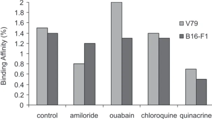
I-EPO showed negligible binding afinity (<2%) for all cell lines tested, although this was slightly higher in V79 (Figure 2).
With regard to different modifiers used, two of these seemed to have a negative effect on cell binding afinity. Quinacrine, which led to lower binding rates, proved to be cytotoxic for both types of cell lines at the concentration used. This effect was visible under optical microscope. Amiloride appeared to have an inhibiting effect on the binding afinity of 125I-EPO in V79 cells,
since it was the only modifier that presented higher binding percentages in B16-F1. Chloroquine showed a similar pattern to that seen in the control group, which may be indicative that this drug exerts no effect on the studied binding process. Finally, ouabain seemed to have a positive strengthening effect on V79 binding afinity.
Thus, this study demonstrates that 125I-EPO had a
weak binding afinity in the normal (V79) and tumoral (B16-F1) cell lines studied. These results may indicate that none of the tested cells expressed EPOr or that there may be a possible loss of biological activity caused by the incorporation of iodine in the molecule.
FIGURE 1 – Radiochemical purity of 125I-EPO by radioiodination
time.
FIGURE 2 – Mean binding affinity of 125I-EPO in V79 and
Biodistribution in animal model
After dissection of the mice, the injected dose (ID) per tissue (Eq. 1) and per gram of tissue (Eq. 2), were calculated for each animal.
Eq. 1
Eq. 2
The ixation of radioactivity veriied in mice sacri-iced 4 hours after injection was shown to be lower than that in mice sacriiced after 1 hour (Figure 3A). This is because excretion increases with time after injection. The kidney and liver are the main routes of excretion and so have higher ixation rates. The levels of 125I-EPO in the
blood are also high because of slow clearance of EPO (4h<T1/2<11h) (Kendall, 2001). The high radioactivity
observed in thyroid, an organ that has a natural ability to capture iodine, suggests that, despite the observed in vitro
stability, there may be some in vivo dissociation of 125
I-EPO, since the solution injected in the mice was almost completely free of iodine. The values obtained in the asses-sment of the mice with induced melanoma demonstrated a residual ixation of 125I-EPO in tumour (Figure 3B) while
other organs showed expected level of ixation, based on a previous study with a healthy animal model.
Owing to the suspected in vivo125I-EPO dissociation,
a TLC-SG chromatography of the urine collected from one of the mice sacriiced 4 hours after injection was per-formed. Results revealed the presence of a radiochemical species with an Rf corresponding to iodine (Rf = 0.9).
This further supported the theory of 125I-EPO deiodination
inside the body.
CONCLUSIONS
In this study, EPO radiolabeling with 125I was
perfor-med to evaluate its potential as a diagnostic or therapeutic radiopharmaceutical in molecular imaging. This radioiso-tope of iodine was chosen for having the most appropriate characteristics for research purposes (low gamma emis-sion energy, 35 keV, and a long half-life of 60 days) and because it mimics the use of 131I or 123I for radiotherapy or
diagnostic radiopharmaceuticals, respectively.
The relative ease of radioiodination and puriication of the molecule, the reproducibility of the process and its high stability, demonstrated by radiochemical purity as
around 93% after 20 days of synthesis, are parameters that render 125I-EPO a physicochemically desirable
radio-pharmaceutical for use in Nuclear Medicine laboratories. The biological tests began with an in vitro study on normal and tumoral (V79 and B16-F1) cell lines, in an at-tempt to examine the binding mechanism of radioiodinated
EPO. This procedure revealed that 125I-EPO has a weak
binding afinity to cells, since the binding values were very low (<2%). However, further studies on new cell lines are needed to conirm whether the weak binding afinity is due to the absence of speciic receptors in the cells used or to the biological activity lost during EPO radioiodination.
Studying biodistribution in an animal model is an essential stage in the development of any radiopharma-ceutical. The in vivo behavior of 125I-EPO was investigated
by intraperitoneal injection into healthy mice and animals with induced melanoma. The peritoneal cavity was chosen because it offers a greater absorption surface allowing faster entry into the bloodstream without the losses associated with FIGURE 3 – Biodistribution of 125I-EPO in mice. A. Comparative
intravenous injection in the tail of the animals. The results in healthy mice showed an expected biodistribution pattern in which the organs involved in the excretion mechanism, such as liver and kidneys, had comparatively high ixation rates. Thyroid proved to be the critical organ, which may indicate the in vivo dissociation of 125I-EPO. In the study involving
mice with melanoma, induced by subcutaneous injection of B16-F1 tumour cells, the uptake of the radiation in neoplastic tissue was residual. This result should be expected since this same cell line showed very low levels of binding afinity in the in vitro study. Further biodistribution analysis in animals with different tumours, such as breast cancer, is now warran-ted. This is the best means of ascertaining whether the poor binding afinity to tumours stems from complete in vivo deio-dination, from loss of biological activity of the radioiodinated EPO, or if the tumoral cell line used is not a good example of an EPOr expressor tumour.
In any event, it seems to be worthwhile to continue our efforts toward developing a new radiopharmaceutical based on EPO. This research work was intended as a irst step toward providing new perspectives for the develo-pment of this radiotracer. Numerous future studies will be needed to better understand the biological activity of EPO and its process of binding and internalization in the cells of a range of tumours. Acquisition of scintigraphic images of animals injected with 123I-EPO may also be of
great importance in evaluating the true capacity of this potential diagnostic radiopharmaceutical.
Given the advances in recent years in the ield of mo-lecular imaging, it might also be useful to conduct a study of
11C-labeled EPO as a potential positron emitter
radiophar-maceutical. In this case the radioisotope would be a carbon atom, a natural element to the molecule, whose biological activity would remain unchanged after radiolabeling.
ACKNOWLEDGMENTS
The authors would like to express their sincere thanks to the Unit of Chemistry and Radiopharmaceutical Sciences of the Portuguese Technological and Nuclear Institute (ITN) and to the Nuclear Medicine Department of the Higher School of Health Technology of Lisbon (ESTeSL), whose cooperation and support have made this research work possible.
REFERENCES
ACS, G.; ZHANG, P.; REBBECK, T.; ACS, P.; VERMA, A. Immunohistochemical expression of erythropoietin and erythropoietin receptor in breast carcinoma. Cancer, v.95, p.969-981, 2002.
ACS, G.; CHEN, M.; XU, X.; ACS, P.; VERMA, A.; KOCH, C. Autocrine erythropoietin signalling hypoxia-induced apoptosis in human breast carcinoma cells. Cancer Lett., v.214, p.243-251, 2004a.
ACS, G.; XU, X.; CHU, C.; ACS, P.; VERMA, A. Prognostic significance of erythropoietin expression in human endometrial carcinoma. Cancer, v.100, p.2376-2380, 2004b.
ECCLES, T.; PATEL, A.; VERMA, A.; NICHOLSON, D.; LUKES, Y.; TUTTLE, R.; FRANCIS, G. Erythropoietin and the erythropoietin receptor are expressed by papillary thyroid carcinoma from children and adolescents. Ann. Clin. Lab. Sci., v.33, p.411-422, 2003.
FISHER, J. Erythropoietin: physiology and pharmacology update. Exp. Biol. Med., v.228, p.1-14, 2003.
KENDALL, R. Erythropoietin. Clin. Lab. Haematol., v.23, p.71-80, 2001.
KUMAR, S.; ACS, G.; FANG, D.; HERLYN, M.; ELDER, D.; XU, X. Functional erythropoietin autocrine loop in melanoma. Am. J. Pathol., v.166, p.823-830, 2005
LAPPIN, T. The cellular biology of erythropoietin receptors.
Oncologist, v.8, p.15-18, 2003.
LODISH, H.; BERK, A.; ZIPURSKY, S.; MATSUDAIRA, P.; BALTIMORE, D.; DAMELL, J. Oncogenic mutations affecting cell proliferation. In: __. Molecular cell biology. 4.ed. New York: Freeman, 2001. cap.24.3, p.1069-1070.
MURPHY, M. 125I-Labelling of erythropoietin without loss of
biological activity. Biochem. J., v.159, p.287-292, 1976.
SIGNORE, A.; CHIANELLI, M.; TOSCANO, A.; MONETINI, L.; RONGA, G.; NIMMON, C.; BRITTON, K.; POZZILLI, K.; NEGRI, M. A radiopharmaceutical for imaging areas of lymphocytic iniltration: 123I-interleukin-2. Labelling
procedure and animal studies. Nucl. Med. Commun., v.13, n.10, p.713-722, 1992.
SIGNORE, A.; CHIANELLI, M.; BEI, R.; OVEN, W.; MODESTI, A. Targeting cytokine/chemokine receptors: a challenge for molecular nuclear medicine. Eur. J.Nucl. Med. Mol. Imaging, v.30, p.149-155, 2003.
WEISS, E.; LANG, H.; BERNHARDT, I. Inhibitors of the K+(Na+)/H+ exchange of human red blood cells.
WINTER, S.; SHAH, K.; CAMPO, L.; TURLEY, H.; LEEK, R.; CORBRIDGE, R.; COX, G.; HARRIS, A. Relation of erythropoietin and erythropoietin receptor expression to hypoxia and anemia in head and neck squamous cell carcinoma. Clin. Cancer Res., v.11, p.7614-7620, 2005.
YASUDA, Y.; FUJITA, Y.; MATSUO, T. Erythropoietin regulates tumour growth of human malignancies.
Carcinogenesis, v.24, p.1021-1029, 2003.
Received for publication on 29th April 2010