www.bjorl.org
Brazilian
Journal
of
OTORHINOLARYNGOLOGY
ORIGINAL
ARTICLE
Effects
of
ozone
therapy
on
facial
nerve
regeneration
夽
Isa
Ozbay
a,∗,
Ilker
Ital
b,
Cuneyt
Kucur
a,
Raziye
Akcılar
c,
Aysenur
Deger
d,
Savas
Aktas
e,
Fatih
Oghan
aaDumlupinarUniversity,DepartmentofOtolaryngology,Kutahya,Turkey
bDumlupinarUniversity,DepartmentofAnesthesiologyandReanimation,Kutahya,Turkey cDumlupinarUniversity,DepartmentofPhysiology,Kutahya,Turkey
dDumlupinarUniversity,DepartmentofPathology,Kutahya,Turkey
eMersinUniversity,DepartmentofHistologyandEmbryology,Mersin,Turkey
Received1February2016;accepted23February2016 Availableonline22April2016
KEYWORDS Ozone; Regeneration; Facialnerve
Abstract
Introduction:Ozone may promote moderate oxidative stress, which increases antioxidant endogenous systems.There areanumber ofantioxidants thathave beeninvestigated the-rapeuticallyforimprovingperipheralnerveregeneration.However,nopreviousstudieshave reportedtheeffectofozonetherapyonfacialnerveregeneration.
Objective:Weaimedtoevaluatetheeffectofozonetherapyonfacialnerveregeneration.
Methods:FourteenWistaralbinoratswererandomlydividedintotwogroupswith experimen-talnervecrushinjuries:acontrolgroup,whichreceivedsalinetreatmentpost-crush,andan experimentalgroup,whichreceivedozonetreatment.Allanimalsunderwentsurgeryinwhich theleftfacialnervewasexposedandcrushed.Treatmentwithsalineorozonebeganonthe dayofthenervecrush.Leftfacialnervestimulationthresholdsweremeasuredbeforecrush, immediatelyaftercrush,andafter30days.Aftermeasuringnervestimulationthresholdsat30 dayspost-injury,thecrushedfacialnervewasexcised.Allspecimenswerestudiedusinglight andelectronmicroscopy.
Results:Post-crushing, the ozone-treated group had lower stimulation thresholdsthan the salinegroup.Althoughthisdidnotachievestatisticalsignificance,itisindicativeofgreater functionalimprovement inthe ozonegroup. Significant differences were found invascular congestion,macrovacuolization,andmyelinthicknessbetweentheozoneandcontrolgroups. Significant differences were also found in axonal degeneration and myelin ultrastructure betweenthetwogroups.
夽 Pleasecitethisarticleas:OzbayI,ItalI,KucurC,AkcılarR,DegerA,AktasS,etal.Effectsofozonetherapyonfacialnerveregeneration. BrazJOtorhinolaryngol.2017;83:168---75.
∗Correspondingauthor.
E-mail:[email protected](I.Ozbay).
PeerReviewundertheresponsibilityofAssociac¸ãoBrasileiradeOtorrinolaringologiaeCirurgiaCérvico-Facial.
http://dx.doi.org/10.1016/j.bjorl.2016.02.009
Conclusion: We found thatozone therapy exerted beneficialeffect onthe regeneration of crushedfacialnervesinrats.
© 2016 Associac¸˜ao Brasileira de Otorrinolaringologia e Cirurgia C´ervico-Facial. Published by Elsevier Editora Ltda. This is an open access article under the CC BY license (http:// creativecommons.org/licenses/by/4.0/).
PALAVRAS-CHAVE Ozônio;
Regenerac¸ão; Nervofacial
Efeitosdaterapiacomozônionaregenerac¸ãodonervofacial
Resumo
Introduc¸ão: Oozôniopodepromover estresseoxidativo moderado,oqueaumentasistemas endógenos antioxidantes. Há determinadonúmero deantioxidantes sendo investigados ter-apeuticamenteparamelhorararegenerac¸ãodonervoperiférico.Noentanto,nenhumestudo anteriorrelatouoefeitodaterapiacomozônionaregenerac¸ãodonervofacial.
Objetivo: Nossoobjetivofoiavaliaroefeitodaterapiacomozônionaregenerac¸ãodonervo facial.
Método: Aotodo,14ratosalbinosWistarforamdivididosaleatoriamenteemdoisgruposcom lesõesexperimentaisporesmagamentodonervo:umgrupocontrole,querecebeutratamento comsoluc¸ãosalinapós-esmagamento;eumgrupoexperimental,querecebeutratamentocom ozônio. Todos os animais foram submetidos a cirurgia na qual o nervo facial esquerdo foi expostoeesmagado.Otratamentocomsoluc¸ãosalinaouozônioseiniciounodiado esmaga-mentodonervo.Oslimiaresdeestimulac¸ãodonervofacialesquerdoforammedidosantesdo esmagamento,imediatamenteapósoesmagamentoeapós30dias.Depoisdemedirlimiares deestimulac¸ãodonervoaos30diaspós-lesão,onervofacialesmagadofoiexcisado.Todasas amostrasforamestudadaspormeiodemicroscopiaópticaeeletrônica.
Resultados: Apósoesmagamento,ogrupotratadocomozônioapresentoumenoreslimiaresde estimulac¸ãodoqueogrupodasoluc¸ãosalina.Emboraistonãotenhasignificânciaestatística, éindicativodemaiormelhorafuncionalnogrupodoozônio.Foramencontradasdiferenc¸as sig-nificativasnacongestãovascular,macrovacuolizac¸ãoeespessuradamielinaentreosgruposdo ozônioecontrole.Diferenc¸assignificativastambémforamencontradasnadegenerac¸ãoaxonal eultraestruturademielinaentreosdoisgrupos.
Conclusão:Verificou-sequeaterapiacomozônioteveefeitobenéficosobrearegenerac¸ãodos nervosfaciaisesmagadosemratos.
© 2016 Associac¸˜ao Brasileira de Otorrinolaringologia e Cirurgia C´ervico-Facial. Publicado por Elsevier Editora Ltda. Este ´e um artigo Open Access sob uma licenc¸a CC BY (http:// creativecommons.org/licenses/by/4.0/).
Introduction
Peripheral facial palsy is the most frequent cranial
neu-ropathy andmay arisefromdiverse mechanisms ofinjury
totheseventhcranial nerve.Afterinjury,regenerationof
thefacialnerveisproblematic.Nerveinjury,suchaslipid
peroxidation of neurovascularcells, can leadtooxidative
stressasaresultoftheproductionoffreeradicals.1,2
Var-iousmethodshavebeenusedtoenhance peripheralnerve
regeneration.3,4 Itis wellknownthatoxygen freeradicals
influencenerveregeneration,andadditionally,some
stud-ieshavedemonstratedthatantioxidantsreducethelevels
offreeoxygenradicals.5,6
Ozone(O3),apowerfuloxidant,isnon-persistentwitha
half-lifeofapproximately20minatnormaltemperatures.7
Itdecomposesanddispersesinwatereasily.O3canrestrain
inflammatory cell factors, activate cyclooxygenase, and
decreasethe stress reactiontohistiocytic oxidation,
aug-mentingthehistiocyticabilityofresistingoxidationandfree
radicals.7 It can alsoscavenge the free radicals resulting
fromchronicinflammation,canserveasapainkillerandis
anti-inflammatory.8
The concept of using ozone to improve the healing
ofinfected wounds,necrotic,or poorlyoxygenatedtissue
hasbeen exploredin orthopedics,dentistryand withskin
wounds.9 However,noprevious study hasreportedonthe
effectofozonetherapyonfacialnerveregeneration.
There-fore,weinvestigatedtheeffectofozonetherapyonfacial
nerveregenerationin rats.To thebestof ourknowledge,
this is the first study to evaluate ozone therapy in this
context.
Methods
Studydesign
Fourteen Wistar albino rats with a mean (SD) weight of
250---300g were housed in groups for 7---14 days under
standardenvironmentalconditions,withfreeaccesstofood
Figure1 Facialnervecrushsurgery.Thetruncusofthefacial nerve was dissected from the adjacent tissue and was then placedbetweentwopairsofhemostaticmosquitoclamps.
identified as control and ozone: the ozone group (n=7)
received an ozone dose of 1.1mg/kg/d intraperitoneal
(IP) for 30 days, and the controls (n=7) received
1.1mg/kg/d IP of saline for 30 days. All animal
pro-cedures were performed in accordance with the
Euro-pean Communities Council Directive of 1986 and with
approval gained by the local Animal Ethics Committee
(2015.02.03).
Facialnervecrush
RatsweresedatedwithIPinjectionsofketamine(80mg/kg)
andxylazine(5mg/kg). Skinovertheleftfacialnervewas
shavedandcleanedwithiodine.Theleftfacialnervewas
exposed withan oblique incisioninferior to the auricule,
with visual identification of the main trunk of the facial
nerve(Fig.1).Thefacialnervetrunkwasthencrushedfor
1min withahemostatic mosquitoclamp at the firstlevel
withoutcuttingtheaxon.
Ozoneapplication
Ozonewasgenerated withanozonegenerator(Humazon®
ProMedic-HumaresGmbH,Germany).TheO3flowratewas
keptconstantat 3L/min, representinga concentrationof
50g/mL and approximately 3% of the O3/O2 gas
mix-ture. Ozone resistant Tygon polymer tubes and single-use
silicon treated polypropylene were used throughout the
experiment to ensure containment of O3 and consistency
of concentrations. The ozone given to each animal was
adjustedtoafinaldose of1.1mg/kg(1)andwasgivenIP
oncedailyfor30days.
Electrophysiologicalthresholdassessment
Thefacialnervestimulationthresholdwasmeasured using
aNerve IntegrityMonitor(NIM-2;MedtronicXomed,
Jack-sonville, FL). Before crushing the nerve, the stimulation
thresholdof the facial nerve wasmeasured in miliamper
(mA)units.Aftercrushingthenerve,thestimulation
thresh-oldofthefacialnervewasre-measured,andthewoundwas
closedinasinglelayerwith4-0vicryl(Ethicon,Germany).
After30daysofozonetherapy,thefacialnervesintherats
wereonceagainexposed,andthenervestimulation
thresh-oldsweremeasured.Theresultswerecomparedwiththose
ofthecontrolgroup.
Pathologicalevaluation
Lightmicroscopicassessment
The leftfacialnervewasdissected fromtheadjacent
tis-suesaftermeasuringnervestimulationthresholds,andthe
crushedpartofthefacialnervewasthenexcised.All
speci-menswerefixedin10%formaldehydewithtamponade.After
fixation,specimenswereembeddedinparaffinblocks,and
4m sectionswerecollected. Allspecimens werestained
withhematoxylin-eosinandtoluidineblue,andwere
exam-ined by a pathologist via light microscopy. All specimens
were investigated for the degree of
macrovacuoliza-tion, vascular congestion, and myelin sheath thickness.
Macrovacuolization and vascular congestion were graded
as none, mild, moderate, or severe. Thickness of the
axonalmyelinsheathwascategorizedasverythin,thin,or
normal.
Electronmicroscopicassessment
Excised facial nerves were fixed in 2.5% glutaraldehyde
(Electron MicroscopySciences,FortWashington, PA,USA),
postfixedin1%osmiumtetroxide(ElectronMicroscopy
Sci-ences)andprocessedroutinelyforelectronmicroscopyand
embedded in resin (Electron Microscopy Sciences).
Ultra-thinsections(50---70nm) werecut withan ultramicrotome
(LeicaMicrosystemsGmbH,Wien,Austria),contrastedwith
uranylacetate-leadcitrate,andexaminedwithanelectron
microscope(JEOL-JEM1011,JeolLtd.,Tokyo,Japan).
Sam-pleswerephotographedwithadigitalcamera(MegaviewIII,
Olympus SoftImagingSolutionsGmbH, Münster,Germany)
attachedtothemicroscope.
Previouslydescribedgrading systemswereusedfor the
ultrastructuralevaluationofmyelinsheathsandaxons
dam-agein thenervefibers.10,11 Myelinatedaxonsweregraded
ultrastructurally as grade0 (normal), grade 1 (separation
in myelin configuration), grade 2 (interruption in myelin
configuration),grade3(honeycombappearance),orgrade4
(collapsedmyelinformingovoids).Axonalultrastructurewas
scoredaccordingtodamageas0(nodamage)1+(low
dam-age),2+(milddamage),or3+(highdamage).Sevensamples
fromeachgroupwereanalyzedbythisquantitative
evalua-tion.Duringthesegradingprocedures,50myelinatedaxons
fromeachsamplewereevaluated.
Statisticalanalysis
StatisticalanalysesofthedatawereconductedusingSPSS
ver.15.0.NonparametricWilcoxonsigned-ranktestwasused
forthecomparisonoftwodependentgroups.
Nonparamet-ric Mann---WhitneyU test wasused for the comparison of
independent groups. Student’s t-tests were used for the
evaluationofelectronmicroscopy.p-Values<0.05were
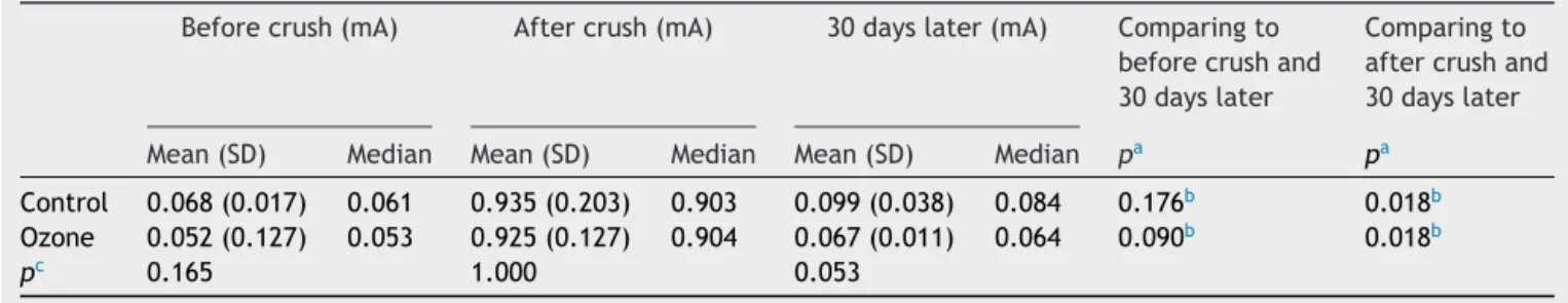
Table1 Comparisonoffacialnervestimulationthresholdsbeforeandaftercrushingand30dayslater.
Beforecrush(mA) Aftercrush(mA) 30dayslater(mA) Comparingto beforecrushand 30dayslater
Comparingto aftercrushand 30dayslater
Mean(SD) Median Mean(SD) Median Mean(SD) Median pa pa
Control 0.068(0.017) 0.061 0.935(0.203) 0.903 0.099(0.038) 0.084 0.176b 0.018b
Ozone 0.052(0.127) 0.053 0.925(0.127) 0.904 0.067(0.011) 0.064 0.090b 0.018b
pc 0.165 1.000 0.053
a Wilcoxonsigned-ranktest. b p<0.05.
c Mann---WhitneyUtest.
Results
Therewasnostatisticallysignificantdifferencein
stimula-tionthresholdsbetweentheozoneandsalinegroups after
crushing(p=1.000),indicatingthattheseverityofthenerve
crush injury was similar in both groups. Although
stimu-lation thresholds were significantly lower from pre-crush
thresholds in both the ozone and saline groups (p=0.018
and0.018) after30 daysof treatment, theozone-treated
group had lower stimulation thresholds than the saline
groupwhencomparedtopost-crushinglevels.Althoughthis
did not reachthe level of statistical significance, it
indi-cates greater functional improvementin the ozone group
(p=0.053).Improvementwasalsoseeninthesalinegroup
butthis wasthoughttobearesultof spontaneous
recov-eryofthefacialnerve.After30daysoftreatment,neither
groupreachedpre-crushingamplitudelevels(Table1).
Significant differences were found in vascular
conges-tion, macrovacuolization, and myelin thickness between
theozoneandcontrolgroupsbylightmicroscopy(Table2;
Fig.2A---D). Severe degenerationtodifferent degreeswas
observed in almost all of the myelinated axons in the
control group by electron microscopy. In slightly
dam-agedmyelinatedaxons,whilethemyelinsheathshadmild
delamination,thecytoplasmic structureofthemyelinated
axonswasnormal.Inseverelydamaged myelinatedaxons,
severe delamination and disintegration were observed in
myelinsheaths. Mitochondrialswellingandloss ofcristae,
and disorganization of microtubules and microfilaments
wereobservedin theaxonalcytoplasm,In addition,some
axons were darkened and contained myelin ovoid bodies
(Figs.3Aand4A,B).
Intheozonegroup,mostofthemyelinatedaxonswere
normal in structure underelectron microscopy.
Mitochon-drial ultrastructure and organization of microtubules and
microfilamentsinaxonalcytoplasmwerenormalin
myelin-ated axons. However, in a few of the myelinated axons,
separation of the myelin configuration and disintegration
ofmyelin sheathswasobserved.Furthermore,therewere
swollen mitochondria withdamaged cristae, disorganized
microtubules,andmicrofilamentsandmyelinovoidbodiesin
themyelinatedaxoncytoplasm(Table3;Figs.3Band4C,D).
Discussion
Manydrugs areused in thetreatment of traumatic facial
paralysis. The most commonly used is corticosterone,
Table 2 Comparison of histopathology variations in the facialnervebetweenozoneandsalinegroupsafter30days vialightmicroscope.
Control(n=7) Ozone(n=7)
n(%) n(%) p Vascularcongestion 0.017a
None 0(0) 3(42.8) Mild 2(28.6) 3(42.8) Moderate 2(28.6) 1(14.3) Severe 3(42.8) 0(0)
Median 2 1
Macrovacuolization 0.002b
None 0(0) 2(28.6) Mild 0(0) 4(57.1) Moderate 5(71.4) 1(14.3) Severe 2(28.6) 0(0)
Median 2 1
Myelinthickness 0.053
Verythin 6(85.7) 2(28.6) Thin 1(14.3) 2(28.6) Normal 0(0) 3(42.8)
Median 2 1
Mann---WhitneyUtest.
a p<0.05. b p<0.01.
whichdecreasescapillarypermeabilityandreducesedema
around the facial nerve. Corticosterone is also thought
to decrease degeneration of the axon while increasing
regeneration.2,12---14TheeffectofvitaminEonnerve
regen-erationaftertraumahasalsobeeninvestigated.15 Vitamin
Eisapowerfulfat-solubleantioxidant,whichcan prevent
formationoffreeradicals,protectingcellsagainstoxidative
stressand lipidperoxidation.Taskale etal.15 investigated
theeffectsofvitaminEandvitaminEpluscorticosteroneon
facialnervehealinginrats.TheyfoundthatvitaminEhad
apositiveeffectonnervehealing;thiseffectwasenhanced
bytheadditionofcorticosterone.Liebermanetal.16
inves-tigatedtheeffectsofcorticosteroidsonfunctionalrecovery
andneuronsurvivalafterfacialnerveinjuryinmice.They
foundthatcorticosteroidtreatmentslowsfunctional
recov-eryanddisturbsneuronsurvivalfollowingfacialnervecrush
injuryinadultmice.Theyalsoclaimedthatthedegreeof
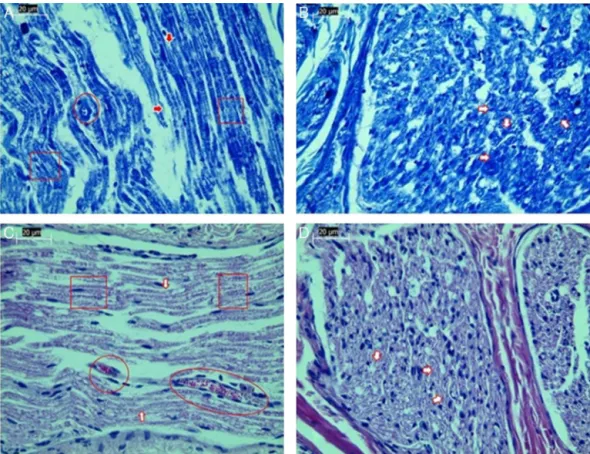
Figure2 Examinationofaxonalstructurepost-injurybylightmicroscopy.(A)Toluidinebluestainingofcontrolgroup.(B)Toluidine bluestainingofozonegroup.(C)H&Estainingofcontrolgroup.(D)H&Estainingofozonegroup.Magnificationwas40×;arrows indicatemyelin sheath,circles indicateareas ofvascularcongestion,andsquaresindicate macrovacuolization. Representative imagesareshown.
Figure3 Transmissionelectronmicrographsofmyelinatedaxonspost-injury.(A)Inthecontrolgroup,delaminationand defor-mationwere seeninmyelinsheathsofmostofthe axons(boldarrows), inadditiontotheappearanceofmyelinovoidbodies (thinarrows)anddarkenedaxonalcytoplasm(arrowheads).Veryfewnormalmyelinatedaxons(asterisks)wereseen.(B)Inthe experimentalgroup,numerousnormallymyelinatedaxonsareseen(asterisks).Delaminationanddeformationinmyelinsheaths (boldarrows)andmyelinovoidbodies(thinarrow)areshowninonlyafewmyelinatedaxons.Magnificationforbothimages:4000×, representativeimagesareshown.
juvenilemice,crushinjuryresultsinoverallpoorfunctional
recoveryandprofoundcelllossinthefacialmotornucleus.
In another study, Toros et al.17 evaluated the effects
of Hyperbaric Oxygen (HBO), methylprednisolone and
combined HBO---methylprednisolone treatments on
trau-maticfacialnerveregenerationin rats.HBOis thoughtto
decreaseinjury-relatededema,decreasetheconcentration
ofoxygenfreeradicalsafterischemiawithreperfusionand
increase localtissueoxygenlevels.18 They concludedthat
combination therapy with methylprednisolone and HBO
might be beneficial for treating the ischemia and edema
thatbothresultfromthefacialnerveinjurycascade.
Spontaneous nerve regeneration with good functional
Figure4 Transmissionelectronmicrographsofcytoplasmofmyelinatedaxons.(A)Inthecontrolgroup,swollenmitochondria withdegeneratedcristaestructure(thinarrows)wereseeninthecytoplasm.Microtubulesandmicrofilamentsweredispersed het-erogeneously.Insomeareas,adecreasedamountofmicrotubulesandmicrofilamentswasobserved(arrowheads).(B)Inthecontrol group,swollenanddamagedmitochondriawereseen(thinarrows),andmicrotubuleandmicrofilamentarraysweredisorganizedand disrupted(arrowheads).(C)Intheexperimentalgroup,theultrastructureofmitochondriawasnormal(boldarrows).Microtubules andmicrofilamentsweredispersedhomogenouslythroughoutthecytoplasm(asterisks).(D)Intheexperimentalgroup, longitudi-nallyalignedmicrotubulesandmicrofilamentswerenormal(arrowheads),andmitochondriaexhibitednormalultrastructure(bold arrows).Magnificationforallimages:3000×,AandCarecrosssectionalviews,BandDarelongitudinalsections,andrepresentative imagesareshown.
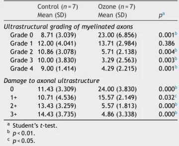
Table 3 Comparison of histopathology variations in the facialnervebetweenozoneandsalinegroupsafter30days viaelectronmicroscope.
Control(n=7) Ozone(n=7)
Mean(SD) Mean(SD) pa
Ultrastructuralgradingofmyelinatedaxons
Grade0 8.71(3.039) 23.00(6.856) 0.001b
Grade1 12.00(4.041) 13.71(2.984) 0.386 Grade2 10.86(3.078) 5.71(2.138) 0.004b
Grade3 10.00(3.830) 3.29(2.563) 0.003b
Grade4 9.00(1.414) 4.29(2.215) 0.001b
Damagetoaxonalultrastructure
0 11.43(3.309) 24.00(3.830) 0.000b
1+ 10.71(4.536) 15.57(2.149) 0.032c
2+ 13.43(3.259) 5.57(1.813) 0.000b
3+ 14.43(3.735) 4.86(3.338) 0.000b a Student’st-test.
b p<0.01. c p<0.05.
nerve.19 This kind ofnerveinjury istreated with
pharma-cological agents, instead of surgery. The healing process
aftercrushinjuryislargelyimpairedduetoincreased
pro-duction of free radicals, rather than neuroinflammation
andedema.20 Antioxidantmaterialsscavengefreeradicals
andcontributetonerveregeneration.Antioxidantenzymes,
such as superoxide dismutase and catalase, protect cells
fromthetoxiceffectsoffreeradicals.Freeradicalsinduce
traumaticcelldamage causing celldeath. Crush injury to
nervesleadstooxidative stress,suchaslipidperoxidation
ofneurovascularcells,bycreatingfreeradicals.21,22
Anumberof antioxidantsthathave beenexamined for
theirabilitytoimproveregenerationofperipheralnerves.
Jangetal.23investigatedtheeffectofginkgobilobaextract
onrecoveryafterfacialnervecrushinjuryintherat.They
found that the intraperitoneal injection of ginkgo biloba
extractwaseffectiveinpromotingregenerationofthenerve
inan experimental facialnervecrush rat model.Another
antioxidantthathasbeenstudiedforuseinsupportingthe
regenerationofcrushedfacialnerveiscoenzymeQ.24
Coen-zymeQ was also found to be effective in promoting the
regenerationof thenerveinan experimental facialnerve
crushratmodel.
Ozone may promote a moderate oxidative stress that,
inturn,increasesendogenousantioxidantsystems.25,26The
protective mechanism mediated by ozone may involve
protein synthesis. Increased reactive oxygen species can
induce antioxidant gene expression in many cells. A
major mechanism of redox homeostasis is reactive
increase expression of antioxidants.27 Therefore, since
ozoneincreasesantioxidantlevels,leadingtoadecreasein
freeradicals,we investigatedtheeffectofozonetherapy
ontheregenerationofcrushedfacialnerves.
In the current study, regeneration of the facial nerve
wasevaluatedbyassessingelectrophysiological thresholds
andbyhistopathologicalexamination.Thereareanumber
ofnerveintegritymonitoring devicesavailable toidentify
andprecludepersistentnervedamage.28,29Inthisstudy,we
usedthe Nerve IntegrityMonitor facial electromyography
technique described by Delgado et al.29 and used in
sev-eralpreviousstudies15,24torecordthecontractionoffacial
muscles. In the present study, although not reaching
sta-tisticalsignificance,therewasimprovementinfacialnerve
functionintheozonegroupwhenassessedusingthreshold
levels.Thereareseveralstudiesintheliteraturereporting
histopathological evaluation of the degree of
macrovac-uolization,vascularcongestion,andmyelinsheaththickness
toassessnervedamage.17,24 Inthepresent study,we used
similar parameters to assess nerve structure; significant
differenceswere found in vascular congestion,
macrovac-uolization, and myelin thickness between the ozone and
controlgroups.
The strengthof ourstudy wasin theevaluation ofthe
degreeoffacialnerveregenerationnotonlybylight
micro-scope, but also by electron microscopy. A large number
ofmyelinatedaxonsfromeachsample (n=50)andatotal
of350myelinated axonsfromeachtreatment groupwere
evaluatedby electron microscopy. In this study, electron
microscopyconfirmedthefindingsoflightmicroscopy.A
lim-itationofourstudywastheshortdurationoffollow-upafter
thetreatments. We explored facialnerves 1 month
post-treatment. This wouldneed tohave been muchlongerin
ordertohaveobservedfunctionalrecovery.Thismaybeone
ofthereasonsourfindingsdidnotreachstatistical
signifi-cance.Therefore, further studies areneeded withlonger
follow-up after treatment. We hope that this preliminary
studywillencourageotherlargerstudiestobeundertaken.
Conclusion
Inconclusion,ourstudyisthefirstofwhichweareaware
toinvestigatethe effects ofozone therapy onthe
regen-eration of crushed facial nerves. Our data suggests that
ozonetherapymayhavebeneficialeffectsonthe
regenera-tionofcrushedfacialnerves.Itspositiveeffectswereseen
especiallyonthepathologicevaluation.Electronmicroscopy
confirmed the results of light microscopy. Therefore, we
conclude that ozone therapy may be a promising avenue
toexplorefor thetreatment of acutefacialparalysisand
peripheralnerveregeneration. Furtherstudiesareneeded
toconfirmourfindings.
Conflicts
of
interest
Theauthorsdeclarenoconflictsofinterest.
References
1.WilsonAD,HartA,BrännströmT,WibergM,TerenghiG.Delayed acetyl-l-carnitine administration and its effect on sensory
neuronalrescueafterperipheralnerveinjury.JPlastReconstr AesthetSurg.2007;60:114---8.
2.Al-Bishri A, Dahlin L, Sunzel B, Rosenquist J. Systemic betamethasone accelerates functional recovery after a crush injury to rat sciatic nerve. J Oral Maxillofac Surg. 2005;63:973---7.
3.SubbannaPK,PrasannaCG,GunaleBK,TyagiMG.Acetylsalicylic acidaugmentsfunctionalrecoveryfollowingsciaticnervecrush inmice.JBrachialPlexPeripherNerveInj.2007;2:3.
4.Lenaz G, Genova ML. Mobility and function of coenzyme Q (ubiquinone)in themitochondrial respiratorychain.Biochim BiophysActa.2009;1787:563---73.
5.ThomasDA,RenK,BesseD,RudaMA,DubnerR.Applicationof nitricoxidesynthaseinhibitor,Nomega-nitro-l-argininemethyl
ester,oninjurednerveattenuatesneuropathy-inducedthermal hyperalgesiainrats.NeurosciLett.1996;210:124---6.
6.PapucciL,SchiavoneN,WitortE,DonniniM,LapucciA, Tem-pestiniA,etal.CoenzymeQ10preventsapoptosisbyinhibiting mitochondrialdepolarizationindependentlyofitsfreeradical scavengingproperty.JBiolChem.2003;278:28220---8.
7.Lin Q, Chen H, Lu C, Wang B, Zhang Y, He X, et al. Effects ofozoneon sciaticnerve in rat.Interv Neuroradiol. 2011;17:281---5.
8.BocciV.Ozone-Anewmedicaldrug.Dordrecht:Springer;2005.
9.ErginelB,ErgineelT,AksoyB,DokucuA˙I.EffectofOzone Ther-apy (OT)on healing of colonic anastomosisin rat model of peritonitis.BalkanMedJ.2014;31:249---53.
10.KaptanogluE,PalaogluS,SurucuHS,HayranM,BeskonakliE. Ultrastructuralscoringofgradedacutespinalcordinjuryinthe rat.JNeurosurg.2002;97:49---56.
11.Erdine S, Bilir A, Cosman ER, Cosman ER Jr. Ultrastructural changesinaxonsfollowingexposuretopulsedradiofrequency fields.PainPract.2009;9:407---17.
12.LambertsSW,BruiningHA,deJongFH.Corticosteroidtherapy insevereillness.NEnglJMed.1997;337:1285---92.
13.MelcangiRC,CavarrettaIT,BallabioM,BallabioM,LeonelliE, SchenoneA,etal.Peripheralnerves:atargetfortheactionof neuroactivesteroids.BrainResBrainResRev.2005;48:328---38.
14.NasserRM,ChenLE,SeaberAV,UrbaniakJR.Protectiveeffect of21-aminosteroidpretreatmentinperipheralnervelow-load crush injury in mature and immature rats. J Orthop Res. 1996;14:823---9.
15.Tas¸kaleP,Topalo˘gluI.ThehealingeffectsofvitaminEwith cor-ticosteroidandvitaminEonnervehealinginratswithtraumatic facialpalsy.KulakBurunBo˘gazIhtisDerg.2010;20:255---9.
16.LiebermanDM,JanTA,AhmadSO,MostSP.Theeffectsof corti-costeroidonfunctionalrecoveryandneuronsurvivalafterfacial nerveinjuryinmice.ArchFacialPlastSurg.2011;13:117---24.
17.TorosSZ,KaracaC¸T,Günes¸P,OysuC¸,ErtugayC¸K,Naibo˘gluB, etal.Hyperbaricoxygenversussteroidinfacialnerveinjury:an experimentalanimalstudy.AmJOtolaryngol.2013;34:530---6.
18.Santos PM, Zamboni WA, Williams SL, Covey JF, Kienstra MA. Hyperbaric oxygen treatment after rat peroneal nerve transection and entubulation. Otolaryngol Head Neck Surg. 1996;114:424---34.
19.ThomasDA,RenK,BesseD,RudaMA,DubnerR.Applicationof nitricoxidesynthaseinhibitor,Nomeganitro-l-argininemethyl
ester,oninjurednerveattenuatesneuropathy-inducedthermal hyperalgesiainrats.NeurosciLett.1996;210:124---6.
20.BagdatogluC,SarayA,SurucuHS,OzturkH,TamerL.Effect oftrapidilinischemia/reperfusioninjuryofperipheralnerves. Neurosurgery.2002;51:212---20.
21.Lundborg G, Myers R, Powell H. Nerve compression injury and increased endoneurial fluid pressure: a ‘‘miniature compartment syndrome’’. J Neurol Neurosurg Psychiatry. 1983;46:1119---24.
neuronalrescueafterperipheralnerveinjury.JPlastReconstr AesthetSurg.2007;60:114---8.
23.JangCH,ChoYB,ChoiCH.Effectofginkgobilobaextracton recoveryafterfacialnervecrushinjuryintherats.IntJPediatr Otorhinolaryngol.2012;76:1823---6.
24.Yildirim G, Kumral TL, Berkiten G, Saltürk Z, Sünnetc¸i G, Öztürkc¸üY,etal.TheeffectofcoenzymeQ10onthe regenera-tionofcrushedfacialnerve.JCraniofacSurg.2015;26:277---80.
25.LeónOS,MenéndezS,MerinoN,CastilloR,SamS,PerezL,etal. Ozoneoxidativepreconditioning:aprotectionagainstcellular damagebyfreeradicals.MediatInflamm.1998;7:289---94.
26.Candelario-Jalil E, Mohammed-Al-Dalain S, Fernandez OS, Menendez S, Perez-Davison G, Merino N, et al. Oxida-tive preconditioning affords protection against carbon
tetrachloride-inducedglycogendepletionandoxidativestress inrats.JApplToxicol.2001;21:291---301.
27.DrögeW.Freeradicalsinthephysiologicalcontrolofcell func-tion.PhysiolRev.2002;82:47---95.
28.KoekkoekSK,DenOudenWL,PerryG,HighsteinSM,DeZeeuw CI.Monitoringkineticandfrequency-domainpropertiesof eye-lid responses in mice with magnetic distance measurement technique.JNeurophysiol.2002;88:2124---33.