FOURTH VENTRICLE COMPUTED
TOMOGRAPHY INDEXES
Standardisation and characteristics in neurocysticercosis
Svetlana Agapejev
1ABSTRACT - Objectives: to propose standardisation of fourth ventricle dimensions and to study its characteristics in neurocysticercosis. Method: a control group (CG) constituted by 114 individuals with normal CT, and 80 patients with neurocysticercosis composed the group with neurocysticercosis (GN). Measures of the inner cranial diameter (Cr), fronto-polar distance between both lateral ventricles (FP), antero-posterior (AP) and latero-lateral (LL) fourth ventricle width based the standardisation of six indexes. Results: AP/Cr, AP/LL and AP/ FP were the more discriminative indexes, presenting in CG the mean values of 0.063, 0.267 and 0.582, respectively. The indexes in GN had values statistically higher than in CG. From GN, 51patients had increased indexes values above 2 standard deviation of the CG mean. AP/Ll was > 1 in 95% of patients with ventricular shunting and in 88% with depression. It also occurred in 73% patients with satisfactory follow-up and in everybody who died. Conclusion: AP/Cr, AP/LL and AP/FP may represent fourth ventricle dimensions. KEY WORDS: fourth ventricle, indexes, computed tomography, standardisation, neurocysticercosis.
Índices do IV ventrículo em tomografia computadorizada de crânio: padronização e características na neurocisticercose
RESUMO - Objetivos: propor padrão de normalidade das dimensões do IV ventrículo e estudar suas características em neurocisticercose. Método: em um grupo controle (GC) constituído de 114 indivíduos com tomografias normais e em outro grupo composto de 80 doentes com neurocisticercose (GN), mediram-se a distância fronto-polar de ventrículos laterais (FP) e os diâmetros craniano interno (Cr), ântero-posterior (AP) e látero-lateral (LL) do IVo ventrículo para a padronização de seis índices. Resultados: AP/Cr, AP/LL e AP/FP foram os índices mais discriminatórios e apresentaram, em GC, valores médios de 0,063, 0,267 e 0,582, respectivamente. Em GN os valores foram estatisticamente superiores a GC. Selecionaram-se 51 doentes do GN com índices > 2 desvios-padrão da média em GC. Neles, AP/Ll foi > 1 em 95% doentes com derivação liquórica e em 88% com depressão, ocorrendo em 73% com evolução satisfatória e todos que faleceram.
Conclusão: AP/Cr, AP/LL e, principalmente, AP/FP são representativos das dimensões do IV ventrículo. PALAVRAS-CHAVE: IVventrículo, índices, tomografia computadorizada, padronização, neurocisticercose.
Departamento de Neurologia e Psiquiatria (DPN) da Faculdade de Medicina de Botucatu da Universidade Estadual Paulista (UNESP), Botucatu SP, Brasil: 1Professora Adjunta do DNP da FMB-UNESP, Botucatu.
Received 27 June 2001, received in final form 24 October 2001. Accepted 7 November 2001.
Dra. Svetlana Agapejev - Departamento de Neurologia e Psiquiatria, Faculdade de Medicina, UNESP - 18618-000 Botucatu SP - Brasil. E-mail: [email protected]
The isolated fourth ventricle (IVthv), also referred in the literature as occluded, trapped, sequestered, encysted, disproportionately large, double compart-ment hydrocephalus, or communicant IVthv, is a clini-cal-pathological entity firstly described by Foltz & Shurtleft1, in 1966. Although pathogenesis remain in the field of controversies, this picture could hap-pen as before as after the installation of ventricular shunting1,2. Clinical manifestations are variable, hap-pening from an acute posterior fossa syndrome, sometimes associated with abrupt alterations of the humour3, until a progressive dilation of IVthv as a
Cysticercosis is the infection with the metacestode larval form of the parasite Taenia solium, characteri-sed by an accentuated tropism for the central ner-vous system provoking local reactions and/or far from its installation site. The beginning or not of symp-toms depends on host-parasite interaction, expressed through an immune response, mediated by immu-noglobulins and cytokines, and as a result of mul-tiple associated mechanisms, generating a clinical and laboratory polymorphism. The more frequent clinical manifestations are represented by epilepsy and intracranial hypertension (ICH), associated or not with psychic disturbances and meningoencephali-tis. This last has a fundamental role on the forma-tion of hydrocephalus the most important sequel due to the fibrosis in the basal cistern. The epilepsy is characterised by more than one type of crisis and a difficult control of the seizures12,13, and could be the first manifestation of hypertensive hydrocepha-lus, diffuse cerebral oedema, or the effect mass of giant cysts. ICH is a frequent and immediate conse-quence of the inflammatory reaction, as in the sub-arachnoid space14,15, as in the parenchyma16,17, or as a direct response to the physical presence of para-sites. Psychic disturbances in neurocysticercosis, could float from mild symptoms like apathy, cogni-tive disturbances, alterations in the humour and me-mory, to more severe psychiatric pictures as demen-tia, depression and schizophrenia18-22. Neurological manifestations are generally associated12,19,21, and could be one of the signs as of ICH12,19,23, as of hyper-tensive or normal pressure hydrocephalus19,24,25.
The objective of this paper is to propose a stan-dardisation for representative indexes of the fourth ventricle dimensions, using traditional CT exams, and to study its characteristics in neurocysticercosis.
METHOD
This project of research evaluated by the Commission of Ethics in Research of the School of Medicine of Botucatu-UNESP received favourable concept (Of. 086/98 - CEP).
Standardisation of fourth ventricle CT indexes (Control Group = CG)
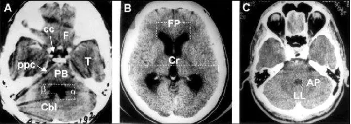
Inclusion criteria: a hazard selection of individuals with normal CT of skull which didn’t present in diagnosis nor follow-up evaluation exam any structural lesion (surgical or not) was done. The measures of the lateral ventricles maximum fronto-polar distance (FP), and the largest in-ner cranial diameter (Cr) in the same slice of the measu-red FP were in all exams performed (Fig 1B). The largest IVthv antero-posterior (AP) width, as well as the latero-lateral (LL) in the same slice of the measured AP (Fig 1C), in all selected exams were measured. When the IVthv pre-sented characteristics similar as shown in Figure 1A, the considered value of AP resulted from the arithmetic aver-age of the obtained measures of a and b. If the patient had more than one CT, the final value of the indexes based on the arithmetic average of all values obtained in all CT of the same patient. Based on the statistical analyst sug-gestion, only one person using common plastic transpar-ent rule and glass magnifying performed all the measures. To put into practice the measures proposed above, for AP and LL selected were those slices where the IVthv ap-peared larger and showed (in the same level) the
intracranial lesions after a trial with ABZ + DCP, were employed.
The presence of at least one of the three IVthv repre-sentative indexes with increased values defined the IVthv involvement. In that way, it was possible to select 51 pa-tients from GN that constituted the group of neurocysti-cercosis with involvement of IVthv (GN-IVthv). Age, sex, clini-cal manifestations, depression and association with rep-resentative indexes of IVthv were the parameters studied in the three forms of clinical evolution: good (GEv), with sequel (SEv) and death (DEv).
Exclusion criteria: patients that filled the same exclu-sion criteria previously mentioned for CG, and had not the above mentioned diagnostic criteria for neurocysticer-cosis were excluded from this study. In other words, pre-senting nothing but associated subcutaneous cysticerco-sis, just intracranial calcifications on CT, impossibility to perform CSF exams or immunologic tests, did not partici-pate in this study.
Standardisation of terms: Index increase = index with obtained value higher than two standard deviation of the statistical average found in the studied controls. Depres-sion = presence almost daily, during more than two weeks, of four or more of the following manifestations: discour-agement or depression, apathy, decrease or increase of appetite, voluntary isolation, floating and silent cry, increa-se or loss of sleep. Parinaud’s syndrome = paralysis or paresis of the upper gaze with alteration in the ocular convergence but conservation of the eyes of doll phenom-enon. Good evolution = patients that came back to the normality of their habitual and personal activities, with-out need of any type of help, using or not symptomatic medication. Evolution with sequel = patients that stayed with partial or total dependence for the execution of their habitual and personal activities, using or not symptom-atic medication.
RESULTS
Tables 1-3 and Figure 2 present the results. In CG, the rate of classification error was 17.54%. The MI2 subgroup showed statistical significant cor-relation (p < 0.05) with AP/Cr, LL/Cr, AP/FP and LL/FP indexes. In MI3, this correlation occurred with FP/Cr (p < 0.05), LL/FP (p < 0.01) and, in FI1, just with LL/ FP (p < 0.01). The indexes didn’t showed significant correlation (p > 0.05) with the MI1, FI2 and FI3 sub-groups. Statistical analysis of the obtained data con-sidered the indexes AP/Cr, AP/FP and AP/LL the most discriminatory.
The studied indexes did not present significant statistical difference between CG and GN, and in the subgroups (F= 0.74; p > 0.10). The before and af-ter periods related to the ABZ + DCP therapy also
tures presented in Figure 1A.It means that frontal lobes, temporal lobes, pre-pons and/or chiasma cisterns, transi-tion pons-bulb and cerebellum close to middle cerebellar peduncle should be saw in the same slice the IVthv was larger. For FP and Cr measures (Fig 1B) selected were those slices where the third ventricle appeared larger and showed (in the same level) the frontal horns of lateral ventricles more distant between itself.
In this way, 114 patients of both sexes (M= masculine and F= feminine), with age varying from 1 to 83 years (I1 = younger than 19 years, I2 = 20 to 39 years, I3 = older than 40 years) were selected. These control patients were gathered in six sex/age subgroups (MI1, MI2, MI3, FI1, FI2, FI3). The analysed CT exams are part of the Service of Com-puted Tomography of the Hospital of the Clinics of the School of Medicine - UNESP files. The exams had performed with CT 9800 Q model General Electric equipment.
Exclusion criteria: patients with absence of CT con-trasted phase and presenting problems in the clearness for the identification of IVthv limits were excluded. CT scans showing asymmetrical head position and presence of any detectable lesion even if all the other CT of the same pa-tient has been normal had also excluded from this study. The uncertainty in the diagnosis of normality expressed by just descriptive report without any conclusion, and di-vergence between the Radiology and Neurology opinion were also considered as exclusion criteria.
To evaluate the IVthv involvement FP/Cr, AP/Cr, LL/Cr, AP/FP, LL/FP, and AP/LL indexes constituted were compared.
Statistical analysis: based on analysis of variance to verify sex (M and F) and age group (I1, I2 and I3) effects. Descriptive statistics to obtain arithmetic average (x), stan-dard deviation (sd), coefficient of variation (CV), median (Md), minimum and maximum values of each studied vari-able, and coefficients of lineal correlation for evaluation between couple of variables were utilised. Application of F statistic to compare groups for the group of variables was executed26.
Group with neurocysticercosis (GN)
Table 3. Fourth ventricle indexes: comparison between control group (CG) and the group with neurocysticercosis (GN) subgroups, before (NB) and after (NA) albendazole plus dextrochlorpheniramine therapy.
Subgroups MI1 MI2 MI3 FI1 FI2 FI3
Indexes
FP / Cr CG = NB CG < NB1 CG = NB CG = NB CG = NB CG < NB1 CG = NA CG < NA3 CG = NA CG = NA CG = NA CG = NA AP / Cr CG < NB1 CG = NB CG < NB2 CG = NB CG = NB2 CG < NB2 CG = NA CG = NA CG < NA1 CG = NA CG < NA2 CG < NA3
LL / Cr CG = NB CG > NB1 CG = NB CG = NB CG = NB CG = NB
CG = NA CG = NA CG = NA CG = NA CG = NA CG = NA
AP / FP CG < NB1 CG = NB CG = NB CG = NB CG = NB1 CG < NB3 CG = NA CG = NA CG = NA CG = NA CG < NA1 CG = NA
LL / FP CG = NB CG > NB1 CG = NB CG = NB CG = NB CG = NB
CG = NA CG = NA CG= NA CG = NA CG = NA CG = NA
AP / LL CG<NB4 CG<NB4 CG < NB1 CG = NB CG < NB1 CG < NB4 CG<NA1 CG<NA1 CG < NA1 CG = NA CG < NA1 CG < NA3 1 p < 0.05; 2 p = 0.05; 3 0.05 < p < 0.10; 4 p < 0.001
Table 1. Fourth ventricle indexes: significant statistical correlation in subgroups of the control group.
Subgroups MI1 MI2 MI3 FI1 FI2 FI3
Indexes (n= 25) (n= 20) (n= 17) (n= 16) (n= 19) (n= 17)
FP/Cr and AP/Cr - - -
-FP/Cr and LL/Cr - - -
-FP/Cr and AP/FP - 0.57 ** - 0.46 * - 0.82 ** - -
-FP/Cr and LL/FP - 0.59 ** - 0.57 ** - 0.84 ** - - - 0.59 *
FP/Cr and AP/LL - - -
-AP/Cr andLL/Cr 0.77 ** 0.83 ** - - 0.62 ** 0.84 **
AP/Cr and AP/FP 0.80 ** 0.93 ** 0.81 ** 0.93 ** 0.93 ** 0.99 **
AP/Cr and LL/FP 0.60 ** 0.74 ** - 0.61 ** 0.62 ** 0.76 **
AP/Cr and AP/LL 0.64 ** 0.57 ** 0.80 ** 0.74 ** 0.76 ** 0.87 **
LL/Cr and AP/FP 0.53 ** 0.87 ** - - 0.52 * 0.87 **
LL/Cr and LL/FP 0.72 ** 0.94 ** 0.56 * 0.83 ** 0.85 ** 0.96 **
LL/Cr and AP/LL - - -
-AP/FP andLL/FP 0.82 ** 0.89 ** - 0.74 ** 0.70 ** 0.84 **
AP/FP andAP/LL 0.63 ** 0.42 * - 0.67 ** 0.69 ** 0.74 ** 0.82 **
LL/FP and AP/LL - - -
-n= number of patients. * p < 0.05; ** p < 0.01.
Table 2. Fourth ventricle indexes: statistical analysis among the obtained values in the control group (CG) and the group with neurocysticercosis (GN).
Indexes CG GN Statistic Significance Comments
x ± sd x ± sd level
FP / Cr 0.244 ± 0.034 0.257 ± 0.041 5.98 p < 0.05 CG < GN AP / Cr 0.063 ± 0.020 0.082 ± 0.037 19.17 p < 0.001 CG < GN LL / Cr 0.109 ± 0.026 0.102 ± 0.034 2.37 p > 0.10 CG = GN AP / FP 0.267 ± 0.100 0.323 ± 0.143 10.19 p < 0.010 CG < GN LL / FP 0.458 ± 0.136 0.409 ± 0.139 5.95 p < 0.025 CG > GN AP / LL 0.582 ± 0.139 0.797 ± 0.213 72.29 p < 0.001 CG < GN
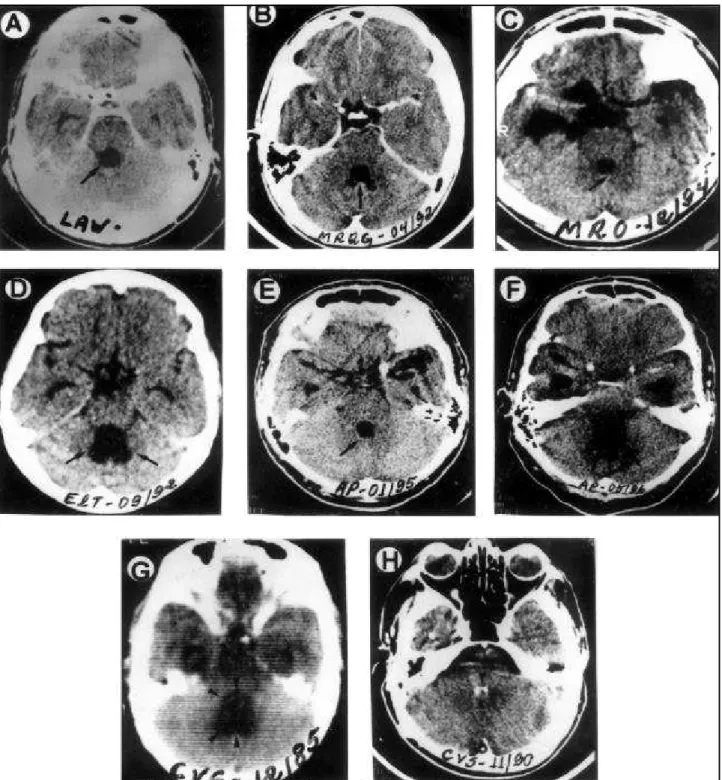
Fig 2. A and B. CT of patients with good evolution showing round aspect IVthv (A) or like “coconut hat” (B). These characteristics
presented normalisation in 27 (80%) patients. Three patients had CT suggestive of IVthv cyst in two of them and, in the lateral ventricle,
in one. C and D. CT of patients with sequel showing round aspect IVthv (C) or like “indigenous canvas” (D). Patients with the
character-istic of C presented normalisation of the aspect associated to improvement of the sequels, in all the patients. Patients with the charac-teristics of D, stayed unaffected, being observed correlation clinic/CT in those with worsening or maintenance of sequels. Two patients had CT suggestive of IVthv cyst. E, F, G and H. Five patients with fatal evolution showed round IVthv on CT (E) with maintenance or
progressive antero-posterior diameter increase (F) of this characteristic. In the other four patients, CT showed accentuated increase of IVthv before the cysticidal therapy (G), followed by regression (H) of its size after albendazole treatment plus ventricular shunting.
Patients with the characteristics of H had another IVthv increase 2 months to 2 years after therapeutic schedule. All the nine patients
did not present difference (FP/Cr: p > 0.50, AP/Cr: p > 0.50, LL/Cr: p > 0.40, AP/FP: p > 0.50, LL/FP: p > 0.50, AP/LL: p > 0.30). Except for the indexes LL/Cr and LL/FP, the others (AP/Cr, AP/FP and AP/LL) showed more higher values in GN that in GC (F = 13.26; p < 0.001).
In the GN-IVthv, the patient age varied from 12 to 72 years, with prevalence of men (57%) and age group of 21-40 years (49%). ICH (55%), meningoen-cephalitis (49%), hydrocephalus (45%), epilepsy (41%), vascular disorders (12%), chronic cerebral oe-dema (8%), mental disorders (8%) and spinal cord involvement (8%), were the isolated (10%) or asso-ciated (90%) presentation of clinical manifestations. Had good evolution 34 (67%) patients, 8 (15%) de-veloped or stayed with sequels, and 9 (18%) died one to 19 months after the end of the therapeutics with ABZ. Epilepsy (56%), ICH (38%) and meningoen-cephalitis (35%), in this order, were the more fre-quent clinical pictures presented by patients with GEv. ICH (75%), hydrocephalus (62%) and menin-goencephalitis (50%) were more common in patients with SEv. ICH associated to hydrocephalus happened in everybody who died.
Depression was verified in 30 (59%) patients, 17 (57%) men and 13 (43%) women. It was associated to Parinaud’s syndrome in 18 (60%), committing 13 (43%) patients with GEv and everyone with SEv or DEv. In 14 (27%) patients, all presenting GEv, de-pression and Parinaud’s syndrome did not occur. The depressive picture presented own characteristics de-pending on the type of evolution. Patients with GEv showed floating manifestations, revolt feeling for the medication need, spontaneous and easy exter-nalisation of the sentiments, besides a good response to the common anti-depressive drugs. Patients with SEv presented more sporadic although more linger-ing periods of depression characterised by negative reaction to the presence of sequel and being veri-fied an association between improvement of the sequel and regression of the depression. Connota-tion of bitterness, sensaConnota-tion of irreversibility and a nostalgia feeling was characteristics of the depres-sion of patients who died, besides any therapeutic response to anti-depressive drugs. This kind of de-pression appeared 2-8 weeks before death, when the manifestations become sudden and progressively more intense and was followed by the installation of severe infectious processes.
Patients with GEv showed a discreet and tempo-rary Parinaud’s syndrome, although in four of them it acquired characteristic of periodicity and consisted of paresis of the upper vertical gaze. The same
hap-pened in those with SEv. Parinaud’s syndrome veri-fied in those who died happened as paralysis of the upper vertical gaze, associated to ophtalmoplegia during the episodes of ICH exacerbation.
Twenty-one (41%) patients had ventricular shunt-ing. Depression and/or Parinaud’s syndrome were present in four (19%) patients with GEv even before this surgical procedure. However, in 17 (81%) cases, depression and/or Parinaud’s syndrome appeared only after surgery, predominantly in those patients who died.
Two (6%) patients with GEv, one (12%) with SEv, and six (67%) with DEv had the typical CSF syndrome in neurocysticercosis. Normal CSF exam was verified in 10 (29%) patients with GEv, in one (12%) with SEv, and in none with DEv.
The exclusive increase of the AP/LL index was ob-served in 22 (65%) patients with GEv, in five (62%) with SEv and in seven (78%) with DEv. The increase of the FP/Cr index, suggestive of supratentorial hydro-cephalus, was associated to the increase of the dis-criminatory indexes in one (3%) patient with GEv, in another (12%) with SEv and in seven (78%) with DEv. Just one of the three indexes was increased in 40 (78%) patients, two in 20 (32%), and all the three in 16 (31%). This increase was exclusively due to AP/LL in 34 (67%) cases and associated with AP/Cr and/or AP/FP in 17 (33%). All the three discriminatory in-dexes were increased so much before as after cysti-cidal therapy in 23 (45%) patients: in 14 (41%) of them with GEv, in three (37%) with SEv and in six (67%) who died.
The index AP/LL>1 was found in all the patients with clinical picture of benign intracranial hyperten-sion or psychiatric alterations as the first and most important manifestation. It also occurred in 86% pa-tients with ICH and cerebrovascular disturbances, in 74% with hydrocephalus, in 71% with epilepsy, in 64% with meningoencephalitis, and in 50% with migraine-like headache or spinal cord involvement. AP/LL>1 was associated to the depression in 26 (87%) patients and it happened before ABZ + DCP treatment in 37 (72%), after this one in 36 (71%) and, so much before as after, in 13 (25%). From these last 13 cases, eight (23%) of them presented GEv and five (55%) died.
DISCUSSION
normal, those individuals which presented some neurological complaint and whose CT had normal medical reports. It is not possible to accomplish ra-diology exams in people not having mental or neu-rological symptoms without hurting ethical precepts when submitting healthy individuals without any clinical manifestation to an unnecessary exhibition of radiation. Consequently, the concept of normal-ity, adapted inside for the values of the interval of two standard deviation of the statistics average of the studied indexes, is a questionable term and it would be more appropriate to say “acceptable as not-altered”.
Due to the variability in the size of the films and in the head angularity of the accomplished exams impeding the uniformity of direct measures for the study of IVthv, it was necessary to establish indexes. The FP/Cr index just has reference in the literature, with the denomination of index of Evans8, 11, 28-30, presenting similar values to the found in the studied individuals.
The CSF spaces global analysis doesn’t suggest correlation among measures and age9, 31. On the other hand, the IVthv shows a continuous and pro-gressive increase with passing of the years11. Al-though the analysed indexes had not shown correla-tion with sex and age, the individual statistical analy-sis suggests that the AP/Cr, LL/Cr, AP/FP and LL/FP in-dexes increase (p < 0.05) during men lifetime. On the other hand, the LL/FP index decreases (p < 0.01) in men above the 39 years and in women until 19 years. The only index that doesn’t suffer influences of age and of sex it is AP/LL. Possibly volumetric differences in the neuroaxis explain these observations9, 11,28,29.
In other words, it is possible to say that the IVthv involvement but not its isolation manifest through the AP/FP, AP/Cr and AP/LL indexes - the discrimina-tory indexes. The presented data show that the AP/ LL index doesn’t depend on the presence of suprat-entorial hydrocephalus nor of the inner cranial di-ameter, and doesn’t suffer influence of age or sex. Consequently, the AP/LL index can be considered “the fourth ventricle sentinel”, besides being its more representative index.
The searched literature didn’t show any reference on the methods to identify an isolation of fourth ventricle except for the subjective evaluation, nor the manner to characterise its involvement. Even so, sev-eral neurological pathologies are related to IVthv en-largement including neurocysticercosis5, 32, 33.
The ventricular inflammatory process seems to
be reflected in the elevated values of the indexes AP/Cr (p < 0.001), AP/FP (p < 0.010) and AP/LL (p < 0.001) in patients with neurocysticercosis when com-pared to the control group. Even so, the LL/Cr and LL/FP indexes were, respectively, equal (p = 0.10) and smaller (p < 0.025).
Patients with neurocysticercosis presenting in-creased the three discriminatory indexes showed that the fourth ventricle became isolated definitively in those who died, lingering but passenger in those with sequels, and of smaller intensity and fleeting in those who had satisfactory evolution. Although it has no difference with between before and after ABZ + DCP therapy, the indexes values did not alter in patients with good evolution, decreased in those with sequels and increased in the cases who died. The AP/LL ³ 1 showed an association with more se-vere clinical pictures and difficult therapeutic con-trol, and it was present in most of the patients with depressive manifestations. Its value above the unit seems to suggest not only a suffering IVthv but also a premonitory sign of its temporary or definitive iso-lation. In neurocysticercosis it can be considered as an prognostic index when depression is present and AP/FP or AP/Cr have increased values.
The IVthv involvement in neurocysticercosis could take place so much for a possible neurotropism of cysticerci antigens as for local biochemical alterations related to the presence of these antigens. It seems that many factors take part of a context involving complex and multivariable mechanisms perhaps mo-dulated by genetic factors. Depression and immu-nodeficiency have serotonin34 as a common bioche-mical substratum, with serotonergic neurons located on brainstem. Perhaps it can exist, mainly in the se-rious forms of neurocysticercosis, some type of pref-erential lesion for the serotonergic system or for some modulating neurotransmitter system21. Depres-sion and immunodeficiency occurred in the patients with neurocysticercosis who didn’t have good evo-lution.
The presented data suggest be the depression associated with increased indexes a signal to worsen clinical evolution mainly if Parinaud’s syndrome is also present. The reversibility of manifestations ob-served in the patients with good evolution and in some of those with sequels suggest a metabolic dis-turbance due to oedema directly against the ven-tricular system and not to a destructive lesion35.
in-dexes return to normal values. However increased values of indexes, mainly the AP/LL, were more evi-dent in patients who died possibly, due to the fact that severe patients have accomplished more CT, contributing to a more easy detection of increases. The presented data allow to conclude that: 1) The AP/Cr, AP/FP and AP/LL indexes can be considered as discriminatory indexes of the fourth ventricle, being AP/LL the sentinel index. 2) Increased value of one of the discriminatory indexes can suggest a begin-ning or a resolution of a fourth ventricle involvement. The increased values of two of the discriminatory indexes suggest a current involvement or a possible incipient isolation. The increase of the three discrimi-natory indexes is compatible with isolated fourth ventricle and the certainty of its involvement. 3) AP/ LL and FP/Cr when increased together suggest a par-tial isolation of fourth ventricle, or hydrocephalus of double compartment. 4) The isolated fourth ven-tricle can be characterised by the fourth venven-tricle involvement syndrome linked to perseverance of the AP/LL index ³ 1 in two or more exams, associated or not with the increase of AP/Cr and AP/FP indexes. 5) The fourth ventricle involvement syndrome may be defined as an association of hydrocephalus (with or without ventricular shunting), depression, clinical polymorfism, temporary or persistent Parinaud’s syn-drome, typical CSF syndrome and, at least, one of discriminatory indexes in evolutionary CT exams. 6) The incomplete fourth ventricle involvement syn-drome, regression of one or more of its components, and lower number of discriminatory indexes, are fac-tors that suggest a better clinic evolution. 7) The perseverance in the format and the rounded char-acteristic of fourth ventricle as CT evolutionary aspec-ts suggest a better prognostic than iaspec-ts variability.
Acknowledgement - The author thanks Prof Dr Seizo Yamashita (Discipline of Radiology - School of Medicine – UNESP) for allow free consultation to CT exams file served as controls for this study. Also thanks Prof. Dr. Paulo R. Curi for the careful statistical analysis of the results.
REFERENCES
1. Foltz EL, Shurtleff DB. Conversion of communicating hydrocephalus to stenosis or occlusion of the acqueduct during ventricular shunt. J Neurosurg 1966;24:520-529.
2. Oi S, Matsumoto S. Pathophysiology of acqueductal obstruction in iso-lated fourth ventricle after shunting. Childs Nerv Syst 1986;2:282-286. 3. Pupo PP, Pimenta AM. Cisticercose do IV ventrículo: considerações anátomo-clínicas e sobre a terapêutica cirúrgica. Arq Neuropsiquiatr 1949;7:274-291.
4. Foltz EL, De Feo DR. Double compartment hydrocephalus: a new clini-cal entity. Neurosurgery 1980;7:551-559.
5. Colli BO, Pereira CU, Assirati JA Jr, Machado HR. Isolated fourth ven-tricle in neurocysticercosis: pathophysiology, diagnosis and treatment. Surg Neurol 1993; 39:305-310.
6. Arana R, Asenjo A. Ventriculographic diagnosis of cysticercosis of the posterior fossa. J Neurosurg 1945;2:181-190.
7. Reixach-Granés R, Betting LCG. Ventriculografia e mielografia com contrastes iodados hidrossolúveis. Arq Neuropsiquiatr 1975;33:82-90. 8. Evans WA. An encephalographic ratio for estimating ventricular enlar-gement and cerebral atrophy. Arch Neurol Psychiatry 1942;47:931-937. 9. Brassow F, Baumann K. Volume of brain ventricles in man determined
by computer tomography. Neuroradiology 1978;16:187-189. 10. Gomori JM, Steiner I, Melamed E, Cooper G. The assessment of changes
in brain volume using combined linear measurements: a CT-scan study. Neuroradiology 1984;26:21-24.
11. Meese W, Kluge W, Grumme T, Hopfenmüller W. CT evaluation of the CSF spaces of healthy persons. Neuroradiology 1980; 19: 131-136. 12. Camargo-Lima JG. Cisticercose encefálica: aspectos clínicos. Tese de
Livre Docência, Escola Paulista de Medicina. São Paulo, 1966. 13. Wadia NH. Neurocysticercosis. In Shakir RA, Newman PK, Poser CM.
Tropical neurology. London: WB Saunders, 1996:247-273.
14. Salazar A, Sotelo J, Martinez H, Escobedo F. Differential diagnosis be-tween ventriculitis and fourth ventricle cyst in cysticercosis. J Neurosurg 1983;59:660-663.
15. Sotelo J, Marin C. Hydrocephalus secondary to cysticercotic arachnoidi-tis: a long-term follow-up review of 92 cases. J Neurosurg 1987;66:686-689.
16. Braga FM, Ferraz FAP. Forma edematosa da neurocisticercose: registro de 4 casos. Arq Neuropsiquiatr 1981;39:434-443.
17. Del Brutto OH, García E, Talámas O, Sotelo J. Sex related severity of inflammation in parenchymal brain cysticercosis. Arch Intern Med 1988;148:544-546.
18. Almeida W. Contribuição ao estudo clínico da cisticercose cerebral. Arch Bras Psychiatria Neurol Med Legal 1915;11:229-264.
19. Bin SG. Contribuição ao estudo dos aspectos neuropsiquiátricos da neurocisticercose. Dissertação de Mestrado, Faculdade de Medicina de Ribeirão Preto – Universidade de São Paulo. Ribeirão Preto, 1996. 20. Forlenza OV, Vieira AHG Filho, Machado LR, Nóbrega JPS, Barros
NG. Transtornos depressivos associados à neurocisticercose: prevalência e correlações clínicas. Arq Neuropsiquiatr 1998;56:45-52. 21. Tavares AR Jr. Aspectos neuropsiquiátricos da neurocisticercose
humana. Tese de Doutoramento, Escola Paulista de Medicina -Universidade Federal de São Paulo. São Paulo, 1994.
22. Trétiakoff C, Silva ACP. Contribuição para o estudo da cysticercose cerebral em particular das lesões cerebraes tóxicas á distancia n’esta affecção. Mem Hosp Juquery 1924;1:37-66.
23. Agapejev S, Silva MD, Ueda AK. Severe forms of neurocysticercosis: treatment with albendazole. Arq Neuropsiquiatr 1996;54:82-93. 24. Torrealba G, Del Villar S, Tagle P, Arriagada P, Kase CS. Cysticercosis
of the central nervous system: clinical and therapeutic considerations. J Neurol Neurosurg Psychiatry 1984;47:784-790.
25. Scaff M, Tsanaclis AMC, Spina-França A. Hidrocéfalo com pressão nor-mal e neurocisticercose. Arq Neuropsiquiatr 1974;32:223-227. 26. Curi PR. Metodologia e análise da pesquisa em ciências biológicas.
Botucatu: Editora Tipomic, 1997.
27. Del Brutto OH, Wadia NH, Dumas M, Cruz M, Tsang VCW, Schantz PM. Proposal of diagnosis criteria for human cysticercosis and neurocysticercosis. J Neurol Sci 1996;142:1-6.
28. Gyldensted C. Measurements of the normal ventricular system and hemispheric sulci of 100 adults with computed tomography. Neuroradiology 1977;14:183-192.
29. Haug G. Age and sex dependence of size of normal ventricles on com-puted tomography. Neuroradiology 1977;14:201-204.
30. Synek V, Reuben JR, Du Boulay GH. Comparing Evans’ index and com-puterized axial tomography in assessing relationship of ventricular size to brain size. Neurology 1976;26:231-233.
31. Sklar FH, Diehl JT, Beyer CW Jr, Clark WK. Brain elasticity changes with ventriculomegaly. J Neurosurg 1980;53:173-179.
32. Agapejev S. Padronização de índices tomográficos do IVo ventrículo e
suas características em doentes com neurocisticercose. Tese de Livre Docência, Faculdade de Medicina de Botucatu – Universidade Estadual Paulista (UNESP). Botucatu, 1998.
33. Loyo M, Kleriga E, Estañol B. Fourth ventricle cysticercosis. Neurosur-gery 1980;7:456-458.
34. Irwin M. Psychoneuroimmunology of depression. In Bloom FE, Kupfer DJ (eds). Psychopharmacology: the fourth generation of progress. New York: Raven Press, 1995,983-998.