Comparison of the time required for
removal of intraradicular cast posts using
two Brazilian ultrasound devices
Abstract: The aim of this in vitro study was to compare the time required for removal of intraradicular cast posts cemented with zinc phosphate (ZF) or glass ionomer cement (GIC), using two Brazilian ultrasound de-vices (BUD). Seventy two human inferior premolars with single root ca-nals were sectioned transversally at the cementoenamel junction. In each specimen, the root canal was endodontically treated, the post space was prepared to a depth of 9 mm and the canal was molded to obtain a post impression. After the casting procedures, the posts were randomly dis-tributed into 2 groups (n = 36) according to the luting material used: G1 - ZF and G2 – GIC. The tooth and luted post set was then embedded in an acrylic resin block. The groups were then divided into 3 subgroups (n = 12) according to the ultrasound device used: A - Enac (Osada Elec-tric, Japan), used as a control group; B - Proi II Ceramic (Dabi Atlante, Brazil) and C - Jet Sonic Satelec (Gnatus, Brazil). The posts were submit-ted to the vibration process with maximum power set on all surround-ing surfaces. Time of application was recorded with a chronometer until complete post dislodgment, and the data were analyzed by the ANOVA test (p < 0.05). The averages required for post removal in G1 and G2 were respectively 41.42 and 92.03 seconds, with signii cant statistical difference (p = 0.001). No statistical difference was observed among the three ultrasound devices (p = 0.088), and the BUD presented a perfor-mance similar to that of the international gold standard device (Enac). Moreover, the type of luting agent had a greater inl uence on the time required for post removal than the origin of the ultrasonic unit.
Descriptors: Post and core technique; Endodontics; Ultrasonics. Manoel Brito-Júnior(a)
Janir Alves Soares(b)
Suelleng Maria Cunha Santos(c) Carla Cristina Camilo(a)
Gil Moreira Júnior(d)
(a) Professor of Endodontics; (c)MSc student in
Health Sciences – State University of Montes Claros (UNIMONTES), Montes Claros, MG, Brazil.
(b) Professor of Endodontics, Federal University
of the Valleys of Jequitinhonha and Mucuri (UFVJM ), Diamantina, MG, Brazil.
(d) Assistant Professor, Dental School,
University of Itaúna, Itaúna, MG, Brazil.
Corresponding author:
Manoel Brito Júnior
Rua Boa Vontade, 227 - Santa Rita Montes Claros - MG - Brazil Cep: 39400-415
E-mail: [email protected]
Introduction
Intraradicular retainers are a valuable resource in the prosthetic rehabilitation of endodontically treated teeth with extensive coronal destruction, due to the mechanical i xation and increased resistance they provide to artii cial crowns.
Nevertheless, in the case of treatment failure, an endodontic retreatment should be performed through cleaning, shaping, disinfecting and i lling the root canal system since this is a conservative and efi cient way to treat periapical injuries.1-3
. In these
circumstances, intracanal post removal often repre-sents an obstacle to endodontic retreatment.2,4-8
During the removal of intraradicular posts, which is commonly a complicated task for both pa-tient and professional, some factors must be consid-ered such as the remaining volume and integrity of tooth tissue, post retention and the technology used in this procedure. It is necessary to establish an ap-propriate plan to avoid irreversible damages such as root fractures.6,9-15 The techniques and instruments
that have been advocated for post removal include burs to drill the post, devices that grasp the posts so that they can be pulled out of the root and ultra-sounds.1,8
Ultrasonic devices have been used for the re-moval of intraradicular posts for decades, coni rm-ing the efi ciency and safety of this method.4,5,16,17
Ultrasonic vibration transfers intense mechanical waves to the cementing layer between the metallic post and the root canal walls, dislodging the post as a result.4 In addition, lower ultrasonic vibrations
applied to the dental structure during post removal save time and preserve root integrity.5,9,10,13,16,18
Ultrasonic units are either of a magnetostrictive type, in which the electromagnetic energy is con-verted to mechanical energy, or of a piezoelectric type, where the deformation of a crystal is convert-ed to mechanical oscillations.11 The
magnetostric-tive ultrasound has the inconvenience of generating intense heat during use. In contrast, piezoelectric devices generate mechanical waves with high-fre-quency stability,5 transmitting a minimal residual
heat to dental structures.2,3,13,15 This is the reason
why piezoelectric equipment is more widely used in clinical practice.2,3,12
For a long time, Enac-Osada, the Japanese-manu-factured piezoelectric ultrasound, has been the most widely used apparatus for post removal.2,4,13,18-21 In
Brazil, ultrasounds with piezoelectric characteristics were recently introduced by the manufacturers Dabi Atlante and Gnatus. However, studies on their use and performance for the removal of intracanal posts are still scarce.
This in vitro study aimed to compare the time required for removal of intraradicular cast posts cemented with zinc phosphate or glass ionomer ce-ment, using two Brazilian piezoelectric ultrasound devices.
Material and Methods
Seventy-two extracted lower premolars with sin-gle root canals, without endodontic treatment and with well preserved coronal and radicular structures were selected from the tooth bank of the Dental School of the State University of Montes Claros, MG, Brazil. The teeth were previously examined un-der light and x10 magnii cation. Those with cracks or corono-radicular fractures were discarded.
Coronal access was initially performed with a #1557 tapered carbide bur (S.S. White Dental prod-ucts, Rio de Janeiro, RJ, Brazil) at high speed, fol-lowed by a compensatory wear with an Endo-Z bur (Dentsply/Maillefer Instruments, Ballaigues, Switzerland). Manual root instrumentation was per-formed with K-i les (Dentsply/Maillefer, Ballaigues, Switzerland) and #2-4 Gates-Glidden drills (Dent-sply/Maillefer, Ballaigues, Switzerland) using the adapted Oregon technique.19 The root canals were
irrigated with a 2.5% sodium hypochlorite solu-tion (Biodinâmica Produtos Químicos Ltda., São Paulo, SP, Brazil) between each instrument followed by smear layer removal with a 14.3% EDTA solu-tion (pH 7.4; Odahcam-Herpo Produtos Dentários, Petrópolis, RJ, Brazil) for 3 minutes and a new ir-rigation with sodium hypochlorite. After drying, the root canals were i lled with gutta-percha cones (Odous, Belo Horizonte, MG, Brazil) and Pulp Ca-nal Sealer-EWT cement (Kerr Corporation, Orange, CA, USA) using the thermomechanical condensa-tion technique.
cementoenamel junction with a carburundum disc (Dentorium, New York, NY, USA) keeping a root length of 15 mm. Post spaces were subsequently pre-pared with #3 Largo burs (Dentsply/Maillefer, Bal-laigues, Switzerland) to a depth of 9 mm and a diam-eter of 1.1 mm to standardize the length and apical diameter of the post preparation. The root canals were molded with chemically activated acrylic resin (Duralay, Reliance Dental, Worth, IL, USA) and the prosthetic posts were cast in copper-aluminum alloy (Goldent L.A., AJE, São Paulo, SP, Brazil).
Sequentially, the specimens were randomly di-vided into 2 groups of equal size (n = 36) according to the luting materials, which were used following the manufacturer’s instructions: G1 - Zinc phos-phate (S.S. White Dental products, Rio de Janeiro, RJ, Brazil) and G2 - glass ionomer (Vidrion C; S.S. White Dental products, Rio de Janeiro, RJ, Brazil
)
. The roots were isolated with solid vaseline and in-cluded in PVC tubes (4 cm in length and ½ inch in diameter) with self-curing acrylic resin (Clássico, Rio de Janeiro, RJ, Brazil). To check the post di-mensions, each specimen was radiographed in or-thoradial position. The extension of each post was measured in the radiographs with the aid of a mil-limetric scale, magnifying lens, and a light box. The specimens were stored at 37°C and 100% humidity. After 7 days, the coronal portion of the post was abraded with #1557 burs (S.S. White Dental prod-ucts, Rio de Janeiro, RJ, Brazil)
and #3203 tapered diamond drills (KG Sorensen, Rio de Janeiro, RJ, Brazil)
at high speed until the cement line was visu-alized. For appropriate adaptation of the ultrasonic tip, the coronal surface of the core was l atted with a #1092 cylindrical diamond drill (KG Sorensen, Rio de Janeiro, RJ, Brazil).The groups (G1 and G2) were then divided into 3 subgroups (n = 12) according to the ultrasound de-vice used: A - Enac (Osada Electric Co. Ltd., Tokyo, Japan) with an ST 09 tip (control group); B - Proi II Ceramic (Dabi Atlante, Ribeirão Preto, SP, Brazil) and C - Jet Sonic Satelec (Gnatus, Ribeirão Preto, SP, Brazil) (experimental groups). All ultrasonic de-vices were used with maximum power under water cooling by the same calibrated operator. During post removal, the ultrasonic tip of each device was
maintained in the coronal region of the post with moderate compressive force and successively run over the buccal, mesial, lingual, distal, and incisal surfaces.
The time required for complete dislodgement of each post was recorded with a digital progres-sive chronometer (Tecnbrás Indústria e Comércio de Equipamentos Eletrônicos Ltda., São Paulo, SP, Brazil). The roots were removed from the acrylic resin and inspected under light and magnii cation to detect cracks and/or fractures. The values obtained were analyzed by the ANOVA test (p < 0.05).
Results
The averages of the mesio-distal and cer-vico-incisal radiographic measures were: post length = 9.1 mm, core length = 4.3 mm, cervical core diameter = 4.9 mm and post cervical diameter = 2.8 mm.
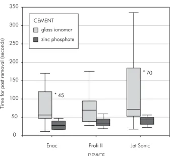
Overall, the posts were removed faster with Enac and Proi II in both groups. For the Enac groups (control), the time ranged between 17 and 120 onds (mean 35.75 s) in G1A, and 12 and 170 sec-onds (mean 76.17 s) in G2A. For Proi II, the time ranged between 20 and 60 seconds (mean 36.67 s) in G1B, and 28 and 175 seconds (mean 76.25 s) in G2B. Jet Sonic Satelec presented the largest ampli-tude of time variation, ranging from 23 to 180 sec-onds (mean 51.83 s) in G1C and 19 to 334 secsec-onds (mean 123.67 s) in G2C.
No statistically signii cant difference (p = 0.088) was observed between the control and the two ex-perimental ultrasound devices (Table 1, Graph 1). However, the time required for post removal var-ied according to the luting agent used. The average times for G1 and G2 were 41.42 and 92.03 seconds, respectively, with signii cant difference between the groups (p = 0.001) (Table 1, Graph 1).
Discussion
extracted human premolars were used for post re-moval by ultrasonic vibration. Although in vitro tests are not always able to reproduce in vivo con-ditions, they can offer comparative values that may guide clinical procedures.
The use of metallic cast intraradicular posts is beyond question due to the propagation of occlusal forces to the radicular structure that they provide, while the use of prefabricated posts reinforced with either glass-i ber or carbon and cemented with adhe-sive materials has gained popularity as they present favorable biomechanical properties and an elasticity modulus which is close to that of dentine.15,20
Never-theless, in the case of extensive coronal destruction, mainly of pillar teeth of partially i xed or removable prostheses, metallic cast posts are still recommend-ed.15
It is generally agreed that post retention is a major factor in the survival of restorations.8,15,19,20 Several
factors may affect post retention such as post type17
(custom made or prefabricated), post design3,10,13
(parallel, tapered, smooth, serrated, threaded), post adaptation to the root canal, cemented depth and cementing medium.1,4,7-9,20 Regarding the latter
con-dition, a previous study22 did not i nd difference in
retentive force when intracanal posts cemented with zinc phosphate- or glass ionomer-based sealers were compared. Another investigation6 demonstrated
that ultrasonic vibration for 10 minutes reduced the retention of zinc phosphate and glass ionomer seal-ers in 39% and 33%, respectively.
Considering the time necessary to dislodge
intra-radicular posts, some studies5,21 showed that posts
cemented with zinc phosphate required a rather short period of time (up to 3 minutes) to be dis-lodged. Conversely, 3 minutes of vibration was not enough to completely release cast posts cemented with glass ionomer cement.17
The results of the present study showed that all the cast metallic posts were successfully removed in a short time interval (mean time up to 2 minutes) with the glass ionomer cement having a signii cant retentive advantage. Considering that the efi ciency of an ultrasound device is related to its capacity to break the juncture between root canal walls and post, a similar behavior could be expected for both luting agents. However, glass ionomeric cements present adhesive properties that can increase its re-tentive abilities when they are used for post cemen-tation.23 Besides, the viscoelastic nature attenuates
the vibrations and absorbs ultrasonic energy trans-mitted to the posts.4,20
In the present study, the performance of two Brazilian ultrasonic units (Proi II Ceramic and Jet Sonic Satelec) was compared to that of the Japanese gold standard (Enac). It was observed that the time required for complete removal of the intraradicular posts ranged from 12 to 334 seconds. Proi II and Enac had a similar behavior, despite the recent
man-Table 1 - Mean time (seconds) required for post removal in the groups 1 and 2.
Group Subgroups n Mean* Standard
Deviation
1-Zinc phosphate cement
1A- Enac 12 35.75 ab 28.63
1B- Profi II 12 36.67ab 13.09
1C- Jet Sonic 12 51.83ab 41.62
2-Glass ionomer cement
2A- Enac 12 76.17ac 50.79
2B- Profi II 12 76.25ac 45.06
2C- Jet Sonic 12 123.67ac 107.84
*Similar letters indicate statistically similar values (p > 0.05). Different letters indicate statistically different values (p < 0.05).
Graph 1 - Time required (in seconds) for post removal for groups and subgroups of the study.
T
im
e
fo
r
po
st
remo
val
(s
eco
nds
)
Profi II
DEVICE
Jet Sonic Enac
0 50 100 150 200 300
250 350
* 45
* 70 CEMENT
glass ionomer
ufacturer’s 20% increase in the power of the Enac. Although Jet Sonic Satelec required a longer time for post removal, mainly when the glass ionomer ce-ment was used, there were no differences among the ultrasound devices. Large standard deviations have clearly contributed to this result.
Considering that this experiment was rigorously controlled, the differences in structure and composi-tion of the dentine of the individual roots, as previ-ously stated,20 might have contributed to this
varia-tion. This variability may also be explained due to variables relative to the tested devices, such as the thickness of the quartz crystal plate and the degree of crystal purity.8,17,19 Moreover, an oscillation in
time was also observed in another study5 that
evalu-ated the performance of two ultrasonic units in the removal of intraradicular posts.
In this study and according to Johnson et al.12
(1996), Gomes et al.6 (2001), and Dixon et al.5
(2002), the ultrasound devices were applied to the intraradicular retainer at maximum power on the surrounding and incisal surfaces. Thus, the ultra-sonic tip was continuously moved 360° around the post to induce resonance. An attempt was made to adjust the ultrasound tip to emit an audible sound level to maximize the energy that was transferred to
the post.17 Comparatively to other devices, the Enac
appeared to have a greater audible sound, mainly on the incisal surface, corroborating an earlier investi-gation.5
Another approach of this study concerns the in-existence of radicular damage after the use of ultra-sounds at maximum power, which so far coni rms the safety of the ultrasonic technique and reduced clinical occurrence of radicular fracture.1,9 Further
research is necessary to coni rm the effect of ultra-sonic times longer than those used in this study.
Conclusions
The ultrasonic units Prof II Ceramic and Jet Son-ic Satelec presented performances similar to that of the international Gold standard Enac in the removal of intraradicular cast posts, although a large vari-ability in the length of time was observed.
All the metallic cast posts cemented with glass ionomer or zinc phosphate sealers were successfully removed in a short time interval (mean time up to 2 minutes).
The luting agents exerted a greater inl uence on the time required for post removal than the origin of the ultrasonic units.
References
1. Castrisos T, Abbott PV. A survey of methods used for post removal in specialist endodontic practice. Int Endod J. 2002;35(2):172-80.
2. Chalfin H, Weseley P, Solomon C. Removal of restorative posts for the purpose of nonsurgical endodontic retreatment: report of cases. J Am Dent Assoc. 1990;120(2):169-72. 3. Lopes HP, Siqueira Jr JF, Elias CN. Retratamento endodôntico.
In: Lopes HP, Siqueira Jr JF. Endodontia: Biologia e Técnica. 2ª ed. Rio de Janeiro: Medsi; 2004. p. 727-85.
4. Buoncristiani J, Seto BG, Caputo AA. Evaluation of ultra-sonic and ultra-sonic instruments for intraradicular post removal. J Endod. 1994;20(10):486-9.
5. Dixon EB, Kaczkowski PJ, Nicholls JI, Harrington GW. Com-parison of two ultrasonic instruments for post removal. J Endod. 2002;28(2):111-5.
6. Gomes APM, Kubo CH, Santos RA, Santos DR, Padilha RQ. The influence of ultrasound on the retention of cast posts ce-mented with different agents. Int Endod J. 2001;34(2):93-9.
7. Smith BJ. Removal of fractured posts using ultrasonic vibra-tion: an in vivo study. J Endod. 2001;27(10):632-4. 8. Stamos DE, Gutmann JL. Survey of endodontic
retreat-ment methods used to remove intraradicular posts. J Endod. 1993;19(7):366-9.
9. Abbott PV. Incidence of root fractures and methods used for post removal. Int Endod J. 2002;35(1):63-7.
10. Garrido AD, Fonseca TS, Alfredo E, Silva-Sousa YT, Sousa-Neto MD. Influence of ultrasound, with and without water spray cooling, on removal of posts cemented with resin or zinc phosphate cements. J Endod. 2004;30(3):173-6.
11. Imura N, Zuolo ML. Remoção de retentor intra-radicu-lar com aparelho de ultra-som. Rev Assoc Paul Cir Dent. 1997;51(3):262-7.
12. Johnson WT, Leary JM, Boyer DB. Effect of ultrasonic vibra-tion on post removal in extracted human premolar teeth. J Endod. 1996;22(9):487-8.
me-tálicos, através de instrumentos rotatórios. Rev Bras Odontol. 1993;50(3):20-5.
14. Stabholz A, Friedman S. Endodontic retreatment - case selec-tion and technique. Part 2: Treatment planning for retreat-ment. J Endod. 1988;14(12):607-14.
15. Vasconcellos AB, Lopes HP. Retentores intra-radiculares. In: Lopes HP, Siqueira Jr JF. Endodontia: Biologia e Técnica. 2ª ed. Rio de Janeiro: Medsi; 2004. p. 696-706.
16. Berbert A, Tanomaru Filho M, Ueno AH, Bramante CM, Ishikiriama A. The influence of ultrasound in removing in-traradicular posts. Int Endod J. 1995;28(1):54-6.
17. Silva MR, Biffi JC, Mota AS, Fernandes Neto AJ, Neves FD. Evaluation of intracanal post removal using ultrasound. Braz Dent J. 2004;15(2):119-26.
18. Krell KV, Jordan RD, Madison S, Aquilino S. Using ultra-sonic scalers to remove fractured root posts. J Prosthet Dent. 1986;55(1):46-9.
19. De Deus QD. Endodontia. 5ª ed. Rio de Janeiro: Medsi; 1992.
20. Hauman CH, Chandler NP, Purton DG. Factors influencing the removal of posts. Int Endod J. 2003;36(10):687-90. 21. Oliveira MRS, Biffi JCG, Mota AS, Maniglia CAG. Avaliação
da remoção de pinos intra-radiculares pré-fabricados através de técnica ultra-sônica. Rev Assoc Paul Cir Dent. 1999;53(5):372-7.
22. Chan FW, Harcourt JK, Brockhurst PJ. The effect of post adaptation in the root canal on retention of posts cemented with various cements. Aust Dent J. 1993;38(1):39-45. 23. Tjan AH, Tjan AH, Greive JH. Effects of various cementation