Assessment
in vitro
of brushing on dental
surface roughness alteration by laser
interferometry
Abstract: Noncarious cervical lesions (NCCLs) are considered to be of multifactorial origin, normally associated with inadequate brushing. This study assessed the inluence in vitro of simulated brushing on NCCL formation. Fifteen human premolars were submitted to brushing in the cementoenamel junction region, using hard-, medium- and soft-bristled brushes associated with a toothpaste of medium abrasiveness under a 200 g load, at a speed of 356 rpm for 100 minutes. The surface topog-raphy of the region was analyzed before and after brushing, by means of a laser interferometer, using “cut-off” values of 0.25 and considering roughness values in Pm. The initial roughness (Pm) results for dentin (D / bristle consistency: 1 – soft, 2 – medium and 3 – hard) were as fol-lows: (D1) 1.25r0.45; (D2) 1.12r0.44; (D3) 1.05r0.41. For enamel (E / bristle consistency: 1 – soft, 2 – medium and 3 – hard), the initial results were: (E1) 1.18r0.35; (E2) 1.32r0.25; (E3) 1.50r0.38. After brushing, the following were the values for dentin: (D1) 2.32r1.99; (D2) 3.30r0.96; (D3) Over 500. For enamel, the values after brushing were: (E1) 1.37r0.31; (E2) 2.15r0.90; (E3) 1.22r0.47. Based on the results of the ANOVA and Tukey statistical analyses (D= .05) it was concluded that soft, medium and hard brushes are not capable of abrading enamel, whereas dentin showed changes in surface roughness by the action of medium- and hard-bristled brushes.
Descriptors: Tooth abrasion; Dentifrices; Toothbrushing.
Alessandra Miranda de Azevedo(a)
Heitor Panzeri(b)
Célio Jesus do Prado(c)
José Daniel Biasoli De-Mello(d)
Carlos José Soares(e)
Alfredo Julio Fernandes-Neto(f)
(a)DDS, Graduate Student; (b)PhD, Department
of Dental Materials and Prosthodontics – School of Dentistry of Ribeirão Preto, University of São Paulo.
(c)PhD, Assistant Professor, Department
of Removable Prosthodontics; (e)PhD,
Department of Restorative Dentistry; (f)PhD,
Professor and Chairman, Department of Occlusion, Fixed Prosthodontics, and Dental Materials – School of Dentistry, Federal University of Uberlândia.
(d)PhD, School of Mechanical Engineering,
Federal University of Uberlândia.
Corresponding author:
Alessandra Miranda de Azevedo
Faculdade de Odontologia de Ribeirão Preto Universidade de São Paulo (USP)
Departamento de Materiais Dentários e Prótese Av. do Café, s/n Ribeirão Preto - SP - Brazil CEP: 14040-904
E-mail: [email protected]
Introduction
Noncarious cervical lesions (NCCLs), generi-cally denominated tooth abrasion, present a variety of forms, and can affect vestibular, lingual and/or proximal surfaces, commonly being of multifactori-al origin.1-4 Factors as acids, and occlusal and
abra-sive forces may interact or act separately, thus con-tributing to the appearance of cervical lesions.3,5-8
These lesions may be classiied as: erosive, attritive, abfractive and abrasive.9 Dental erosion is tooth
structure loss by nonbacterial chemical action;2,5,6,10
attrition is wear of one surface against another and abfraction is a wedge-shaped lesion, located at the cementoenamel junction, caused by stress generated by biomechanical force.1,7,11
Clinically, the term abrasion refers to pathologi-cal wear by objects repeatedly in contact with the teeth.2 Brushing with dentifrice is an example of a
triple-body abrasion process, in which disaggregated particles slide between the tooth and brush bristles, the size of the abrasive particles and pressure being important factors in the speed at which the surface undergoes abrasion.12 However, deinition of this
process as an isolated etiologic factor for cervical le-sions is still controversial. There are studies in which toothbrushing without dentifrice is apparently in-capable of abrading enamel and dentin,7,9,13-17 as the
toothpaste abrasiveness may be caused by a combina-tion of its erosive effect and the mechanical effect of the toothbrush bristles,18 while other studies showed
that toothbrushing without dentifrice may induce abrasion.19,20 Frequency and toothbrushing technique
are also factors related to tooth abrasion.21
In view of this context, an hypothesis is set forth that brushing and the type of toothbrush result in topographic alteration of human enamel and dentin, characterizing this process as an etiologic factor of noncarious cervical lesions. In order to conirm this hypothesis, this study assessed the topography, in vitro, of human enamel and dentin before and af-ter brushing with soft-, medium- and hard-bristled brushes associated with dentifrice.
Material and Methods
Tooth obtainment
To conduct this study, 15 healthy human
pre-molars were selected because they presented high incidence of abrasive lesions,3,22 but did not present
any type of lesion on the vestibular enamel and root dentin faces, and had been indicated for extraction due to periodontal problems or orthodontic purpos-es. Teeth that presented any damage resulting from forceps during extraction were excluded.20 This
study was approved by the Research Ethics Com-mittee, Federal University of Uberlandia (Protocol No. 224/04).
Sample obtainment
The selected teeth were embedded in polystyrene resin (Aerojet, São Paulo, SP, Brazil), in the propor-tion of 12% monomer to 2% catalyzer. The teeth were placed horizontally with the vestibular face penetrating approximately 1 mm into a utility wax slide, and afterwards, enveloped by a rectangular 25 x 10 x 10 cm aluminum matrix. Polystyrene resin was poured in till it was full, and when the resin was completely polymerized, the set was detached from the wax and the matrix, removed, with the result that the tooth, except for the vestibular face, was embedded in resin. The samples received a inishing procedure to remove excess resin and to clean them of wax. The samples were identiied and stored in distilled water at a temperature of 37qC in an oven and then randomly divided into three groups: 1 – use of soft-bristled brushes; 2 – use of medium-bris-tled brushes, and 3 – use of hard-brismedium-bris-tled brushes. To deine the type of substrate, D was designated to identify Dentin and E, to identify enamel.
Initial surface topography determination
Initially, the samples were metal-coated (Emi-tech K550, Emi(Emi-tech Technologies Ltd., Kent, Eng-land), by deposit of a thin layer of gold, equivalent to 10-6mm, in order to increase surface
analyzed by speciic software (Mountains Map
3, Besançon, France), enabling this surface to be characterized with regard to shape and undulation and to calculate surface roughness parameters, us-ing “cut-off” values of 0.25.23 The roughness
pa-rameter assessed for numerical characterization of the surface was as follows: Sq, standard devia-tion of the distribudevia-tion of surface peak and val-ley heights,24,25 associated with assessment of the
functional parameters: Ssk, symmetry coeficient, the parameter used to measure the symmetry of a proile in relation to the mean plane, and Sk, lat-tening coeficient, which describes the form of topography height distribution. The values found for each parameter were statistically analyzed, the parameter Sq being submitted to the parametric ANOVA and Tukey tests (D =.05) and Sk and Ssk expressed in frequency.
Simulated brushing
After determining initial topography, the sam-ples were washed under running water to remove the gold layer. Next, they were placed inside an ultrasonic vibration device (Thornton, Vinhedo, SP, Brazil) containing distilled water and stayed there for 10 minutes. Then they were washed with soap and water, alcohol and distilled water and then ixed horizontally in the receptacles by means of modeling compound. To perform the abrasion tests, a brushing machine was used comprised of six stainless steel compartments to put the samples in. The test specimen is placed on the internal base of the receptacle, ixed to a metal plate by means of modeling compound (DFL, Rio de Janeiro, RJ, Brazil). The appliance has a support to which the toothbrush is ixed, aligned parallel to the plate,
regulated by screws positioned on the sides and top. The machine was set to run a horizontal course of 3.8 cm, applying a 200 g load at a speed of 356 rpm for 100 minutes, corresponding to 2 years of normal standard tooth brushing. Similar toothbrushs (Tek, Johnson & Johnson, São José dos Campos, SP, Bra-zil), with small, oval-headed and round-tipped syn-thetic bristles of soft, medium and hard consistency were ixed to the supports and adjusted so that a largest number of bristles would come into contact with the sample. Fifteen milliliters of a suspension prepared with 70 ml of distilled water and 70 g of dentifrice of medium abrasiveness (Contente, Uber-lândia, MG, Brazil) were poured into each tray con-taining the sample, in order to perform brushing for 100 minutes.
Final surface topography determination
When the brushing ended, the samples were washed under running water and then submitted to ultrasonic vibration (Thornton, Vinhedo, SP, Brazil) for 10 minutes to remove the abrasive particles. The samples were metal-coated again and the surface to-pography parameters were obtained again, in accor-dance with the same measuring methodology used initially.
Results
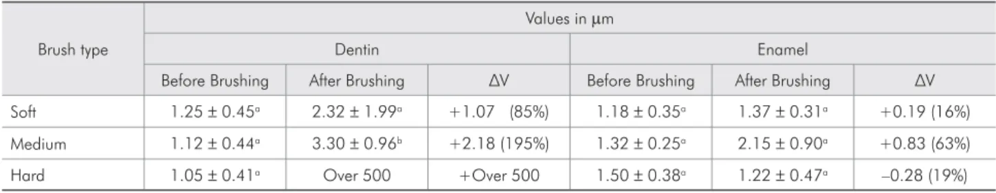
Mean and standard deviation values of the pa-rameter Sq for the human enamel and dentin are presented in Table 1. The data were submitted to the analysis of normality and homogeneity and were shown to present normal and homogenous distribution for the parameter Sq. Therefore, sta-tistical analysis was carried out by means of a two-way ANOVA. Post hoc comparisons among
Table 1 - Mean and standard deviation values of the parameter Sq and statistical categories – Tukey Test (P < 0.05).
Brush type
Values inPm
Dentin Enamel
Before Brushing After Brushing ¨9 Before Brushing After Brushing ¨9 Soft 1.25r0.45a 2.32r1.99a +1.07 (85%) 1.18r0.35a 1.37r0.31a +0.19 (16%)
Medium 1.12r0.44a 3.30r0.96b +2.18 (195%) 1.32r0.25a 2.15r0.90a +0.83 (63%)
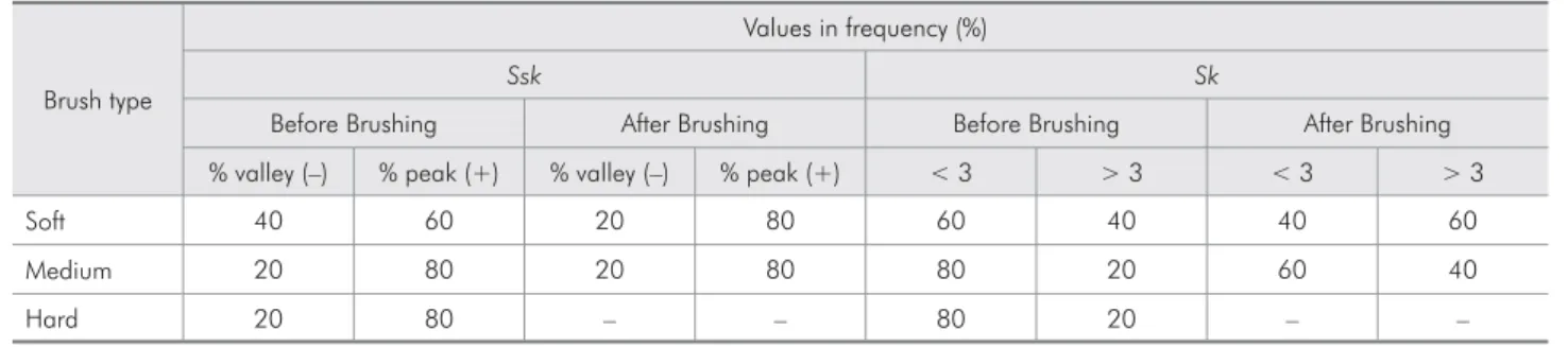
groups were done using the Tukey HSD test. Sta-tistical signiicance was set at .05. The values Sk and Ssk were presented in the form of frequency, as they presented variation limits in positive and negative values. There was no statistically signii-cant difference for the substrate enamel before and after simulated toothbrushing, irrespective of the toothbrush hardness, for all parameters analyzed, as well as for dentin with a soft toothbrush. How-ever, the results of parameter Sq found for the me-dium brush showed a signiicant increase in dentin surface roughness after brushing. Dentin abrasion with the hard brush could not be analyzed because it was deined as being over 500Pm, in excess of the laser interferometer reading capacity. For the parameter Ssk, enamel presented predominantly negative values, indicating a larger number of val-leys before and after brushing; on the other hand, dentin presented predominantly positive values, indicating a larger number of peaks (Tables 2 and 3). The graphic representation of surface roughness along the analyzed area is represented in Figures 1 (A and B) and 2 (A and B), axonometric images that allow relief to be seen.
Discussion
The hypothesis tested in this study was accepted only for dentin. The type of brush only inluenced the dentin substrate topography and did not harm the enamel surface. Human tooth enamel behavior was similar for the three types of toothbrushes, and presented no signiicant variation for the parameter Sq among the groups, before and after simulated brushing. Dentin presented statistically similar re-sults to those of enamel for the soft brush, but for the medium brush, there was increased surface rough-ness after brushing. Tooth structure abrasion with the hard-bristled brush was higher than 500Pm, thus it was not possible to assess the roughness pa-rameters of this structure by the methodology ap-plied.
After the brushing procedure, the enamel surface was not abraded. Because of its highly mineralized content, enamel is extremely hard.26 However, when
fracture occurs, it is reported as a result of enamel prism disorganization due to stress concentration in the cervical region of the tooth. The action of den-tifrices and brush could result in fracture expansion only.1,10,11
Table 2 - Values in frequency of Ssk/Sk for dentin according to type of brush used.
Brush type
Values in frequency (%)
Ssk Sk
Before Brushing After Brushing Before Brushing After Brushing % valley (–) % peak (+) % valley (–) % peak (+) < 3 > 3 < 3 > 3
Soft 40 60 20 80 60 40 40 60
Medium 20 80 20 80 80 20 60 40
Hard 20 80 – – 80 20 – –
Table 3 - Values in frequency of Ssk/Sk for enamel according to type of brush used.
Brush type
Values in frequency (%)
Ssk Sk
Before Brushing After Brushing Before Brushing After Brushing % valley (–) % peak (+) % valley (–) % peak (+) < 3 > 3 < 3 > 3
Soft 80 20 100 0 80 20 60 40
Medium 80 20 80 20 100 0 40 60
With regard to surface form characterization, in this study, dentin presented a symmetry coeficient with a predominance of peaks, and it was more sus-ceptible to abrasion in comparison with the enamel
surface, in which valleys were predominant.
Abrasion on the dentin surface was observed in an abrasion test with soft-bristled brushes,20 in
con-trast with the result obtained in the present study,
Alpha = 45o
1.55 mm
1.1 mm
1.36 mm 1.22 mm
16.9Mm
Mm
15 16
14 13 12 11 10 9 8 7 6 5 4 3 2 1 0
7.5 8
7 6.5 6 5.5 5 4.5 4 3.5 3 2.5 2 1.5 1 0.5 0
Mm
8.3Mm
Beta = 30o
Alpha = 45o Beta = 30o
Alpha = 45o
1.42 mm 1.28 mm
1.28 mm 1.39 mm
13.6Mm
Mm
13 12 11 10 9 8 7 6 5 4 3 2 1 0
20 22
18 16 14 12 10 8 6 4 2 0
Mm
23.5Mm
Beta = 30o
Alpha = 45o Beta = 30o
A
B
Figure 1, A and B - Surface Topography - x, y and z (length, width and height) of analyzed area.A - Dentin roughness before brushing, and
B - Increase of dentin roughness after brushing with medium-bristled tooth brush.
Figure 2, A and B - Surface Topography - x, y and z (length, width and height) of analyzed area.A - Enamel roughness before brushing, and
B - Enamel roughness presented no significant variation before and after simulated brushing.
in which this type of bristle did not result in abra-sion of this structure. However, these authors used a load of 300 g while, in the present study, the load applied was 200 g. In an abrasion test by means of human dentin brushing,14,16,27 there was no
signii-cant difference in the abrasion of this structure with regard to toothbrush bristle hardness. On the other hand, a reduction in abrasion was reported when hard bristles were used.19 This differs from the
re-sults of the present study, in which it was noted that hard-bristled brushes presented greater abrasion in dentin than the other types of bristles.
Abrasion test studies did not observe enamel structure abrasion,13 a result in agreement with that
obtained in the present study, when soft-, medium- and hard-bristled brushes were used on this same substrate. Other studies related enamel abrasion and abrasion by brushing.20 However, in those
re-searches, the abrasion tests were related to exposure to acid and lateral forces, respectively.
To many authors, the abrasive effect of dentifrice on dentin and enamel structure abrasion is related more to abrasive concentration and is hardly inlu-enced by bristle-hardness.15-17 Nonetheless, abrasion
may be caused by the corrosive effect of the denti-frice combined with the mechanics of the toothbrush bristles.2,8,13 As there was no variation in the type of
dentifrice used in the present study, it was not pos-sible to relate abrasion and abrasive concentration. However, the results showed that there was no enam-el abrasion during the brushing procedure. On the other hand, in dentin, abrasion was observed with
the use of medium and hard-bristled brushes using dentifrice of medium abrasiveness for both groups, which does show the inluence of the type of brush.
Regarding the topography analysis, the rough-ness parameters can be calculated using two-dimen-sional (2D) or three-dimentwo-dimen-sional (3D) study.24 2D
parameters are used for proile analysis. However, digital techniques of surface analysis in 3D make possible the study of a three-dimensional area of the surface without contacting it. The accomplishment of digital analyses associated to a reading without contact by means of optical instruments in this study made possible the attainment of data with-out distortions or damages to the surface of enamel and dentine structures,25 but the optical reading was
sensitive, preventing the attainment of focus in the dentine surface after brushing with hard-bristled brushes due to a resultant wear superior to 500Pm.
Conclusion
In accordance with the methodology used and based on the analysis of the data obtained in this study, it was possible to conclude that: Brushing with the use of soft-, medium- and hard-bristled brushes and dentifrice of medium abrasiveness is not capable of abrading human enamel. In dentin, medium- and hard-bristled brushes caused increased surface roughness.
Acknowledgements
The authors are indebted to the inancial support granted by FAPEMIG (Grant no. 160/2004).
References
1. Grippo JO, Simring M. Dental “Erosion” Revisited. J Am Dent Assoc. 1995;126(5):619-28.
2. Imfeld T. Dental erosion. Definition, classification and links. Eur J Oral Sci. 1996;104(2):151-5.
3. Levitch LC, Bader JD, Shugars DA, Heymann HO. Non-car-ious cervical lesions. J Dent. 1994;22(4):195-207.
4. Smith BGN. Toothwear: Aetiology and Diagnosis. Dent Up-date. 1989;16(5):204-12.
5. Attin T, Knöfel S, Buchalla W, Tütüncü R. In situ Evalu-ation of Different RemineralizEvalu-ation Periods to Decrease Brushing Abrasion of Demineralized Enamel. Caries Res. 2001;35(3):216-22.
6. Attin T, Siegel S, Buchalla W, Lennon ÁM, Hannig C, Becker K. Brushing Abrasion of Softened and Remineralised Dentin: An in situ Study. Caries Res. 2004;38(1):62-6.
7. Braem M, Lambrechts P, Vanherle G. Stress-induced cervical lesions. J Prosthet Dent. 1982;67(5):718-22.
8. Eisenburger M, Shellis RP, Addy M. Comparative Study of Wear of Enamel Induced by Alternating and Simultaneous Combinations of Abrasion and Erosion in vitro. Caries Res. 2003;37(6):450-5.
10. Lee WC, Eakle WS. Possible role of tensile stress in the eti-ology of cervical erosive lesion of teeth. J Prosthet Dent. 1984;52(3):374-80.
11. Edwin D, Meyer G, Schwartz P. The etiology of wedge-shaped defects: A morphological and function-oriented investigation. J Gnathol. 1991;10(1):49-56.
12. Kliemann C. Lesões Cervicais Não-Cariosas por Abrasão (Esco-vação Traumática). J Bras Clin Odontol Integr. 2002;6(1):204-9.
13. Addy M, Hunter ML. Can tooth brushing damage your health"
Effects on oral and dental tissues. Int Dent J. 2003;53(3):177-86.
14. Bjorn H, Lindhe J, Grondahl HG. The abrasion of dentine by commercial dentifrices. Odontol Revy. 1966;17(2):109-20. 15. Cohen RB. Toothpaste abrasion. J Am Dent Assoc.
2004;135(11):1520-2.
16. Manly RS, Brudevold F. Relative abrasiveness of natural and synthetic toothbrush bristles on cementum and dentin. J Am Dent Assoc. 1957;55(6):779-80.
17. Radentz WH, Barnes GP, Cutright DE. A survey of factors possibly associated with cervical abrasion of tooth surfaces. J Periodontol. 1976;47(3):148-54.
18. Svinnseth PN, Gjerdet NR, Lie T. Abrasivity of toothpastes: An in vitro study of toothpastes marketed in Norway. Acta Odontol Scand. 1987;45(3):195-202.
19. Dyer D, Addy M, Newcombe RG. Studies in vitro of abrasion by different manual toothbrush heads and standard tooth-paste. J Clin Periodontol. 2000;27(2):99-103.
20. Litonjua LA, Andreana S, Bush PJ, Tobias TS, Cohen RE. Wedged cervical lesions produced by toothbrushing. Am J Dent. 2004;17(4):237-40.
21. Bergström J, Lavstedt S. An epidemiologic approach to tooth-brushing and dental abrasion. Community Dent Oral Epide-miol. 1979;7(1):57-64.
22. Kitchin PC. The prevalence of tooth root exposure, and the relation of the extent of such exposure to the degree of abra-sion in different age classes. J Dent Res. 1941;20(6):565-81. 23. Bastos FS, Godoy GCD, Las Casas EB, Buono VTL. Análise
da topografia do esmalte dentário. In: 59º Congresso Anual Internacional da ABM. São Paulo. Anais. 2004. [Cd-Rom]. 24. Gadelmawla ES, Koura MM, Maksoud TMA, Elewa IM,
Soli-man HH. Roughness parameters. J Mater Process Technol. 2002;123(1):133-45.
25. Hutchings IC. Tribology: friction and wear of Engineering Materials. London: Edward Arnold; 1992.
26. Tyldesley WR. The mechanical Properties of Human Enamel and Dentine. Br Dent J. 1959;106(1):269-78.