In vitro
evaluation of human dental enamel surface roughness
bleached with 35% carbamide peroxide and submitted to
abrasive dentifrice brushing
Avaliação
in vitro
da rugosidade superficial do esmalte dental
humano clareado com peróxido de carbamida a 35% e
submetido à escovação com dentifrícios abrasivos
Claudia Cia Worschech* José Augusto Rodrigues*
Luis Roberto Marcondes Martins** Gláucia Maria Bovi Ambrosano***
ABSTRACT:The aim of thisin vitrostudy was to evaluate the surface roughness of human enamel bleached with 35% carbamide peroxide at different times and submitted to different superficial cleaning treatments: G1 - not brushed; G2 - brushed with fluoride abrasive dentifrice; G3 - brushed with a non-fluoride abrasive dentifrice; G4 - brushed without dentifrice. Sixty fragments of human molar teeth with 4 x 4 mm were obtained using a diamond disc. The specimens were polished with sandpaper and abrasive pastes. A perfilometer was used to measure roughness average (Ra) values of the initial surface roughness and at each 7-day-interval after the beginning of treatment. The bleaching was performed on the surface of the fragments for 1 hour a week, and the surface cleaning treatment for 3 minutes daily. The samples were stored in individual receptacles with artificial saliva. Analysis of variance and the Tukey test revealed significant differences in surface roughness values for G2 and G3, which showed an increase in roughness over time; G1 and G4 showed no significant roughness differences. The bleaching with 35% carbamide peroxide did not alter the enamel surface roughness, but when the bleaching treatment was performed combined with brushing with abrasive dentifrices, there was a significant increase in roughness values.
DESCRIPTORS:Tooth bleaching; Peroxides; Dentifrices; Toothbrushing.
RESUMO:O propósito deste estudoin vitrofoi avaliar em diferentes tempos a rugosidade superficial do esmalte dental humano clareado com peróxido de carbamida a 35% e submetido a diferentes tratamentos superficiais de limpeza: G1 - não escovado; G2 - escovado com dentifrício fluoretado abrasivo; G3 - escovado com dentifrício não fluoretado abrasivo; G4 - escovado sem dentifrício. Sessenta fragmentos de molares humanos com 4 x 4 mm foram obtidos atra-vés do seccionamento com discos diamantados. Os espécimes foram polidos com lixas e pastas abrasivas. Um perfilô-metro foi utilizado para determinar os valores da média de dureza Ra (“roughness average”) iniciais e a cada intervalo de 7 dias após o início do tratamento clareador. O clareamento foi realizado na superfície dos fragmentos por 1 hora se-manalmente, e os tratamentos superficiais, por 3 minutos diariamente. No restante do tempo, os espécimes eram ar-mazenados em receptáculos individuais com saliva artificial. A análise de variância e o teste de Tukey revelaram dife-renças estatísticas significantes na rugosidade superficial em função do tempo. G2 e G3 demonstraram um aumento nos valores de rugosidade; G1 e G4 não apresentaram diferenças estatísticas. O clareamento com peróxido de carba-mida a 35% não altera a rugosidade superficial do esmalte humano, mas, quando associado ao tratamento superficial com abrasivos, ocorre um aumento significante da rugosidade superficial.
DESCRITORES:Clareamento de dente; Peróxidos; Dentifrícios; Escovação dentária.
* MS, Department of Restorative Dentistry; **ScD, Professor, Department of Statistics; ***PhD, Professor, Department of Commu-nity Dentistry – School of Dentistry of Piracicaba, State University of Campinas.
INTRODUCTION
Bleaching procedures have gained popularity with patients and dentists as conservative tech-niques to lighten natural teeth in order to improve
the harmony of the smile. In-offic vital bleaching has been used to introduce the patient to the bleaching process, to improve post-bleaching cases or to function solely as a bleaching treatment8
perox-ide or hydrogen peroxperox-ide in concentrations higher than those used for at-home bleaching, and is car-ried out with rubber dam for 1 to 2 hours8
.
However, the exposure of tooth hard tissues to bleaching agents can result in microstructural changes in the enamel surface2,3,8,13,18
. Some au-thors, through scanning electron microscopy (SEM) evaluation, have demonstrated demineral-ization, surface defects, and degradation of sound enamel3,8,13,18
. Fluoride therapy is strongly indicated to avoid these side effects1
, and one of the most useful methods of fluoride application is the use of dentifrices.
On the other hand, abrasives dentifrices play an important role in the cleaning process by re-moving extrinsic stains, and patients commonly use them during the bleaching treatment. Some complicating factors may be explained by acknowl-edging that cleaning effectiveness may not be solely related to abrasion, and there is concern that some abrasives may contribute to excessive tooth wear7,11
.
The alteration of the external enamel structure after bleaching treatment may be worse after cleaning with abrasive dentifrices as they might re-move the degraded enamel an enhance the phe-nomena of erosion and wear.
However, to maintain oral health, teeth need to be daily brushed and there is a lack of evidence on the effects of bleaching agents combined with tooth brushing on the enamel surface as well as on its influence on enamel roughness. The goal of this study was to investigate thein vitroeffects induced
by an in-office bleaching regimen combined with a brushing treatment with abrasive dentifrices, with and without fluoride, on the enamel surface roughness.
MATERIAL AND METHOD
Experimental design
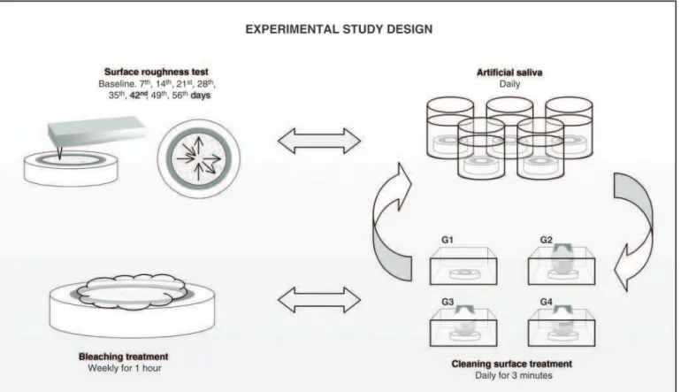
The factors under study were cleaning surface treatment in four levels: G1 not brushed; G2 brushed with fluoride abrasive dentifrice; G3 -brushed with abrasive dentifrice without fluoride; G4 - brushed without dentifrice; and time in nine levels: baseline, 7, 14, 21, 28 days of bleaching treatment and 7, 14, 21, and 28 days after the end of bleaching (post-treatment period).
The experimental units were 60 sound human enamel fragments, randomly assigned to treat-ment groups (n = 15). Six repeated measuretreat-ments of surface roughness in roughness average (Ra)
values were recorded on the surface of every speci-men at each 7-day interval (Figure 1).
Enamel fragment preparation
Since this study was designed to use human teeth, it was submitted to the Ethical Committee in Research, Piracicaba Dentistry School, State Uni-versity of Campinas (UNICAMP) and it was ap-proved in compliance with resolution CNS #196/96 of the National Committee of Health, Health Department (Brazil).
Thirty freshly extracted human non-erupted third molars were used. Immediately after extrac-tion, the teeth were stored in a 1% thymol solution (pH = 7). The roots were discarded and the crowns were longitudinally sectioned with double-faced diamond disks (KG Sorensen, Barueri, SP, Brazil) using a low-speed handpiece (Kavo do Brasil, Joinville, SC, Brazil) to produce 60 enamel frag-ments with 4 x 4 mm from the middle of the buccal or lingual aspect. The fragments presenting stains or cracks were not used. After completing the sec-tioning, the specimens were soaked in distilled and deionized water at 37ºC.
The enamel fragments were embedded individ-ually in a self-curing polyestyrene resin (BL 41110, Cromex, São Paulo, SP, Brazil) in a polyvinyl chlo-ride ring mold with 2.0 cm in diameter, allowing the external surface of the enamel to be exposed.
The molds were removed, and in order to shape a plane enamel surface for roughness testing, the fragments’ surfaces were leveled with a water-cool-ing mechanical grinder (Maxgrind/Solotest, São Paulo, SP, Brazil). Aluminum oxide disks were used in a sequential granulation of 400, 600, and 1,000 (Carborundum, 3M do Brasil, Sumaré, Brazil), and, after that, the specimens were pol-ished with felt cloth and abrasive pastes with gran-ulation of 6, 3, 1, and ½mm mixed with a mineral oil coolant (Arotec Ind. e Com. Ltda., São Paulo, Brazil).
Bleaching treatment
A 35% carbamide peroxide bleaching agent (Opalescence Quick, Ultradent Co., South Jordan, UT, USA) was selected. The enamel fragments were exposed to the bleaching agent for one hour a week, during a 28-day period.
For the bleaching agent treatment, a graduated syringe was used to apply 0.02 ml of bleaching agent on the enamel surface of each specimen6,12,15
at 37ºC.
After 1 hour of exposure, the bleaching agent was washed out under running distilled and deionized water for 5 seconds. Then the specimens received the surface cleaning treatment and were placed in a freshly prepared remineralization solu-tion that was changed daily. The remineralizasolu-tion solution was similar to natural saliva in terms of Ca and P contents, as proposed by Featherstoneet al.5 (1986) and modified by Serra and Cury in
199216
, and was composed of 1.5 mmol/L of Ca, 50 mmol/L of KCl, 0.9 mmol/L of PO4, and
20 mmol/L of tri-hydroxymethil-aminomathan at pH = 7.0.
After 28 days, at the end of the bleaching treat-ment (4 applications), all groups were kept in a
daily changed remineralization solution over 28 days still receiving the surface cleaning treat-ments.
Surface cleaning treatment
Everyday, after the bleaching treatment, the specimens were submitted to a surface cleaning treatment according to each group’s specifica-tions, as follows: Group 1, control group, was not submitted to a cleaning treatment; Groups 2 and 3 received a surface cleaning treatment with a comercial dentifrice containing calcium carbon-ates as abrasives, respectively with fluoride and without fluoride (Table 1); Group 4 received the brushing treatment without dentifrice.
Brushing procedures were performed for 3 min-utes in a brushing machine, set at a usual brush force of 200 g and 250 cycles per minute. The brush heads were from the same brand of nylon multi-tufted toothbrushes, and each product was brushed onto specimens with individual brush heads.
The dentifrices were freshly prepared in slurries (1 part of dentifrice to 3 parts of deionized and dis-tilled water, in weight). The slurries were agitated for 20 minutes before use.
At the end of the surface cleaning treatment, the samples were removed from the machine’s re-cipients, washed out with distilled water and maintained in individual receptacles at 37ºC until the next brushing cycle.
Surface roughness test
Before the bleaching treatments, a perfilometer (Surf-Corder mod. 1700, Kosaka, Tokio, Japan) was used to measure the initial surface roughness (baseline). Three different directions were used to perform six measurements on the surface of each specimen, with a cut off (lc) of 0.25 mm at a
veloc-ity of 0.1 mm/s (ISO 4228). The mean Ra value (mm) was determined for each specimen, at each 7-day interval.
RESULTS
Statistical analysis involved a parametric method using repeated measures analysis of vari-ance (ANOVA) followed by Tukey’s HSD hoc analy-sis (a = 0.05). The mean values of roughness are shown in Table 2, and the behavior of enamel roughness may be observed in Graph 1.
Baseline data were obtained in order to verify the initial surface smoothness (p < 0.05) and to contrast differences between the groups. At the baseline values and at the 7th day all groups
showed statistically similar means. The groups submitted to surface cleaning treatments pre-sented a statistically significant increase in the surface roughness with time, as compared to the control group (G1), that showed statistically simi-lar means of surface roughness at different time intervals.
Group 2 showed a statistically significant in-crease in surface roughness from the baseline to the 14th
day and from the 14th
day to the 21st
day. After the 21stday the values were statistically
simi-lar until the end of the treatment, with the excep-tion of the 42ndday, which showed the highest
me-dian value.
TABLE 2 -Means of surface roughness (Ra) and standard deviation (SD) values for each treatment agent at different time intervals.
Groups
G1 - non brushed
G2 - brushed with fluoride abrasive
dentifrice
G3 - brushed with abrasive dentifrice
G4 - brushed without dentifrice
Period Means SD Means SD Means SD Means SD
Baseline 0.089 A a 0.033 0.100 A d 0.031 0.085 A d 0.031 0.096 A c 0.027
7th
day 0.096 A a 0.037 0.125 A cd 0.030 0.116 A c 0.031 0.094 A c 0.029
14th
day 0.098 B a 0.035 0.148 A c 0.045 0.138 AB c 0.052 0.101 AB bc 0.021
21st
day 0.101 B a 0.037 0.177 A b 0.053 0.176 A b 0.063 0.114 B abc 0.032
28th
day 0.107 B a 0.038 0.176 A b 0.064 0.185 A ab 0.082 0.108 B abc 0.033
35th
day 0.105 B a 0.033 0.201 A ab 0.074 0.190 A ab 0.074 0.129 B a 0.039
42nd
day 0.109 B a 0.040 0.203 A a 0.067 0.203 A a 0.087 0.122 B ab 0.031
49thday 0.108 B a 0.044 0.198 A ab 0.053 0.188 A ab 0.081 0.120 B abc 0.044
56thday 0.111 B a 0.051 0.190 A ab 0.054 0.187 A ab 0.067 0.119 B abc 0.041
TABLE 1-Ingredients used to prepare the dentifrices by the Proderma Pharmacy.
Compounds
Abrasive dentifrice with
fluoride (G2)
Abrasive dentifrice
without fluoride (G3)
Micronized
calcium carbonate 52.5% 52.5%
Glycerin 25% 25%
Natrosol gel 18% 18%
Sodium lauryl
sulfate 2% 2%
Sodium fluoride 0.16%
-Distilled water qsp qsp
The mean values of roughness in Group 3 showed a statistical increase from baseline to the 7th and 14th days which were not different from
each other. At the 21st
day the roughness mean values increased and were statistically different from the baseline, 7th
and 14th
day values. At the 28thday the roughness presented an increase until
the 42nd
day, which differed statistically from that of the 21stday, and the roughness values showed a
decrease but the values did not differ statistically from the 28th
day until the 56th
day.
Group 4 showed statistically similar roughness values until the 28th
day. The 35th
day showed the highest roughness median value of this group, which statistically differed from those of the base-line, 7th
, and 14th
days. After the 35th
day, these val-ues decreased, and the value of the 42nd
day statis-tically differed from those of the baseline and 7th
day. The values of the 49th
and 56th
days did not dif-fer from all the values and also from the baseline values.
DISCUSSION
Patients who submit themselves to a tooth bleaching treatment generally are patients that brush their teeth 3 or 4 times a day to achieve health and beauty. Dentifrices are sometimes rec-ommended for specific purposes, like cleaning or abrasion, in order to improve the bleaching pro-cess by removing superficial stains and polishing teeth. The most common abrasives these days are hydrated silica, dicalcium phosphate dihydrate,
and calcium carbonate21
. However, their abrasive-ness may be responsible for superficial tooth wear and other complicating factors11
, and abrasion may be more severe when associated to bleaching treatment regimens.
Several studies have evaluated the effects of bleaching agents on tooth structure mainly using scanning electron microscopy. The use of 10% carbamide peroxide has produced slight surface modifications and the use of highly concentrated agents indicated for in-office treatment has caused more severe alterations on the enamel micro-structure10,13,24
. The microhardnessin vitrostudies
have demonstrated contradictory results, present-ing mineral loss with the extended use of low pH agents, or increase in the mineral content due to demineralization and remineralization phenomena caused by the use of remineralization solutions and agents with neutral pH1,15
.
Profilometric analyses are also conflicting. Titleyet al.20(1988) found an apparent increase in
surface porosities on enamel after the use of a 35% solution of hydrogen peroxide; McGuckin et al.13
(1992) observed a slight increase in surface rough-ness after the use of 30% hydrogen peroxide, whereas Wanderaet al.22(1994) and Gürganet al.9
(1997) reported no surface alterations after bleaching with at-home agents as observed in the present study in G1.
However, the protocol employed in this study tried to be similar to an in-office bleaching treat-ment performed in the oral cavity with the
bleach-GRAPH 1 -Mean roughness of enamel fragments bleached and submitted to superficial
ing agent application followed by brushing and im-mersion in a solution similar to artificial saliva. The use of artificial saliva may decrease superficial erosion and it might favor remineralization of the enamel surface17
, a possible reason for the results observed in G1. Nevertheless, the sole use of 35% carbamide peroxide for 1 hour weekly did not cause surface roughness alterations.
The aim of this study was to evaluatein vitrothe
effect of in-office bleaching associated to abrasive dentifrices brushing. G1 may be considered a con-trol group because it was not brushed and did not show alterations in enamel roughness during the experimental treatment times. G4 was a second control group whose specimens were not brushed with abrasive dentifrices and did not differ statisti-cally from G1, demonstrating that the brushing had no effect on the bleached enamel surface roughness. However, the roughness tests were performed in the samples before and during exper-imental times, so that each specimen in a group could be considered as its own control. G4 had a non-statistical increase from baseline to the 28th
day, which was statistically significant at the 35th
day, suggesting a slight effect of the brushing dur-ing the bleachdur-ing treatment; however, such effect was not significant at the 49th
and 56th
days. The enamel surface roughness was increased after the surface cleaning treatment with abrasive dentifrices in G2 and G3. Although the roughness of these groups had been similar during all experi-ment times, the increase occurred from the 14th
day, for G2, and from the 21st
day, for G3, up to the 42nd
day; after that day the roughness kept high until the 56th
day, revealing higher roughness val-ues than the baseline ones, G1, and G4.
Our results indicate that the fluoride present in G2 was not able to prevent the increase in the sur-face roughness, and it may be supposed that some erosion and loss of enamel had occurred. Attinet al.1
(1997) have shown that the application of a flu-oride solution cannot prevent, but may reduce, the loss of mineral from enamel during at-home bleaching treatment with 10% carbamide perox-ide. According to Neveset al.14
(2002) the appear-ances of enamel surfaces brushed with either a fluoridated or non-fluoridated dentifrice were quite similar. The same study showed that the control group, brushed with toothbrush and water,
showed a smooth surface caused by the tooth brushing treatment.
Nevertheless, the G4 roughness values were statistically similar to those of G1 (control group) at all the studied times; G4 presented a surface roughness that ranked between the groups brushed with abrasive dentifrices (G2 and G3) and the control group (G1), strengthening the possible erosive effect of the brushing without the denti-frices.
On the other hand, this study evaluated only one surface roughness parameter, the Ra. This pa-rameter describes the overall roughness of a sur-face and it can be defined as the arithmetic average value of all absolute distances of the roughness profile from the centerline within the measuring length. Although the Ra parameter is the most used parameter to evaluate roughness, other pa-rameters may be used as complementary data to obtain more information about the profile shape23.
This study has been one of the first to evaluate the effects of abrasive dentifrices on enamel rough-ness after in-office bleaching and adverse effects were observed. Further research using other roughness parameters and in vitro and in vivo
models are necessary to observe these adverse ef-fects on the enamel surface.
The oxidative process involved in and the low pH of tooth-bleaching products have been consid-ered as the main source of adverse effects on min-eralized tissues during bleaching treatments15.
Low concentrations of carbamide peroxide pro-mote varying degrees of surface porosity and structural change, depending on the bleaching agent4,10,13,24. The use of abrasive-containing
denti-frice might result in enamel microwear14,21. The
slight abrasion in this enamel may remove the su-perficial degraded layer and promote a “new” sur-face, even though, with high roughness values14.
CONCLUSION
This in vitro investigation showed that 35%
carbamide peroxide did not alter enamel surface roughness, but when the bleaching treatment was
performed with cleaning treatments, through brushing with abrasive dentifrices, a significant increase in roughness values was observed.
REFERENCES
1. Attin T, Kielbassa AM, Schwanenberg M, Hellwig E. Effect of fluoride treatment on remineralization of bleached enamel. J Oral Rehabil 1997;24:282-6.
2. Basting RT, Rodrigues AL, Serra MC. The effect of 10% carbamide peroxide bleaching material on microhardness of sound and demineralized enamel and dentin in situ. Oper Dent 2001;26:531-9.
3. Bitter NC. A scanning electron microscopy study of the ef-fect of bleaching agents on enamel: a preliminary report. J Prosthet Dent 1992;67:852-5.
4. Ernest CP, Marroquin BB, Willershausen-Zönnchen B. Ef-fects of hydrogen peroxide-containing bleaching agents on the morphology of human enamel. Quintessence Int 1996;27:53-6.
5. Featherstone JDB, O’Really MM, Shariati M, Brugler S. Enhancement of remineralization in vitroandin vivo.In: Leach SA. Factors relating to demineralization and remineralization of the teeth. 3rded. Oxford: IRL; 1986. p. 23-34.
6. Freitas PM, Basting RT, Rodrigues AL, Serra MC. Effects of two 10% peroxide carbamide bleaching agents on dentin microhardness at different time intervals. Quintessence Int 2002;33:370-5.
7. Gerlach RW, Barker ML, Hyde JD, Jones MB. Effects of a tartar control whitening dentifrice on tooth shade in a pop-ulation with long-standing natural stain. J Clin Dent 2001;12:47-50.
8. Gultz J, Kaim J, Scherer W, Gupta H. Two in-office bleach-ing systems: a scannbleach-ing electron microscope study. Compend Contin Educ Dent 1999;20:965-70.
9. Gürgan S, Bolay S, Alacan R. Adherence of bacteria to bleached or unbleached enamel surfaces. J Oral Rehabil 1997;24:624.
10. Hegedüs C, Bistey T, Flora-Nagy E, Keszthelyi G, Jenei A. An anatomic force microscopy study on the effect of bleaching agents on enamel surface. J Dent 1999; 27:509-15.
11. Isaacs RL, Bartizek RD, Owens TS, Walters PA, Gerlach RW. Maintenance of tooth color after prophylaxis: compari-son of three dentifrices. J Clin Dent 2001;12:51-5.
12. McCracken MS, Haywood VB Demineralization effects of 10 percent carbamide peroxide. J Dent 1996;24:395-8. 13. McGuckin RS, Babin JF, Meyer BJ. Alterations in human
enamel surface morphology following vital bleaching. J Prosthet Dent 1992;68:754-60.
14. Neves NA, Castro RA, Coutinho ET, Primo LG. Microstruc-tural analysis of demineralized primary enamel afterin vi-trotooth brushing. Pesqui Odontol Bras 2002;16:137-43. 15. Rodrigues JA, Basting RT, Rodrigues Jr AL, Serra MC.
Ef-fects of 10% carbamide peroxide bleaching materials on enamel microhardness. Am J Dent 2001;14:67-71. 16. Serra MC, Cury JA. Thein vitroeffect of glass-ionomer
ce-ment restoration on enamel subjected to a demineraliza-tion and remineralizademineraliza-tion model. Quintessence Int 1992; 24:39-44.
17. Silverstone, LM. Remineralization phenomena. Caries Res 1997;11(Supp1):59-84.
18. Smidt A, Weller D, Roman I. Effect of bleaching agents on microhardness and surface morphology of tooth enamel. Am J Dent 1998;11:83-5.
19. Tames D, Grando J, Tames DR. Alterações do esmalte den-tal submetido ao tratamento com peróxido de carbamida 10%. Rev Assoc Paul Cir Dent 1998;52:145-9.
20. Titley K, Torneck CD, Smith D. The effect of concentrated hydrogen peroxide solutions on the surface morphology of human tooth enamel. J Endod 1988;14:69-74.
21. Viscio D, Gaffar A, Fakhry-Smity S, Xu T. Present and fu-ture technologies of tooth whitening. Compend Contin Educ Dent 2000;21:36-43.
22. Wandera A, Feigal RJ, Douglas WH, Pintado MR. Home-use tooth bleaching agents: an in vitro study on quantitative effects on enamel, dentin and cementum. Quintessence Int 1994;25:541-6.
23. Whitehead SA, Shearer AC, Watts DC, Wilson NHF. Com-parison of methods for measuring surface roughness of ce-ramic. J Oral Rehabil 1995;22:421-7.
24. Zalking M, Arwaz JR, Goldman A, Rotstein I. Surface mor-phology changes in human enamel, dentin and cementum following bleaching: a scanning electron microscopy study. Endod Dent Traumatol 1996;12:82-8.