Radiographic prevalence of root canal
ramifications in a sample of root canal
treatments in a Brazilian Dental School
Prevalência radiográfica de ramificações
do canal radicular em uma amostra de
tratamentos endodônticos em uma Faculdade
de Odontologia Brasileira
Abstract: The aim of this study was to radiographically investigate the presence of root canal ramiications found after endodontic treatment, and to determine any relationship between their presence and the type of the auxiliary chemical substance used. The study evaluated 1,470 endodontic treatments performed by inal year undergraduate students at the Dental School of Piracicaba, State University of Campinas (UNICAMP), SP, Brazil, during the period from 1998 to 2000. The X-rays taken during treatment were evaluated in order to establish the presence of ramiications of the root canal system. The initial X-ray did not show the presence of any canal ramiications. After illing, X-rays showed only 3 ramiication types: 3.06% of lateral canals, 2.99% of apical deltas, and 0.1% of interradicular canals. The maxillary premolars showed the highest number of lateral ca-nals (n = 13), followed by mandibular premolars (n = 10) and maxillary incisors (n = 10). Apical deltas were mostly found in mandibular molars (n = 14), followed by maxillary in-cisors (n = 9). Only mandibular molars had interradicular canals. The detection of ramii-cations increased with the use of EDTA. However, no statistically signiicant relationship was found between the type of auxiliary chemical substance used and the number of root canal ramiications detected after root canal illing. It was concluded that the frequency of root canal ramiications found radiographically was low in treatments performed by undergraduate students.
Descriptors: Endodontics; Root canal; Dental records; Education, dental.
Resumo: O objetivo deste estudo foi investigar radiograicamente a presença de ramii-cações do canal radicular encontrada depois do tratamento endodôntico, e determinar qualquer relação entre a presença destas e do tipo de substância química auxiliar usada. O estudo avaliou 1.470 tratamentos endodônticos executados pelos estudantes do últi-mo ano da Faculdade de Odontologia de Piracicaba, Universidade Estadual de Campinas (UNICAMP), SP, Brasil, no período de 1998 a 2000. Foram avaliadas as radiograias fei-tas durante o tratamento para veriicar a presença das ramiicações dos sistemas de canais radiculares. A radiograia inicial não mostrou a presença de qualquer ramiicação. Depois da obturação as radiograias mostraram apenas 3 tipos de ramiicação: 3,06% de canais laterais, 2,99% de deltas apicais e 0,1% de canais interradiculares. Os pré-molares supe-riores mostraram o maior número de canais laterais (n = 13), seguidos pelos pré-molares inferiores (n = 10) e incisivos superiores (n = 10). Deltas apicais foram encontrados princi-palmente em molares inferiores (n = 14), seguidos por incisivos superiores (n = 9). Apenas molares inferiores apresentaram canais interradiculares. A detecção de ramiicações au-mentou com o uso do EDTA. Entretanto, nenhuma relação estatisticamente signiicante foi encontrada entre o tipo de substância química auxiliar usada e o número de ramiica-ções visualizadas após a obturação dos canais radiculares. Foi concluído que a freqüência de ramiicações do canal radicular encontrada radiograicamente é baixa em tratamentos executados por estudantes universitários.
Descritores: Endodontia; Canal radicular; Registros odontológicos; Educação em odontologia.
Iadasa de Quadros(a)
Alexandre Augusto Zaia(b)
Caio Cezar Randi Ferraz(b)
Francisco José de Souza Filho(b)
Brenda Paula Figueiredo de Almeida Gomes(b)
(a) MSc in Endodontics; (b)Associate Professors of Endodontics – School of Dentistry of Piracicaba, State University of Campinas.
Corresponding author:
Brenda Paula Figueiredo de Almeida Gomes Faculdade de Odontologia de Piracicaba – FOP/UNICAMP
Departamento de Endodontia
Av. Limeira, 901 - Piracicaba - SP - Brazil CEP: 13414-900
E-mail: [email protected]
Introduction
The main objective of endodontic therapy is to promote the chemo-mechanical cleansing of the entire pulp cavity and to perform its complete ob-turation with an inert illing material. The chemo-mechanical preparation plays an important role in removing pulpal (organic) and dentinal debris (in-organic), as well as microorganisms from the root canal system. The preparation is mechanically per-formed using endodontic iles and thoroughly ir-rigating the canal with an inert solution in order to remove debris. In addition, the canal must be chemically prepared using an auxiliary chemical substance or a combination of substances.9 It is also
well known that smear layer removal provides effec-tive decontamination and improves the seal of root illings, as the sealer penetrates into the open den-tinal tubules, which decreases microleakage.2,18
The obturation of the entire root canal system, from crown to apex, is meant to eliminate empty spaces and to preserve the decontamination per-formed in the course of the chemo-mechanical cleansing step.11
Although it has been reported that non-micro-bial factors might be implicated in root canal treat-ment failure, the literature suggests that persistent interradicular or secondary infections are the major causes of failure of root canal treatment.17
More-over, persistence of microorganisms is more related to the anatomical complexity of root canals than to operator inadequacies.7 An inadequate
chemo-me-chanical preparation and root canal illing associ-ated with untreassoci-ated and unilled canal ramiication and isthmus contribute to the persistence of infec-tion and, consequently, failure of the root canal treatment.
A canal is often left untreated because the dentist fails to recognize its presence. The dentist must have a thorough knowledge of root canal morphology be-fore being able to successfully perform endodontic treatment.13,20
Root-canal morphology studies have used freshly extracted teeth and clearing techniques.5,10 Previous
studies have suggested that radiographic images are not reliable for the detection of multiple canals and lateral canals, and could not distinguish centrally
placed apical foramina from those eccentrically located. The clearing technique, which makes the teeth somewhat transparent, has considerable value in studying the anatomy of the root-canal system. This is because, unlike radiographic images, it pro-vides a three-dimensional view of the pulp cavity in relation to the exterior of the tooth and allows a thorough examination of pulp chambers and root canals. However, the clearing technique remains useful only as a teaching/research tool with little or no clinical applicability.
Furthermore, transverse anastomosis in molars, lateral canals and apex deltas are important com-munication pathways between the pulp and the periodontium. In their studies, Rubach, Mitchell15
(1965); Bender, Seltzer3 (1972); Ross14 (1972) and
Gutmann8 (1978) indicated that toxins and
inlam-matory products can pass between these two struc-tures through the lateral, accessory and apex ca-nals.
Vertucci20 (1984), using the clearing technique,
made a detailed study of two thousand and four hundred permanent teeth. The results showed great variation in root canal anatomy. The most variable root canal anatomy was found among maxillary second premolars. The rate of lateral canals in max-illary molars was around 50% in the mesiobuccal roots, and they were mostly found at the apex. In the mandibular molars (mainly irst molar), lateral canals were found in great numbers in the furcation region.
The aim of this study was to radiographically investigate the presence of root canal ramiications found after endodontic treatment was performed by inal year undergraduate students and to determine any relationship between their visualization and the type of auxiliary chemical substance used.
Material and Methods
All treatments were done with rubber dam af-ter asepsis. The instrumentation proceedings were done employing the hybrid technique according to Valdrighi et al.19 (1991). The coronal two-thirds of
each canal were prepared initially using iles up to size 35. A size 2 Gates-Glidden bur (GG) (Dentsp-ly-Maillefer Co., Ballaigues, Switzerland) was then used with gentle force up to the length of the coronal two-thirds of the canal followed by a size 3 GG bur up to a length 1 mm shorter than the previous one. A size 10 ile (Dentsply-Maillefer Co., Ballaigues, Switzerland) was used to run through the canal 1 mm beyond the length of the coronal two-thirds between each ile and bur, in order to maintain the passage of canal patency. A size 15 K-ile (Dentsply Maillefer, Ballaigues, Switzerland) with rubber stop was introduced carefully into each canal in order to determine the work length. Apical instrumentation commenced with a straight ile of the same size as the apical foramen and the canal was enlarged up to 3 ile sizes larger. The instrument was used with a half turn reaming action until the ile became loose within the canal. The canal was instrumented with an increasing succession of iles until the master api-cal ile was reached. The root canals were further instrumented following a step-back enlargement with 1 mm increments up to three sizes larger than the master apical ile. The master ile used to pre-pare the apical stop and a size 10 ile was used to maintain the foramen patency.
The available auxiliary chemical substances were 0.5% and 1% NaOCl; 17% EDTA or 2% chlorhexi-dine (CHX) gel. The NaOCl and EDTA were pre-pared in the same drugstore (Proderma Farmácia de manipulação LTDA., Piracicaba, SP, Brazil). The EDTA was used before placement of intraca-nal medication or root illing. It was removed with a inal lush with 0.89% sterile saline solution. The chlorhexidine gel (EndogelTM, Endosupport,
Ita-petininga, SP, Brazil) consisted of a water-soluble gel base (1% natrosol) and CHX gluconate.
The root canals were illed using standardized gutta-percha points (Tanariman, Manaus, AM, Brazil) and a zinc oxide sealer (Endométhasone, Septodont, Ain-Maur, France) during the lateral condensation technique. Excess gutta-percha was
seared off with a hot instrument (Paiva’s plugger, Dentsply Indústria e Comércio Ltda., Petrópolis, RJ, Brazil). The cervical portion of the warm gutta-percha was vertically condensed by irmly using a Paiva’s plugger # 2. A layer of Coltosol was placed into the canal oriices after removing 2 mm of gutta-percha and sealer from the entrance of the canal.24
If the involved tooth would not receive a prosthetic restoration, a permanent restoration was placed at the end of the root canal treatment, or at least one week after the procedure. If it was known that there would be delay in the prosthetic restoration, a 2 mm thick Coltosol seal was performed followed by the placement of resin.
All the X-rays taken during the treatment were studied in order to check the frequency and to clas-sify the ramiications of the root canal system. The radiographs were examined using an x-ray viewer and magnifying lens (X 3).
The canal ramiications after root canal illings were classiied as: lateral canals, apical deltas and interradicular canals.
All the clinical records and the types of root canal ramiication found before and after the root canal illing procedure were entered into a comput-erized database. The Pearson chi-square test or the one-sided Fisher’s Exact test, as appropriate, were chosen to test the null hypothesis that there was no relationship between the presence of root canal ramiication and the use of auxiliary chemical sub-stance, in the clinical records evaluated.
Results
The initial X-ray, taken with the parallel tech-nique, did not show the presence of any root canal ramiications.
Table 1 shows the frequency of each teeth group with endodontic treatment. In the 3-year study, the most treated tooth was the mandibular molar.
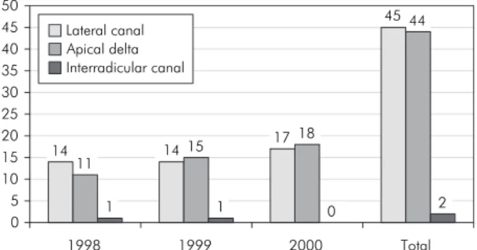
ca-nals were found in 1998 (0.32%, n = 1) and in 1999 (0.17%, n = 1) (Graph 1).
The maxillary premolars showed the highest rate of lateral canals (0.88%, n = 13); followed by man-dibular premolars (0.68%, n = 10) and maxillary
incisors (0.68%, n = 10). Apical deltas were mostly found in mandibular molars (0.95%, n = 14), fol-lowed by maxillary incisors (n = 9). Only the man-dibular molar group presented interradicular canals (Graph 2).
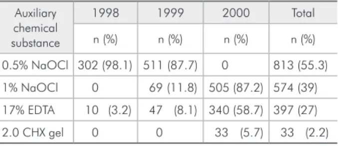
During 1998, the mostly used auxiliary chemical substance was 0.5% NaOCl. In 1999, some of the ca-nals were irrigated with 1% NaOCl and, in some of the cases, 17% EDTA was used as a inal auxiliary chemical substance. In 2000, some cases were irrigat-ed with 2% chlorhexidine gel. The use of EDTA (after using the auxiliary chemical substance) increased in 2000, as shown in Table 2, as well as the detection of root canal ramiications in the same period (Graph 1). However, no statistically signiicant relationship was found between the type of auxiliary chemical sub-stance used and the number of root canal ramiica-tions detected after root canal illing (p > 0.05).
Table 1 - Total number of tooth types treated in each year.
1998 1999 2000 Total
n (%) n (%) n (%) n (%)
Maxillary incisor 69 (22.4) 136 (23.3) 132 (22.7) 337 (22.9)
Maxillary canine 30 (9.7) 36 (6.1) 32 (5.5) 98 (6.6)
Maxillary premolar 52 (16.8) 93 (15.9) 93 (16.0) 238 (16.1)
Maxillary molar 46 (14.9) 66 (11.3) 70 (12.0) 182 (12.4)
Mandibular incisor 6 (1.9) 26 (4.4) 18 (3.1) 50 (3.4)
Mandibular canine 3 (0.9) 23 (3.9) 22 (3.7) 48 (3.2)
Mandibular premolar 28 (9.0) 64 (10.9) 79 (13.6) 171 (11.6)
Mandibular molar 74 (24.0) 139 (23.8) 133 (22.9) 346 (23.5)
Total 308 (21.0) 583 (39.6) 579 (39.4) 1470 (100.0)
Graph 1 - Total number of ramification types in each pe-riod.
14 14 17
45
11
1 1
15 18
44
0 2
0 5 10 15 20 25 30 35 40 45 50
Lateral canal Apical delta Interradicular canal
1999 2000 Total
1998
Graph 2 - Total number of ramifications related with tooth
type. Lateral canalApical delta
Interradicular canal 10
3
13
10
3
6 9
6
5
6
14
2
0 2 4 6 8 10 12 14 16
A B C D E F G H
2 2
0 0 0 0 0 0 0 0 0 0
A - Maxillary incisors B - Mandibular incisors
C - Maxillary canines D - Mandibular canines
E - Maxillary premolars F - Mandibular premolars
Discussion
Conventional radiography can render only a two-dimensional image, and therefore does not allow a three-dimensional study of the root canal system and its ramiications. However, the X-ray is the only clinical method to investigate root canal ramiica-tions. Unfortunately, most of the time we can detect the existence of root canal ramiications only after illing, or when there is a previous image of a perira-dicular radiolucency, which can suggest a communi-cation between the main canal and the periodontal ligament. Due to the radiographic superimposition, the small dimension of these canals and the limita-tions of radiographs, in most cases, these ramiica-tions cannot be observed.7
Filling of the teeth was done by lateral conden-sation with gutta-percha, using Endométhasone as sealer in all cases. The number of ramiica-tions clinically found after obturation (inal X-ray) (6.19%, n = 91/1,470) was lower than that found in an in vitro study of root canal systems (27.45%, n = 313/1,140).6 It should be noted that in vitro
studies have evaluated the three-dimensional pic-ture of cleared teeth, whereas in vivo studies employ a radiographic technique.
Vertucci’s classiication considers as a lateral ca-nal all caca-nals that connect the main caca-nal with the periodontal ligament, independently of root localiza-tion.20 In the present study, the following
ramiica-tion types were found: lateral canal, apical delta and interradicular canal. Premolars were the tooth type which showed the highest number of lateral canals, and molars, the highest number of apical deltas. This proportion agrees with that of a previous study.6
NaOCl followed by 17% EDTA was used to
re-move the smear layer. When it is present, the smear layer interferes with the diffusion of antimicrobial agents used during the chemo-mechanical prepara-tion.1,4,21 Moreover, it also interferes with the sealer
penetration into the dentinal tubules, consequently interfering with the bonding.22,23 Smear layer
degra-dation by proteolytic bacteria enzymes leads to the formation of hollow spaces between the root canal illing material and the root canal wall, which might allow microleakage.12,16
In the present work, the detection of root ca-nal ramiications increased from 1998 to 2000, as did the use of a substance for smear layer removal (EDTA) during the same period. Since the introduc-tion of 1% NaOCl (1999) and 2% chlorhexidine gel (2000) as auxiliary chemical substances in un-dergraduation clinics, the use of EDTA has strongly been emphasized. However, no statistically signii-cant association was found between type of auxil-iary chemical substance and frequency of root canal ramiication. It is important to note that the number of canals irrigated with EDTA could be greater than that stated in the clinical records, since students are orientated to use it before intracanal medication and root canal illing. Unfortunately, many of the students’ treatment records (n = 50) did not state the auxiliary chemical substance used. For this rea-son, the total number of cases presented in Table 2 (n = 1,420), without counting the number of treat-ments with EDTA (as it was not used alone), was not similar to that presented in Table 1 (n = 1,470).
Further clinical trials involving not only dental schools but also specialist clinics are necessary in order to obtain stronger evidence of the number of ramiications illed after root canal treatment. In such studies, other factors such as illing technique should also be evaluated for their effectiveness in illing the root canal ramiications.
Conclusions
It was concluded that the frequency of root canal ramiications radiographically found was not high in treatments performed by undergraduate students, although the clinically found number will always be expected to be smaller than that found in in vitro
studies.
Table 2 - Auxiliary chemical substance distribution related with treatment number in each studied year.
Auxiliary chemical substance
1998 1999 2000 Total
n (%) n (%) n (%) n (%)
0.5% NaOCl 302 (98.1) 511 (87.7) 0 813 (55.3)
1% NaOCl 0 69 (11.8) 505 (87.2) 574 (39)
17% EDTA 10 (3.2) 47 (8.1) 340 (58.7) 397 (27)
Acknowledgements
The authors would like to express their sin-cere gratitude to Professor Harald Eriksen for valuable advice. We would also like to thank Mr.
Adailton dos Santos Lima for technical support. This work was supported by the Brazilian agen-cies FAPESP (04/05743-2; 04/07057-9), and CNPq (304282/2003-0 & 140113/03-7).
References
1. Baumgartner JC, Mader CL. A scanning electron microscopic evaluation of four root canal irrigation regimens. J Endod. 1987;13(4):147-57.
2. Behrend GD, Cutler CW, Gutmann JL. An in vitro study of smear-layer removal and microbial leakage along root canal fillings. Int Endod J. 1996;29(2):99-107.
3. Bender IB, Seltzer S. The effect of periodontal disease on the pulp. Oral Surg Oral Med Oral Pathol. 1972;33(3):458-77. 4. Byström A, Sundqvist G. The antibacterial action of sodium
hypochlorite and EDTA in 60 cases of endodontic therapy. Int Endod J. 1985;18(1):35-40.
5. Çaliskan MK, Pehlivan Y, Sepetcioglu F, Turkun M, Tuncer SS. Root canal morphology of human teeth in a Turkish popu-lation. J Endod. 1995;21(4):200-4.
6. De Deus QD. Frequency, location, and direction of lateral secondary and accessory canals. J Endod. 1975;1(11):361-6. 7. Gomes BPFA, Rodrigues HH, Tancredo N. The use of a mod-elling technique to investigate the root canal morphology of mandibular incisors. Int Endod J. 1996;29(1):29-36. 8. Gutmann JL. Prevalence, location, and patency of accessory
canals in the furcation region of permanent molars. J Peri-odontol. 1978;49(1):21-6.
9. Harrison JW. Irrigation of the root canal system. Dent Clin North Am. 1984;28(4):797-808.
10. Karagoz-Kucukay I. Root canal ramifications in mandibular incisors and efficacy of low-temperature injection thermoplas-ticized gutta-percha filling. J Endod. 1994;20(5):236-40. 11. Lopes HP, Siqueira JF Jr, Elias CN. Scanning electron
micro-scopic investigation of the surface of gutta-percha cones after cutting. J Endod. 2000;26(7):418-20.
12. Meryon SD, Brook AM. Penetration of dentin by three oral bacteria in vitro and their associated cytotoxicity. Int Endod J. 1990;23(4):196-202.
13. Peikoff MD, Perry JB, Chapnick LA. Endodontic failure at-tributable to a complex radicular lingual groove. J Endod. 1985;11(12):573-7.
14. Ross IF. The relation between periodontal and pulpal disor-ders. J Am Dent Assoc. 1972;84(1):134-9.
15. Rubach WC, Mitchell DF. Periodontal disease, accessory ca-nals and pulp pathosis. J Periodontol. 1965;36:34-8. 16. Sen BH, Wesselink PR, Türkün M. The smear layer: a
phenom-enon in root canal therapy. Int Endod J. 1996;28(3):141-8. 17. Siqueira Junior JF. Aetiology of root canal treatment failure:
why well-treated teeth can fail. Int Endod J. 2001;34(1):1-10.
18. Taylor JK, Jeansonne BG, Lemon RR. Coronal leakage: effects of smear layer, obturation technique and sealer. J Endod. 1997;23(8):508-12.
19. Valdrighi L, Biral RR, Pupo J, Souza Filho FJ. Técnicas de ins-trumentação que incluem instrumentos rotatórios no preparo biomecânico dos canais radiculares. In: Leonardo MR, Leal JM, editors. Endodontia – Tratamento de canais radiculares. São Paulo: Panamericana; 1991 p. 290-9.
20. Vertucci FJ. Root canal anatomy of the human permanent teeth. Oral Surg Oral Med Oral Pathol. 1984;58(5):589-99. 21. Vivacqua-Gomes N, Gomes BPFA, Zaia AA, Teixeira FB,
Souza-Filho FJ. Influence of irrigants on the coronal micro-leakage of laterally condensed gutta-percha root fillings. Int Endod J. 2002;35(9):791-5.
22. White RR, Goldman M, Lin PS. The influence of the smeared layer upon dentinal tubule penetration by endodontic filling materials. Part II. J Endod. 1987;13(8):369-74.
23. White RR, Goldman M, Lin PS. The influence of the smeared layer upon dentinal tubule penetration by plastic filling ma-terials. J Endod. 1984;10(12):558-62.